KaiWei Zhao, Ameersing Luximon, Ganesan Balasankar, CheeKooi Chan (2020). A New Representational Method of Human Foot Anatomical Landmark and its Application in Foot Posture Data Acquisition. In: Advances in Applied Digital Human Modeling. AHFE Open Access, vol. 11. https://doi.org/10.54941/ahfe100414
Functions like protection and comfort are essential requirements of the footwear or orthosis. Naturally, the status of foot anatomy features, judged from both inside and skin surface, is of great significance to the effectiveness of the product. In this study, a novel method to estimate the foot anatomy structural deformation from the skin surface in three typical postures is proposed, which can effectively reveal the inner anatomy status without using CT or MRI. Technology of scanning with range sensor is adopted, and it considerably promotes the efficiency of acquisition of the foot texture model. Reverse engineering tools are used to precisely catch the anatomical landmark locations, and the relevant data is shown in the way of comparison.
Three-dimensional localization of anatomical landmarks of the foot is of great significance in medical use. As (Liu, 2004) related, the malleoli, which is a key landmark in lower extremity examination, plays an important role in leg measurement and defining the axis of the upper ankle joint. As indicated in this paper, in terms of kinetic analysis of the lower limb both in static and dynamic status, the exact position of the ankle joint is also of particular importance in motion capture and key dimension determination. However, there are limitations in traditional methods of anatomical landmark marking. Traditionally, optical markers are attached to the skin close to the malleoli, while this method is not so convincing and doubted of its reliability due to skin movements (Cappozzo, 1994). A different approach to this problem is the idea that an anatomical landmark can be defined by particular shape of the skin. It is widely accepted that the underlying anatomical structure is independent of the skin movements (Besl, 1988). Once a constant representation is figured out for the structural shape, the landmark can be determined. Besides, invariant also means that it is neither dependent on the actual coordinate system nor surface parameterization. Curvature data (key of this method) is chosen as constant shape description (Frobin, 1982). A parameterized representation of the foot surface needs to be produced ahead and coordinates, derivatives and curvature maps are available (Barros RML, 2002). Hence the landmarks are characterized and discriminated. Koenderink shape index (Koenderink, 1992), which is a key tool to separate the convexity from the concavity and shape’s color changes according to its deepness and the surface types, is integrated in this method.
(Drerup, 1985) presented a method for the localization of anatomical landmarks on the human body surface. Surface shape with systematic curvature description is analysed in their method. Basic surface coordinates (geometrical mesh info) are acquired through optical methods such as moiré topography or rasterstereography, which provides the conditions for curvature calculation. Reverse engineering technology is highly needed in pre-processing for geometry feature generation and thus characteristic parameters such as curvature and derivative distribution will be calculated. Localization of vertebra prominens is presented as an example in their works and the results are compared with that of conventional method that the anatomical landmarks are manually palpated and marked, which reveals that the former has higher reliability and accuracy.
As illustrated, two lines are defined by the landmarks: the malleoli, the fibular head, the point anterior to lateral tibial condyle and the medial tibial condyle. These two lines form an angle that is generally regarded as measurement of tibial torsion, the mean of which is 20.58°. This is a typical example of measuring the inner structure by surface anatomy characteristics, which has a promising and broad application prospect.
(Subburaj, 2008) described a computer-aided method used for extracting anatomical landmarks from a three- dimensional digital model generated from multiple CT images. In the experiment, an accurate three-dimensional reconstructed pelvis model was adopted for landmark location. Similar as the method in the literature mentioned above, landmark identification through curvature analysis were used instead of conventional manual palpation way after three-dimensional geometrical feature characterization hence regions like peaks, ridges, pits and ravines on the surface are identified and classified automatically. The result can be applied on significant dimension measurement, pre-operative planning that are essential for the surgeries requiring anatomical landmarks on skin or skeletal tissue, like resecting deceased tissue and positioning custom implants or mega endoprostheses.
Not only is how to identify the anatomical landmarks important but also what to mark must be studied. (Agić, 2006) studied this subject and suggested some geometric descriptors. In terms of the foot functionality, the bone inertial tensor descriptor plays a significant role. Eigenvalue of the inertial tensor can describe the bone shape, thus contributing to the definition of a coordinate system in bone centroid. The medial longitudinal arch also plays an important role among the structural characteristic descriptors in foot shape and kinematics analysis (Razeghi, 2002). Besides, the principal component analysis was applied by to the foot structure and three principal components that respectively reflecting the characteristics of size, shape and comfort were concluded.
The methods mentioned above have a common serious defect: low efficiency. To calculate the curvature map and then get the convex and concave regions through landmark detecting algorithm is considerably time-consuming and equipment-demanding. With the development of range sensing technology, a novel three dimension scanning tool, Microsoft Kinect, which is characterized by low cost and high efficiency of data acquisition, has come out. It can finish scanning an object together with its color and texture within 1 minute. Basing on Kinect and its supporting suites, a new method of accurately locating the human foot landmarks is presented in this paper, together with its preliminary application in foot posture data acquisition.
METHOD FOR LOCATING AND DIGITALIZING THE FOOT ANATOMICAL LANDMARKS Determination of Landmarks for Representation
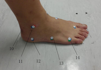
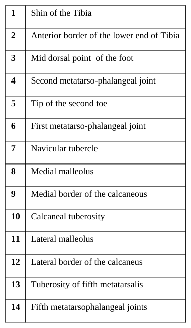
The first metatarsal bone is most apt to get fractured among the multiple anatomical structures within human foot, whichever posture he or she is in, e.g. standing under normal pressure, raising the heel to a medium or a higher height and even jumping. The second metatarsal bone follows the first one in this respect. The calcaneous part always bears large quantity of compressive stress and hence is quite inclined to get fatigue fracture. As is known to all, subtalar joint and true ankle joint play an important role in foot and ankle statics, kinetics and kinematics. Hence, the locations of the key functional landmarks, such as the most prominent points on medial and lateral malleolus, can help effectively estimate and evaluate function status of foot anatomy. Accordingly, the locations in terms of the foot surface anatomy were chosen as shown in Figure 1 and Table 1.
Figure 1. Landmarks (white stickers) for the experiment (for better visual effect, zero-heel posture is taken as the example for landmark show).Table 1: Landmarks on foot for test
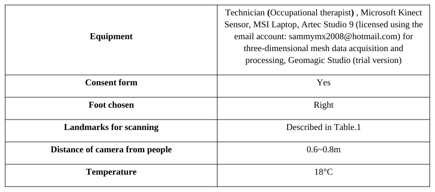
Equipment
Table 2: Experiment equipment Technician (Occupational therapist) , Microsoft Kinect Sensor, MSI Laptop, Artec Studio 9 (licensed using the
Except for Microsoft Kinect, MSI laptop with a CPU of Intel® Core ™ i7-4700MQ and a graphic card of Nvidia GeForce GTX 765M / GDDR5 2GB, together with a RAM of 16GB DDR3L, was adopted. The performance of the computer can have considerable impact on the efficiency and results. Artec Studio 9 from Artec Ltd. was used in the experiment due to its excellent stability, high efficiency and accurate and powerful function for texture mapping, vertex color assignment and mesh data processing. Geomagic Studio 11 (trial version) was chosen for landmark location measurement and preliminary analysis.
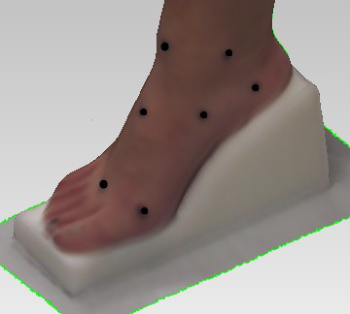
Procedure A 25 years old women subject was selected for this study. The basic demographic data was collected including height (156cm) and weight (49 kg). The original anatomical landmarks were identified through touching detection by an experienced occupational therapist, and then marked out with black stickers (with a diameter of 5mm). First of all, three postures were tested: Normal standing, medium heel and high heel position. The marked foot (right foot) of the subject was scanned by Microsoft Kinect, and the textured models with landmarks are output in the format of obj. Secondly, the exported mesh models were imported into Geomagic Studio (trial version), and the landmarks were enhanced. Finally, the key locations and dimensions represented by the markers were measured interactively. Therefore, the correlations between the anatomical structures of the natural standing status and the other two typical postures were measured and identified. Figure 1 shows the experiment procedure.
Marked foot scanning and
Original landmarks determination
the textured models output
Mesh model processing including landmark enhancing
Key dimensions and locations
Exploration of correlations between the natural standing status
measurement
and the other two postures
Figure 2. Experiment procedure
Figure available in the original PDF paper.
RESULTS
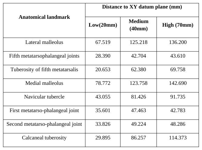
The exported mesh model with texture and color (See Figure 3) were processed through the steps of noise and redundant data removal, smoothing and marker enhancement in Geomagic Studio 11. Meshlab 3.2(64-bit) was used for final check and three-dimensional demonstration. The normal standing, medium heel and high heel postures are the three main categories for measurement. The XY datum plane is determined by 3-point method through randomly picking 3 points far apart to each other and distributed uniformly on the ground mesh data.
Afterwards, the distances from the XY datum plane to each landmark and the linear distances between some of the landmarks in the three postures are measured in Geomagic Studio 11, too. The result can be seen in Table 3.
(a)
(b)
(c)
Figure 3. Three-dimensional mesh model, a: low heel (20mm); b: medium heel (40mm); c: high heel (70mm)Table 3. Distances between the landmarks and the XY datum plane (8 key landmarks of the 14 are chosen) Distance to XY datum plane (mm)
CONCLUSIONS
Through measurements on the textured foot mesh model, we can draw the posture features through key landmarks’ location. Since it is widely accepted that the anatomical landmarks can reflect the inner structure status during different postures, they can be regarded as descriptions of foot postures in terms of their distinguishing status of different anatomical parts, such as tensioning and compressing. What the new representing method presented in this paper contributes to foot anatomy status analysis most is its qualitative conclusion from the quantitative measurements. For instance, through the measured distances of head of 1st metatarsal bone and the medial cuneiform in Table 3, the angles in terms of the 1st metatarsal bone (regarded as a line segment) between its geometrical status from the normal standing to the medium posture, and from medium to high heel posture, can be calculated. Although due to skin movements and touching errors caused by manual operation by the occupational therapist, the angles may be effectively used for analyzing the stress status of the 1st metatarsal bone and even the whole foot arch.
ACKNOWLEDGEMENTS
This study was supported by RGC General Research Fund (B-Q26V) and RGC General Research Fund (B-Q31C).
REFERENCES
Agić, A. (2006). "Foot structure descriptors". Ergonomia IJE&HF, 28(3), 237–248. Barros RML, F. P., Paterniani PES, Brenzikofer R, Filho ECL. (2002). "Three-dimensional reconstruction and analysis of human back surface". Paper presented at the Seventh International Symposium on the 3-D Analysis of Human Movement, Newcastle upon Tyne, England. Besl, P. J. (1988). "Surfaces in range image understanding" (Vol. 27): Springer-Verlag New York. Cappozzo, A., Catani, F., Leardini, A. (1994). "Skin movement artefacts in human movement photogrammetry". Journal of
Biomechanics, 27(6), 755. Drerup, B., Hierholzer, E. (1985). "Objective determination of anatomical landmarks on the body surface: measurement of the vertebra prominens from surface curvature". J Biomech, 18(6), 467-474. Frobin, W., Hierholzer, E. (1982). "Analysis of Human Back Shape Using Surface Curvatures". Journal of Biomechanics, 15(5),
379-390. doi: Doi 10.1016/0021-9290(82)90059-8 Koenderink, J. J., van Doorn, A. J. (1992). "Surface shape and curvature scales". Image and vision computing, 10(8), 557-564. Liu, X., Kim, W., Drerup, B. (2004). "3D characterization and localization of anatomical landmarks of the foot by FastSCAN".
Real-Time Imaging, 10(4), 217-228. doi: DOI 10.1016/j.rti.2004.05.009 Razeghi, M., Batt, M. E. (2002). "Foot type classification: a critical review of current methods". Gait Posture, 15(3), 282-291. Subburaj, K., Ravi, B., Agarwal, M. (2008). "3D shape reasoning for identifying anatomical landmarks". Computer-Aided Design and Applications, 5(1-4), 153-160.
LISTED IN THE AHFE DIGITAL BOOKSTORE CATALOGUE
Explore the complete digital volume
Advances in Applied Digital Human Modeling
Read this individual research paper free through the original publisher, or explore the full digital proceedings book.
ISBN 978-1-4951-2094-7
Explore this ISBN in the AHFE Bookstore ↗Listed in the July 2026 bookstore catalogue; search by ISBN to confirm current availability · Digital book purchase optional