José Antonio Diez - De Pablos, Luis Martí Bonmati, Nicolás Palomares, Ignacio Espíritu García-molina, Fernando Mollá-doménech, Carlos Atienza, Orlando Carles Diaz, Adrian Morales Casas, Jose Laparra (2024). Humanizing X-Ray Services for Children with Cerebral Palsy: A Holistic Approach to Functionality, Usability and Aesthetics. In: Health Informatics and Biomedical Engineering Applications. AHFE Open Access, vol. 142, pp. 40–50. https://doi.org/10.54941/ahfe1005069
The adequate acquisition of X-ray images is crucial for effectively monitoring and treating patients with significant spinal deformities, particularly those with mobility limitations, mainly children. Patients with these considerations include individuals with cerebral palsy, who face additional challenges in doctor-patient interactions due to speech and cognitive restrictions. Moreover, patients with spasticity resulting from paralysis may exhibit uncontrollable limb movements.In the absence of suitable devices forcing patients to maintain a stable seated position during imaging, they often adopt inadequate postures, risking misdiagnosis and unnecessary radiation exposure if exam repetition is needed. To address this issue, an X-ray Sitting Support device has been designed to accommodate patients with these pathologies and ensure high-quality radiographic images while prioritizing patient safety and comfort.The development of the X-Ray Sitting Support device was based on a Human Factors plan and User Experience methodologies, with an iterative process focusing on physical ergonomics, usability, and patient acceptance. Feedback from patients, medical personnel, and caregivers was integrated throughout the design process, from defining requirements to real-world prototype validation. This comprehensive approach ensured that the imaging sitting support met the needs of both patients and medical professionals, enhancing the effectiveness and safety of radiological examinations for individuals unable to stand.
Several children’s conditions are related to spine deformities. One of them is related to infantile cerebral palsy (ICP), involving a permanent disorder
Humanizing X-Ray Services for Children With Cerebral Palsy 41
resulting from a brain injury that affects mobility and posture, significantly limiting their activity (Sadowska et al., 2020). This disability is often accom- panied by sensory and intellectual impairments and spasticity (Rosenbaum et al., 2007).
Additionally, spinal deformities are present in children and evolve with growth (Tsirikos, 2010). For this reason, monitoring spinal deformities through radiographic examinations is crucial in children with spinal defor- mities during their development to define accurate diagnosis and appropriate treatment, considering that it affects more than 125.000 persons in Europe (Sellier et al., 2010).
Routine full spine plain film radiography is typically performed with the patient standing, as this position allows for a more accurate assessment of spinal alignment and curvature under the influence of gravity and stability. When the patient is standing, the spine is loaded with body weight, which can reveal deformities or deviations that might be hidden in other positions, such as lying down or sitting. However, radiographic images are usually taken in a seated position due to the characteristics of children with these spinal deformities.
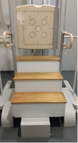
At the same time, there is a lack of suitable devices for patients to main- tain a stable seated posture while obtaining high-quality radiographic images. Consequently, patients often assume inadequate postures, leading to poten- tial misdiagnosis and unnecessary radiation exposure if the acquisition needs to be repeated. Without appropriate equipment, hospitals apply solutions as makeshift aids for patients to sit on during radiographic examinations (Figure 1).
Figure 1: Basic wood support to maintain ICP posture with the help of parents and clinicians.
The consequence of this approach is the low quality of the obtained radio- graphic images. The patient may slip during the test due to a lack of restraints and several other situations that make the examination unpleasant for the patient or their caregivers, leading to the inability to perform the test. Further- more, since this system does not provide patient safety or allow for correct positioning on its own (given its unique characteristics and difficulties in maintaining the proper posture), healthcare personnel and caregivers have to remain in the examination room holding the patient, being also exposed to radiation while the radiographic image is taken.
In response to these constraints, we designed, developed, and validated an imaging sitting support device that allowed entire spine radiographic exams in children with extensive spinal deformities while ensuring high image quality. To achieve this goal, a human factors plan has been developed to guide the product design, ensuring compliance with regulatory requirements, radiographic image quality, functional requirements, ergonomics, comfort, and usability while minimising any potential risks associated with using this product.
HUMAN FACTORS PLAN FOR HUMANIZING X-RAY IMAGING A Human Factors (HF) Plan can described as a systematic plan for developing a product combining HF knowledge and methodologies from User Experi- ence (UX) to guarantee the incorporation of diverse user perspectives. This comprehensive approach not only enhances the usability and functionality of the product but also fosters inclusivity and responsiveness to the needs of all involved parties. Furthermore, incorporating Human Factors through the asset life cycle, particularly early in the design process, provides the most benefits: avoidance of errors due to poor design often requires our human component to function infallibly to avoid problems and incidents, as well as to mitigate and recover from escalating situations (Stanton et al., 2017).
In this specific project, a Human Factors Plan was implemented following the three main phases defined by Morales et al. (2023) and considering the experience of actively involving patients in the co-creative design (Belda Lois et al., 2014; López-Vicente et al., 2016):
Learn: Understanding the intended users’ needs, preferences, and behaviours.
Ideate: generating potential solutions to address the identified user needs and challenges. This stage encourages creative thinking and collaboration among multidisciplinary teams.
Validate testing and evaluating the proposed solutions to ensure they meet user requirements and goals.
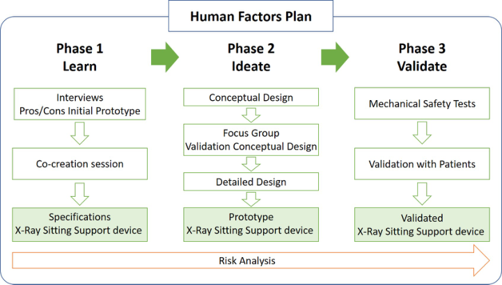
This framework facilitates an iterative process instead of solely depending on single final validations, a practice often observed in many medical product developments. Embracing this approach effectively reduces the time and costs associated with the development process. Figure 2 delineates the phases of the Human Factors Plan implemented and outlines the activities for the study’s development.
Humanizing X-Ray Services for Children With Cerebral Palsy 43
The development of these activities has been undertaken by Eiffage, leveraging their knowledge and experience in electromedical equipment for Diagnostic Imaging, supported by IBV with expertise in Human Factors. This achievement has been made possible through the active involvement of healthcare professionals from the Radiology Service of the Hospital Univer- sitario y Politécnico La Fe (La Fe Hospital) in Valencia (Spain), who possess extensive experience in X-ray procedures for this population. Additionally, input and collaboration have been sought from the collective of patients and professionals related to ICP caregivers in Valencia (Asociación Valenciana de Ayuda a la Parálisis Cerebral - AVAPACE).
Figure 2: Phases of the HF plan and activities for the development of the imaging sitting support.
PHASE 1 - LEARN HOW TO UNDERSTAND THE PROBLEM, PROVIDE CLEAR REQUIREMENTS, AND IDENTIFY KEY RISKS The initial phase’s objective consisted of defining the design specifications for the imaging sitting support and establishing necessary criteria regarding safety, usability, ergonomics, adjustments, patient immobilisation solutions, and acceptance.
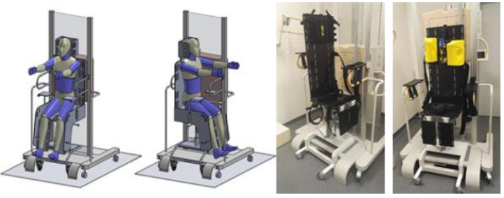
The definition of specifications has been carried out through two activi- ties. Firstly, a focus group assessed a prototype of an imaging sitting support developed in a previous project (Figure 3). This involved a meeting with healthcare professionals and the design team to review previous results and gather the latest feedback from clinicians, patients, and caregivers. The fol- lowing improvements were emphasised: armrest support, backrest rigidity, tilting support, the inclusion of strap adjustments, head and leg immobilisa- tion to prevent spastic movements, better coating materials, and enhanced aesthetics.
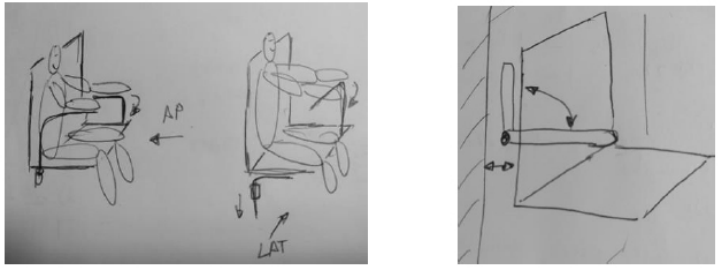
The second activity involved a co-creation session to prioritise the require- ments and specifications identified so far and propose solutions and their features using brain drawing techniques (Figure 4). This session brought together the design team imaging clinicians and ICP caregivers, fostering collaborative participation.
Figure 3: CAD file and prototype of imaging sitting support.
The outcomes of both activities enabled the definition of the require- ments for the new solution: imaging sitting support should allow lateral and anteroposterior support of the patient during the radiological examination. It should enable frontal and lateral radiographic images of patients without requiring a change in their position.
The support should ensure proper patient restraint (trunk, pelvis, head), bearing the patient’s weight and guaranteeing stability during transportation with the patient seated. Additionally, support area materials in the radio- graphic image field should be radiotransparent to avoid interference and observation errors in the X-ray.
Figure 4: Sketches from the cocreation session.
The initial assessment identified potential product risks, and a preliminary risk management plan was developed to mitigate them. This risk management
Humanizing X-Ray Services for Children With Cerebral Palsy 45
plan underwent continuous iteration, with newly unforeseen risks being incorporated as they were detected in later stages of the project, such as during clinical validation. Risk management planning involves identifying generic and product-specific types of hazards. Risk analysis was conducted referencing Regulation (EU) 2017/745 (2017).
Firstly, known or foreseeable generic hazards related to the product were identified, such as biological hazards, environmental hazards, and inappropriate user interface. Secondly, specific product risks were deter- mined after addressing generic risks, considering the general safety and performance requirements and based on product knowledge. Their signif- icance was deduced through analysis following the EN ISO 14971 (2020) standard.
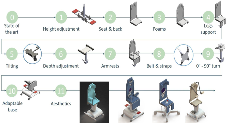
PHASE 2 - IDEATE: FROM SPECIFICATIONS AND CONCEPTUAL DESIGNS TO DIFFERENT PROTOTYPES Based on the established specifications, the goal of this stage was the design and development of the imaging sitting support device. Initially, a concep- tual design was created to meet all the designed specifications, including ergonomic requirements, comfort, and usability. During the conceptual design phase, various solutions were proposed for each comprised component and mechanism (Figure 5).
Figure 5: An iterative process to assess alternatives before developing the final design.
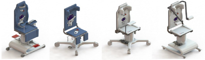
The definition of the conceptual design was achieved through an iterative process with continuous engagement of designers and developers of Eiffage (incorporating technical insights of imaging systems and focusing on ensuring market success and effective deployment), with imaging clinicians and ICP caregivers (Figure 6).
Figure 6: Design alternatives (blue and white) for the X-ray sitting support of Eiffage.
Simultaneously, a search was conducted for commercial elements that could be used for the support accessories (straps, head stabilisers). Further- more, concept testing was performed to enhance functionalities, such as padding options, headrests, and aesthetic improvements. Additionally, an in- depth analysis was conducted to define the material for the backrest, which needed to be radiotransparent to ensure optimal quality of the radiological image of the spine.
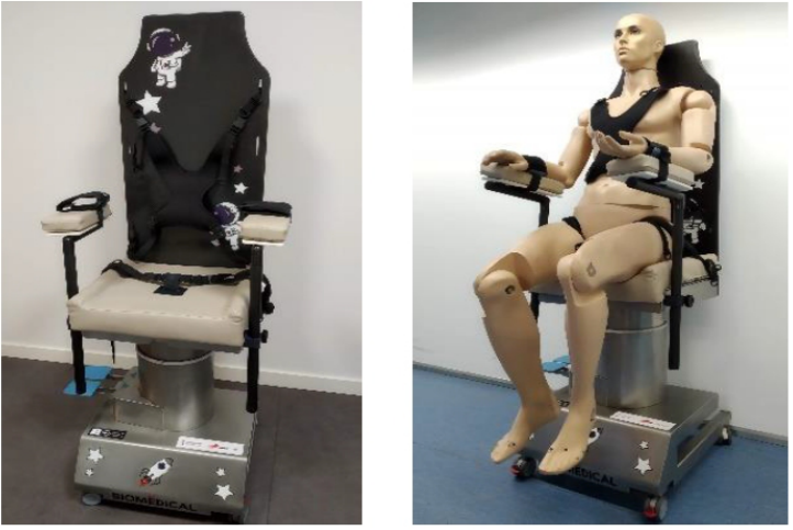
Following the conceptual design, a detailed proposal for the imaging sitting support system was made, considering all the functionalities and adjustments. Once the detailed design was defined, manufacturing a prototype of a sitting support device was initiated. The prototype was developed to be fully func- tional with materials and finishes that ensure patient safety in all age ranges (Figure 7).
Figure 7: Functional prototype of the imaging sitting support device of Eiffage.
Humanizing X-Ray Services for Children With Cerebral Palsy 47
PHASE 3 - VALIDATE: FROM ITERATIVE TEST WITH REDUCED USER SAMPLE TO PRE-CLINICAL VALIDATION The validation of the prototype included the development of mechanical tests to guarantee the safety of patients and its pre-clinical validation in a natural environment with patients. Firstly, mechanical tests were performed to verify basic issues such as stability, overturn in different conditions, loads, entrap- ment and edge risks based on EN 1022 (2019) and EN ISO 21856 (2023) standards. The prototype successfully passed all tests.
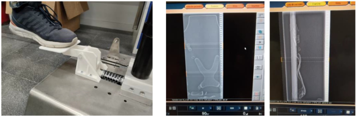
Concurrently, iterative usability tests were conducted to ensure readi- ness for validation under real-world conditions, serving as a preliminary check before clinical validation and market deployment. These tests identified areas for improvement that could be implemented before clinical validation. Indeed, rapid prototypes were developed to evaluate various aspects, ranging from the mechanisms’ functionality to the straps’ opacity (Figure 8), enabling efficient product refinement before proceeding to the next activity.
The final activity involved conducting a study in natural clinical settings to validate the imaging sitting support with volunteer patients, aiming to ver- ify compliance with specifications established in earlier project phases and validate the prototype. The Medical Imaging Clinical Department at Hospi- tal La Fe conducted the clinical study. The clinical study was approved by the Ethical Committee of the Hospital and involved five underage patients with cerebral palsy and significant spinal deformities (scoliosis, neuromuscu- lar scoliosis, ataxia with scoliosis) who were unable to maintain a standing posture. Gender balance was ensured, and four clinicians participated in the validation. Legal representatives provided authorisation for the participa- tion of these volunteer patients, and corresponding informed consent was obtained.
Figure 8: Left: rapid prototype for height adjustment. Right: opacity of the straps.
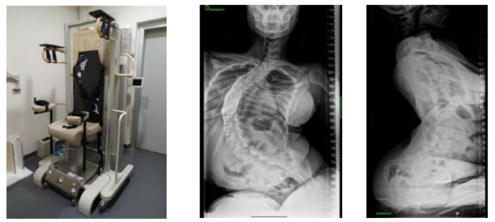
After evaluating the obtained radiographs and patient-related variables, the results demonstrated that the prototype design meets the functional and usability requirements for use with patients during clinical practice (Figure 9). The device’s radiotransparent material ensures satisfactory radi- ological image quality. The width and depth of the seat were adequate for providing the required patient support, contributing to the patient’s comfort
and well-being during the procedure. The available regulations were suit- able and facilitated patient positioning. The restraint systems were adequate for immobilising the patient. The dimensions and rules of the armrests were appropriate. Additionally, the aesthetic design of the prototype, incorporat- ing child-friendly stickers, was well-received and contributed to a visually pleasing appearance. During clinical validation, some areas for improvement were also identified that could optimise the prototype (such as overall weight reduction and incorporating an intermediate hole in the backrest for the thoracic harness).
Figure 9: Clinical validation in natural conditions, including X-ray results.
CONCLUSION
Several key conclusions emerge regarding the imaging sitting support device’s impact on healthcare services and patient well-being.
Firstly, sitting support significantly enhances the quality of healthcare ser- vices during radiology tests by improving the overall experience for patients and their families. This quality improvement extends beyond the clinical setting, positively affecting the quality of life for patients undergoing these procedures and their relatives.
Secondly, the methodology employed in developing and deploying the X-Ray sitting support device has reduced both the time and costs associ- ated with market deployment. These efficiencies are especially advantageous for individuals with extensive spinal deformities and cerebral palsy, as they streamline the process of accessing essential healthcare technologies and services.
Moreover, the results of clinical validation underscore the effectiveness of the imaging sitting support device in meeting the necessary criteria for func- tionality, usability, and user acceptance. These positive outcomes validate the sitting support’s effectiveness and mitigate the need for further significant improvements, ensuring its readiness for widespread use.
Looking ahead, the primary focus shifts towards enhancing market uptake, a task that presents challenges due to the optimization of industrialization
Humanizing X-Ray Services for Children With Cerebral Palsy 49
and the relatively limited market size. Nevertheless, spinal deformities and related situations, such as Infantile Cerebral Palsy, Amyotrophic Lateral Scle- rosis and Muscular Dystrophy of Duchene, represent a large number of patients in Europe who will benefit from this solution.
Despite these challenges, increasing market penetration is crucial to advancing the humanisation of healthcare and preventing the exclusion of populations with disabilities from accessing vital X-ray tests. This under- scores the importance of strategic initiatives to promote awareness, acces- sibility, and affordability of imaging sitting support, ensuring its widespread adoption and maximising its impact on improving healthcare outcomes.
ACKNOWLEDGMENT
The authors would like to acknowledge the Agencia Valenciana de Inno- vación (AVI), which has partially financed these results with the project INNCAD/2021/75.
REFERENCES
Belda Lois, J. M., López-Vicente, A., Laparra Hernández, J., Poveda, R., Ferreras, A.,
2014. User Participation in the Design of an Alternative Communication Sys- tem for Children with Diskinetic Cerebral Palsy Including Emotion Management. pp. 260–263. https://doi.org/10.1007/978-3-319-08596-8_41 Casas, A. M., Vicente, A. L., Solano-García, L., Laparra, J., 2023. 3 sprints from zero to innovative medical device in 16 months: Benefits of combining Human Factors and Agile, in: Production Management and Process Control. Presented at the AHFE (2023) International Conference, AHFE Open Acces. https://doi.org/ 10.54941/ahfe1003047 EN 1022:2019. Furniture - Seating - Determination of stability, 2019. EN ISO 14971:2020 Medical devices - Application of risk management to medical
devices, 2020. EN ISO 21856:2023. Assistive products - General requirements and test methods (ISO 21856:2022), 2023. López-Vicente, A., Artacho-Pérez, C., Jarque-Bou, N., Raya, R., Lloria, M.,
Belda-Lois, J.-M., Rocón, E., 2016. Adaptive inputs in an interface for people with Dyskinetic Cerebral Palsy: Learning and usability. Technology and Disability 28, 79–89. Regulation (EU) 2017/745 of the European Parliament and of the Council of 5 April 2017 on medical devices, amending Directive 2001/83/EC, Regulation (EC) No 178/2002 and Regulation (EC) No 1223/2009 and repealing Council Directives 90/385/EEC and 93/42/EEC (Text with EEA relevance.), 2017., OJ L. Rosenbaum, P., Paneth, N., Leviton, A., Goldstein, M., Bax, M., Damiano, D., Dan,
B., Jacobsson, B., 2007. A report: the definition and classification of cerebral palsy April 2006. Dev Med Child Neurol Suppl 109, 8–14. Sadowska, M., Sarecka-Hujar, B., Kopyta, I., 2020. Cerebral Palsy: Cur- rent Opinions on Definition, Epidemiology, Risk Factors, Classification and Treatment Options. Neuropsychiatr Dis Treat 16, 1505–1518. https://doi.org/10.2147/NDT. S235165 Sellier, E., Cruz, J. de la, Cans, C., 2010. Surveillance of Cerebral Palsy in Europe: the SCPE network. Bulletin Épidémiologique Hebdomadaire 191–194.
Stanton, N. A., Salmon, P. M., Rafferty, L. A., Walker, G. H., Baber, C., Jenkins, D. P.,
2017. Human factors methods: A practical guide for engineering and design. CRC Press. Tsirikos, A. I., 2010. Development and treatment of spinal deformity in patients with cerebral palsy. Indian J Orthop 44, 148–158. https://doi.org/10.4103/0019-5413. 62052
LISTED IN THE AHFE DIGITAL BOOKSTORE CATALOGUE
Explore the complete digital volume
Health Informatics and Biomedical Engineering Applications
Read this individual research paper free through the original publisher, or explore the full digital proceedings book.
ISBN 978-1-964867-18-2
Explore this ISBN in the AHFE Bookstore ↗Listed in the July 2026 bookstore catalogue; search by ISBN to confirm current availability · Digital book purchase optional