Akihiro Kato, Masami Hashimoto, Chiaki Oshiyama, Takuichi Nishimura (2023). System Development of Motor Imagery BCI : An Approach from The Human Side. In: The Human Side of Service Engineering. AHFE Open Access, vol. 108, pp. 216–223. https://doi.org/10.54941/ahfe1003123
Non-invasive brain-computer interface (BCI) uses mainly electroencephalography (EEG) to operate external devices. In particular, BCI is needed for people with severe physical disabilities, such as ALS, because they are unable to move their bodies other than their brains; BCI that uses Motor Imagery (MI) requires the identification of obvious ERPs associated with MI. In this study, a system focused on BCI-training and MI support was developed, and EEG was measured after system use. The system is characterized by the presentation of a simulated hand animation to support MI of left and right hand grasping. As a result, significant ERPs were confirmed in three subjects, however, it is possible that some subjects may not be able to observe ERPs if the number of subjects is increased. Therefore, the BCI-training needs to be carefully developed. In particular, it is necessary to consider teaching methods that facilitate subjects to gain control over their own mental states and internal sensations. However, there is no knowledge on how to teach BCI-training. Therefore, the author has conceived of a system that enables participants to simultaneously train themselves and acquire knowledge about BCI control in BCI-training.
Non-invasive brain-computer interface (BCI) uses mainly electroencephalo- graphy (EEG) to operate external devices (Lebedev and Nicolelis, 2006). BCI is a promising communication method for people with paralysis and physical disabilities. In particular, BCI is needed for people with severe physical disabi- lities, such as ALS, because they are unable to move their bodies except their brains. BCI using potentials associated with Motor Imagery (MI) has been actively studied because of its potential for active control. MI potentials are a type of event-related potentials (ERPs) that appear as EEG fluctuations in specific areas and frequency bands of the cortex due to internal and external events. Desynchronization (ERD) and Event-related Synchronization (ERS), which cause a decrease or increase in amplitude in the alpha (10-14 Hz) and beta (14-18 Hz) bands (Pfurtscheller and Neuper, 2001). Previous resea- rch has demonstrated contralateral localized mu rhythm ERD and occipital localized alpha rhythm ERS in self-paced hand movements (Pfurtscheller and
System Development of Motor Imagery BCI: An Approach From the Human Side 217
Silva, 1999), and the development of a BCI system requires the identification of clear ERPs for MI as in previous studies (Pfurtscheller and Neuper, 2001).
It has been noted that ERD is difficult to detect without prior training (Takahashi et al. 2009). EEG can be self-regulated through training (Hayashi et al. 2020). This study aims to confirm ERP by developing a system focused on BCI-training and MI support.
A SYSTEM TO SUPPORT FIRST-PERSON MI Unique Point
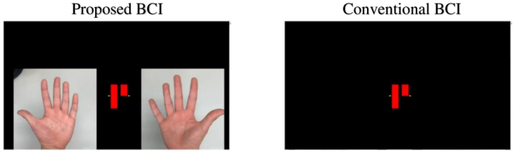
We have developed a BCI system to assist with MI related to left and right hand grasping. First, we describe the execution screen (see Figure 1). The proposed BCI system presents simulated hand animations. This can assist the MI of the corresponding hand.
Details MI-training-section, parameter-adjustment-section, and BCI-training-section are executed in this order (see Table 1).
In the MI-training-section, the participants were instructed to grasp the corresponding left and right hand in accordance with the timing of the grasp animation moving on the screen in order to experience and practice the motor image. At first, the participants were asked to perform actual grasping, and then gradually they were asked to perform only MI.
In the parameter-adjustment-section, one pair of F3-F4, C3-C4, and P3-P4 was selected from the combinations that best represent the difference between the left and right hands moving and not moving. The thresholds used in the next section were also determined here.
Figure 1: System to support first-person MI.Table 1. Phases of the system.
Table available in the original PDF paper.
Section MI-training Parameter-adjustment BCI-training Role Experiencing the MI and become familiar with MI Selection of electrodes and determination of classification thresholds Training to perform MI to increase ERP
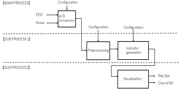
Figure 2: System configuration diagram.
In the BCI training-section, MI is practiced while looking at the EEG, which is visualized in an easily understood graph (see Figure 2). The patient is instructed to practice so that the red bar is lower than the yellow line. Ada- pted from Toyama’s method (Toyama et al. 2008), the BCI training system was implemented using multiprocessing in python. Four threads are proces- sed in parallel: sensing (A/D conversion), preprocessing, indicator generation, and indicator visualization (see Figure 2).
USEFULNESS OF THE SYSTEM Material & Method
The subjects were three healthy males in their 20s. An electroencephalograph (Polymate MINI AP108) was used and the experiment was conducted in a shielded room between 10 am and noon. Subjects performed the task in a resting sitting position approximately 70 cm away from the laptop display, and the subjects’ hands were hidden by a cloth to prevent confusion caused by viewing their real hands.
Evaluation was performed to assess the results of ERPs observations using the above system (see Figure 3). In the Evaluation, the participants were ran- domly instructed to perform “right hand imagery”, “left hand imagery”, and “nothing” 20 times each, and to see if there was a difference between “noth- ing” and “right hand imagery” or “nothing” and “left hand imagery” at this time or between “right hand imagery” and “left hand imagery”. Each mea- surement lasts 7 seconds, and during 3–5 seconds The patient was asked to perform the hand opening and closing movements at equal intervals.
Electrode placement for EEG measurements was based on the extended international 10–20 method (klem, 1999), with six EEG points: F3, C3, P3, F4, C4, and P4. The reference electrode was A1 and the ground electrode was A2. The sampling frequency was 500 Hz. The EEG data were split with 40% overlap and STFT was performed using a Hanning window. The EEG
System Development of Motor Imagery BCI: An Approach From the Human Side 219
Figure 3: Flow of the experiment.
data for 0–7 seconds in the alpha band (8-14 Hz) were then converted to rate of change using the median power in the first 1–2 seconds of measurement in the alpha band (8-14 Hz) as the beginning value (Toyama et al. 2008). To handle sudden noise, a synchronous arithmetic mean was applied to the rate of change for each trial.
RESULT
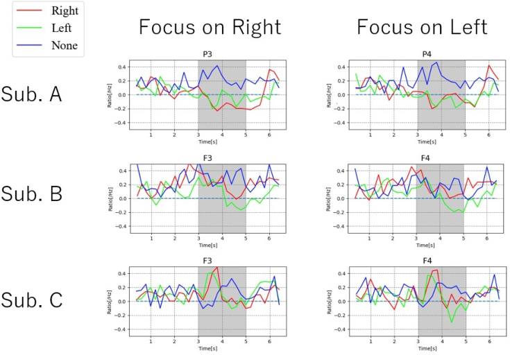
Figure 4 shows the results. Each shows the rate of change after synchronous arithmetic mean over three subjects. The red line indicates MI of the right hand, the green line indicates MI of the left hand, and the blue line indicates EEG when no MI was performed. The left column shows the electrodes atta- ched to the left side of the head and the right column shows the electrodes attached to the right side of the head, based on the fact that ERPs are locally identified on the contralateral side of the hand performing MI. That is, we expect the red line in the left column to have a significant change and the green line in the right column to have a significant change.
Figure 4: Results of evaluation experiments.
In Subject A, at 3–6 seconds, Right and Left have decreased amplitudes on both sides of the head compared to None. In the 0–7 second time window, the amplitude of Right decreased on the left side of the head in the 3–6 second time window. These results indicate that ERD was observed. However, there was no significant difference between Right and Left.
In Subject B, the amplitude of Left decreased on both sides of the head compared to None at 4–6 s. In Right, the amplitude decreased on the left side of the head. In the 0–7 second period, the amplitude of Left decreased on the right side of the head in the 4–6 second period. From the above, it can be said that ERD was clearly observed in the left hand and faintly observed in the right hand. However, there was no significant difference between Right and Left.
Subject C showed an increase in amplitude for Right and Left compared to None at 3–4 seconds, and a decrease in amplitude for Right and Left compa- red to None at 4–5 seconds. In the 0–7 second time window, the amplitudes of Right and Left increased significantly in the 4–5 second time window. From the above, it can be said that the ERS was observed. However, there was no significant difference between Right and Left.
DISCUSSION
The system focused on BCI-training and MI support allowed us to obse- rve significant ERPs. However, the shape of the ERPs showed different characteristics in all three patients. This may be because the training time was very short and the training is not effective. Although we were able to observe ERPs in the three subjects in this study, it is possible that some subjects may not be able to observe ERPs if the number of subjects is increased. Therefore, we need to consider more carefully the design of BCI-training.
Individual differences in MI can be attributed to individual differences in neural substrates (Kotegawa et al. 2021), so it is necessary to teach MI appro- priately for each individual (Mizuguchi and Kanosue, 2017). With regard to the content of instruction, it is necessary to focus on the state of mind and internal sensations of the subject. On these grounds, it has been repor- ted (Alimardani and Gherman, 2022) that relaxation and (visual) fatigue levels cause significant differences in BCI performance among users, and that the basal ganglia-cortical network is important for self-regulation, in other words, “feeling rather than thinking” may better manipulate BCI (Kasahara et al. 2022). By the way, BCI-training is a new concept born in the 21st cen- tury. Therefore, the teaching method and know-how of BCI-training have not been established, and the subject must find the know-how by himself/herself under simple instructions and learn EEG control in self-training. Therefore, we have developed a method to make self-training more efficient and to store the know-how that emerges during self-training as knowledge. In the next passage, we describe the concept of a BCI-training system that allows subjects to find the know-how on their own.
System Development of Motor Imagery BCI: An Approach From the Human Side 221
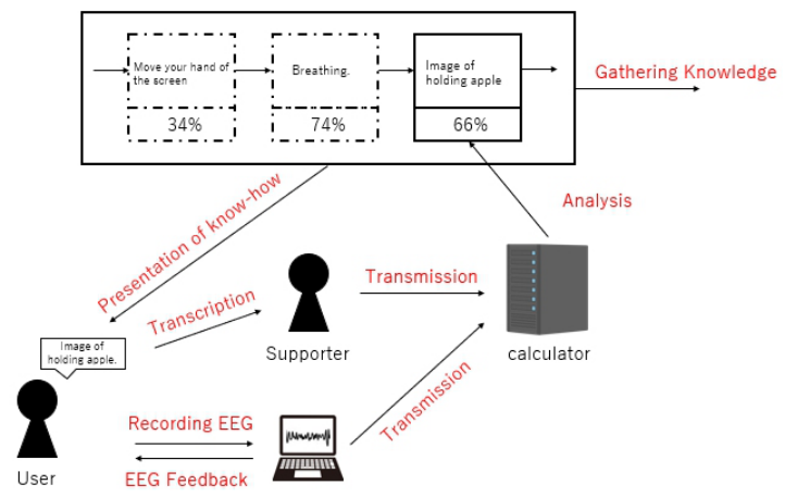
A Knowledge-Acquisition BCI-Training System A system was conceived to acquire knowledge of BCI (see Figure 5). The system records the evaluation of the results in relation to the know-how. Users can browse past know-how to help them in their independent training. This system allows users to practice efficient BCI-training. It is also envisioned that the system will be able to collect knowledge about BCI-training suffi- ciently and efficiently. Specifically, this system is used by repeating steps (1) through (7).
(1) The user declares the know-how to be tried. (2) The user performs an MI task while being aware of the know-how. (3) The EEG during the MI task is measured. The EEG is then fed back to the user. (4) The EEG and the know-how are recorded. (5) The EEG is analyzed and the evaluation index is calculated by calculator. (6) The evaluation index and know-how are linked and recorded. (7) Users can freely view the know-how and use it for independent training.
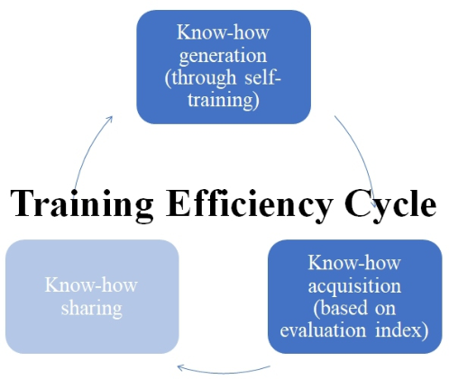
Finally, the flow of making this system useful in practice is described. This system accounts for the know-how generation and know-how acquisition portions of the training efficiency model (see Figure 6). If a lot of know-how is gathered by many users using this system, the know-how can be formalized and someone’s know-how can be shared with someone else. It can be applied not only to BCI-training but also to practice in areas where know-how has not been established.
Figure 5: Conceptual diagram of a knowledge-acquisition BCI-training system.Figure 6: Training efficiency cycle.
CONCLUSION
We developed a BCI system to support MI related to left hand and right hand grasping, and were able to confirm significant ERPs in three subjects. In order to be able to confirm significant ERPs in other subjects, further research on BCI-training is needed. In particular, the author will design BCI-training with a focus on the user side.
In the future, the system in its conceptual stage will be implemented and evaluated. In addition, by operating the system, the author plans to collect know-how on BCI-training.
ACKNOWLEDGMENT
This work was supported by the New Energy and Industrial Technology Development Organization (NEDO) JPNP18002.This work was supported by JST [Moonshot R&D] [Grant Number JPMJMS2237]. We received con- siderable cooperation from Taiga Watanabe. Finally, we would like to thank all the informants, faculty, academic staff, and collaborators who contributed to this study.
REFERENCES
Alimardani, M., & Gherman, D. -e. (2022). Individual Differences in Motor Imagery BCIs: A Study of Gender, Mental States and Mu Suppression. 2022 10th International Winter Conference on Brain-Computer Interface (BCI), 1–7. Hayashi, M., Mizuguchi, N., Tsuchimoto, S., & Ushiba, J. (2020). Neurofeedback of
Scalp Bi-Hemispheric EEG Sensorimotor Rhythm Guides Hemispheric Activation of Sensorimotor Cortex in the Targeted Hemisphere. NeuroImage, 223, 117298.
System Development of Motor Imagery BCI: An Approach From the Human Side 223
Kasahara, K., Dasalla, C. S., Honda, M., & Hanakawa, T. (2022). Basal Ganglia- Cortical Connectivity Underlies Self-Regulation of Brain Oscillations in Humans. Commun Biol, 5, 712. Kotegawa, K., yasumura, A., & Teramoto, W. (2021). Changes in Prefrontal Cortical
Activation during Motor Imagery of Precision Gait with Age and Task Difficulty. Behav Brain Res, 399(113046). Klem, G., Lüders, H. -e., Jasper, H., & Elger, C. (1999). Individual Differences in Motor Imagery BCIs: A Study of Gender, Mental States and Mu Suppression. The International Federation of Clinical Neurophysiology. Electroencephalogr Clin Neurophysiol Suppl, 52, 3–6. Lebedev, N. A., & Nicolelis, M. A. l. (2006). Brain–Machine Interfaces: Past, Present
and Future. Trends Neurosci, 29, 536–546. Mizuguchi, N., & Kanosue, K. (2017). Motor Image and Motor Performance.
Measurement & Control, 56(8), 568–572. Pfurtscheller, G., & Neuper, C. (2001). Motor Imagery and Direct Brain-Computer Communication. In Proceedings of the IEEE, 89(7), 1123–1134. Pfurtscheller, G., & Silva, F. h. lopes da. (1999). Event-Related EEG/MEG Synch-
ronization and Desynchronization: Basic Principles. Clinical Neurophysiology, 110(11), 1842–1857. Takahashi, M., Gouko, M., & Ito, K. (2009). Online Motor Imagery Training Effect for the Appearance of Event Related Desynchronization (ERD)*. Transactions of the Institute of Systems, Control and Information Engineers, 22(5), 199–205. Toyama, J., Ando, J., & Tamura, T. (2008). Basic Research in Event-Related de-
Synchronization and Synchronization (ERD/ERS) of EEG for Developing a Brain- Computer-Interface Driving Simulator. THE INSTITUTE OF ELECTRONICS INFORMATION AND COMMUNICATION ENGINEERS, 108(383), 113–117.
LISTED IN THE AHFE DIGITAL BOOKSTORE CATALOGUE
Explore the complete digital volume
The Human Side of Service Engineering
Read this individual research paper free through the original publisher, or explore the full digital proceedings book.
ISBN 978-1-958651-84-1
Explore this ISBN in the AHFE Bookstore ↗Listed in the July 2026 bookstore catalogue; search by ISBN to confirm current availability · Digital book purchase optional