Yiu Wong, Joanne Yip, Mei-Chun Cheung (2023). Biofeedback Posture Training for Adolescent Idiopathic Scoliosis Patient. In: Human Factors and Systems Interaction. AHFE Open Access, vol. 84, pp. 87–95. https://doi.org/10.54941/ahfe1003593
Scoliosis is an abnormal lateral deviation of the spine. Over 10 degrees of lateral curvature in the anteroposterior plane is already regarded as abnormal and scoliosis by the American Scoliosis Research Society. Scoliosis can be congenital, developmental, or degenerative. However, over 65% of the scoliosis cases were idiopathic. Scoliosis generally develops in the thoracic spine and/or the thoracolumbar area of the spine. One of the factors of scoliosis is the skeletal muscle around the aforementioned area of the spine. If the muscle strength between the left and right paraspinal muscles is imbalanced, internal pressure will develop and cause scoliosis. Though the risk of curve progression is the highest during puberty, adolescents with mild scoliosis (Cobb’s angle between 10 and 19) are generally closely monitored. With the increasing mobile phone usage among adolescents, it is very likely that adolescents with mild scoliosis develop poor posture during their prolonged screen time. This may affect the skeletal muscle development in the spine, causing an imbalance between the left and right paraspinal muscles, further accelerating the curve progression of scoliosis. To slow down or prevent the curve progression, biofeedback posture training has been developed by our research team. Early adolescents sat in front of a computer screen with animated videos as biofeedback to monitor their muscle activities of the paraspinal muscles. They underwent 30 sessions of biofeedback posture training, each session consisting of 3 minutes baseline assessment and 5 trials of 5-minute posture training.Currently, 18 adolescents with mild scoliosis have completed our training. 13 out 18 of them have their spine curve progressed less than 5 Cobb’s angle. In addition, 6 of them even reduced their spine curve by more than 5 Cobb's angle.
Adolescent idiopathic scoliosis (AIS) is very common among adolescents. AIS patients suffer lateral spinal curvature over 10 degrees measured with the Cobb’s method. There is no definite cause of idiopathic scoliosis, but the risk for progression was the highest during puberty (Carlson). After the peak of growth, AIS patients with 20 degrees or more deformity might stay stable (Peterson). However, if the AIS patients worn scoliosis orthosis as prescribed
when their condition was still mild (with the Cobb’s less than 20), their pro- gression could be halted (Peterson). Traditional scoliosis orthosis is made of rigid plastic material, providing correctional pressure at certain body points. There are many problems with the brace such as irritation, intervention with wearers’ social life. Nevertheless, the effectiveness of scoliosis ortho- sis depends on the wearer’s alertness on his or her posture and motivation to straighten his or her spine when poor posture is adopted.
Due to the coronavirus disease 2019 (COVID-19), many schools have tran- sitioned to virtual or online teaching. As a result, children and adolescents switched from transitional face-to-face lessons to using display devices to have online lessons. This provides an opportunity for AIS patients to develop good posture during online lectures.
Electromyographic (EMG) Monitor Biofeedback has been proven effective for psychological, physical and psych- ophysical problems. It is a system involving measurement of a subject’s physical activities and/or psychological activities. After processing the data, the system then provides feedback to the subject based on the data. Electrom- yographic monitor is a popular biofeedback system. This system measures the electrical activities of muscles and shows them on a monitor. A review of the surface electromyography (sEMG) studies on upper extremity dysfu- nction (Lyons) showed that sEMG is a valuable method for increasing upper extremity muscle activity. Moreover, there were studies on applying sEMG on scoliosis treatment. A postural training device called Micro Straight was used in training AIS patients’ posture(Wong). The result was good; subjects with 4-day wear trial had their time with poor posture reduced by around 26%. These research shows the possibility of posture measurement and posture training with EMG monitoring.
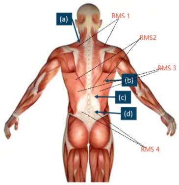
Biofeedback Posture Training In our biofeedback posture training, sEMG will be used to monitor sub- jects’ spinal muscles. The electrodes are placed on subjects’ trapezius(a), latissimus dorsi(b), thoracic erector spinae(c), and lumbar erector spinae(d). The electrodes position for surface sEMG demonstrates as the following (Figure 1).
Each subject is seated in front of a monitor with video playing in the back- ground. An operator will be closely monitoring the subject’s EMG signal with their monitor. A blind is placed between the subject and the operator to avoid distraction. Before the training starts, the operator will correct the subject posture if needed. The training starts after the operator assigns a thre- shold to each EMG channel which corresponds to different spinal muscles. The video will stop playing if anyone of the channel exceeds the assigned threshold. The operator will then instruct the subject to correct their posture.
Each subject will undergo 30 sessions of training, each consists of 5 sets of 5 minutes training. Subjects are required to take one training session every week. Two assessments will be conducted before and after the posture training programme. The assessment consists of 3 sets of 3 minutes sEMG
Biofeedback Posture Training for Adolescent Idiopathic Scoliosis Patient 89
Figure 1: The electrodes position for sEMG.
recording with the subject standing as well as sitting. There is no video pla- ying during sEMG recording and the subject will not be reminded when poor posture is adopted.
X-Ray Radiograph 18 adolescents with mild scoliosis have completed our training. 13 out 18 of them have their spine curve progressed less than 5 Cobb’s angle. In addition, 6 of them even reduced their spine curve by more than 5 Cobb’s angle. The following Table 1 contain 4 subjects with significant improvement in Cobb’s angle.
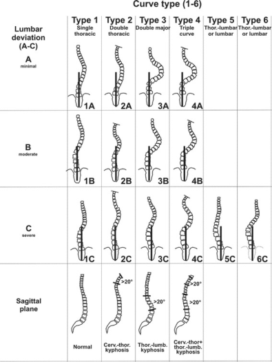
From the X-ray radiograph, we notice that subjects with significant Cobb’s angle improvement are mainly Type 1 AIS (according to Lenke classification). In general, Type 1 AIS subjects show better improvement than other subjects.
Electromyography Results Each subject is required to take an assessment before and after their bio- feedback posture training programme. In each assessment, we record the subject’s spinal muscles activities in their natural posture. The root mean square (RMS) of each sEMG reading is calculated in order to give a rela- tive estimated how much power each spinal muscle is outputting when the subject is sitting and standing. Then the ratio of the RMS between each pair of muscles is calculated. At last, we take the average of all three ratios for each pair of muscles. Note that, the closer the value gets to one, that pair of muscles is more balanced.
Subject EMG005 Cobb’s angle has reduced by 10.3
and their EMG reading reflects that. Their latissimus dorsi and thoracic erector spinae dra- stically improved after finishing our training (Table 2). We noticed that EMG005 was very compliant with our instruction during training and acti- vely thought about their posture even after each training session. On the other hand, their standing EMG reading remains fairly balanced before and after training (Table 3).
Table 1. The X-ray result of the subjects with significant improvement in Cobb’s angle.
Table available in the original PDF paper.
The pre-assessment The Post-assessment EMG005 Cobb Angle (16.3
) EMG005 Cobb Angle (6
)
EMG002 Cobb Angle (17.6
) EMG002 Cobb Angle (12image6.png
Continued
Biofeedback Posture Training for Adolescent Idiopathic Scoliosis Patient 91
Table 1. Continued.
Table available in the original PDF paper.
The pre-assessment The Post-assessment EMG009 Cobb Angle (13.2
) EMG009 Cobb Angle (7.6
)
EMG018 Cobb Angle (17.2
) EMG018 Cobb Angle (6
)
Figure 2: The Lenke classification for adolescent idiopathic scoliosis.Table 2. The RMS ratio average of subject EMG005 sitting, with each spinal muscles are labelled as the following: trapezius (A), latissimus dorsi (B), thoracic erector spinae (C), and lumbar erector spinae (D).
Table available in the original PDF paper.
Pre RMS ratio average (A)
Pre RMS ratio average (B)
Pre RMS ratio average (C)
Pre RMS ratio average (D) 1.36 0.15 3.02 1.02 Post RMS ratio average (A)
Post RMS ratio average (B)
Post RMS ratio average (C)
Post RMS ratio average (D) 1.203 1.13 1.043 0.96
Subject EMG018 Cobb’s angle has reduced by 11.6
. However, their trape- zius, latissimus dorsi, thoracic erector spinae has become less balanced (Table 4). This is due to their poor posture developed before our training. EMG018 had adopted kyphotic posture when sitting. Thus, extra force is needed from
Table 3. The RMS ratio average of subject EMG005 standing, with each spinal muscles are labelled as the following: trapezius (A), latissimus dorsi (B), thoracic erector spinae (C), and lumbar erector spinae (D).
Table available in the original PDF paper.
Pre RMS ratio average (A)
Pre RMS ratio average (B)
Pre RMS ratio average (C)
Pre RMS ratio average (D) 1.091328215 0.097091781 3.310707547 1.148073169 Post RMS ratio average (A)
Post RMS ratio average (B)
Post RMS ratio average (C)
Post RMS ratio average (D) 0.963216246 1.082756427 1.009198578 0.879335114
Biofeedback Posture Training for Adolescent Idiopathic Scoliosis Patient 93
Table 4. The RMS ratio average of subject EMG018 sitting, with each spinal muscles are labelled as the following: trapezius (A), latissimus dorsi (B), thoracic erector spinae (C), and lumbar erector spinae (D).
Table available in the original PDF paper.
Pre RMS ratio average (A)
Pre RMS ratio average (B)
Pre RMS ratio average (C)
Pre RMS ratio average (D) 1.05384722 1.053844691 1.205105963 1.143836554 Post RMS ratio average (A)
Post RMS ratio average (B)
Post RMS ratio average (C)
Post RMS ratio average (D) 0.597026234 0.714644859 2.258138172 1.319314671
thoracic erector spinae. Even after 30 sessions training, EMG018 still had a tendency to adopt kyphotic posture. Similarly, affected by kyphotic posture, they were hunchbacking when standing. After our training, they were aware of their poor posture and continuously correcting their posture (Table 5).
Subject EMG002 Cobb’s angle is reduced by 5.6
which may not be as much as aforementioned subjects. Nevertheless, their thoracic erector spinae has gained better balance than before. Overall, EMG002 spinal muscle bala- nce has been improved during sitting (Table 6). As for their standing posture, the EMG shows that their spinal muscles remain fairly balanced before and after training (Table 7).
Among all four subjects, subject EMG009 shows the least improvement in terms of Cobb’s angle. There is very minimum improvement in spinal muscle balance (Table 8).
We noticed EMG009 needed more time to learn than other aforementioned subjects. Despite that, EMG009 is still one of the fastest learners among all
Table 5. The RMS ratio average of subject EMG018 standing, with each spinal muscles are labelled as the following: trapezius (A), latissimus dorsi (B), thoracic erector spinae (C), and lumbar erector spinae (D).
Table available in the original PDF paper.
Pre RMS ratio average (A)
Pre RMS ratio average (B)
Pre RMS ratio average (C)
Pre RMS ratio average (D) 1.350157307 0.942158805 1.439314721 1.181368001 Post RMS ratio average (A)
Post RMS ratio average (B)
Post RMS ratio average (C)
Post RMS ratio average (D) 0.706580978 0.771666786 1.354316716 0.966756092
Table 6. The RMS ratio average of subject EMG002 sitting, with each spinal muscles are labelled as the following: trapezius (A), latissimus dorsi (B), thoracic erector spinae (C), and lumbar erector spinae (D).
Table available in the original PDF paper.
Pre RMS ratio average (A)
Pre RMS ratio average (B)
Pre RMS ratio average (C)
Pre RMS ratio average (D) 1.18993667 1.190359366 0.5738420237 0.7411585413 Post RMS ratio average (A)
Post RMS ratio average (B)
Post RMS ratio average (C)
Post RMS ratio average (D) 1.205645769 0.9515196548 1.112470898 0.9572505483
Table 7. The RMS ratio average of subject EMG002 standing, with each spinal muscles are labelled as the following: trapezius (A), latissimus dorsi (B), thoracic erector spinae (C), and lumbar erector spinae (D).
Table available in the original PDF paper.
Pre RMS ratio average (A)
Pre RMS ratio average (B)
Pre RMS ratio average (C)
Pre RMS ratio average (D) 1.124071623 0.970153944 1.140446024 1.206524487 Post RMS ratio average (A)
Post RMS ratio average (B)
Post RMS ratio average (C)
Post RMS ratio average (D) 0.963216246 1.082756427 1.009198578 0.879335114
Table 8. The RMS ratio average of subject EMG009 sitting, with each spinal muscles are labelled as the following: trapezius (A), latissimus dorsi (B), thoracic erector spinae (C), and lumbar erector spinae (D).
Table available in the original PDF paper.
Pre RMS ratio average (A)
Pre RMS ratio average (B)
Pre RMS ratio average (C)
Pre RMS ratio average (D) 0.832765226 1.162447603 1.129815429 0.969635097 Post RMS ratio average (A)
Post RMS ratio average (B)
Post RMS ratio average (C)
Post RMS ratio average (D) 0.906957143 1.084853117 1.318368041 0.858032038
Table 9. The RMS ratio average of subject EMG009 standing, with each spinal muscles are labelled as the following: trapezius (A), latissimus dorsi (B), thoracic erector spinae (C), and lumbar erector spinae (D).
Table available in the original PDF paper.
Pre RMS ratio average (A)
Pre RMS ratio average (B)
Pre RMS ratio average (C)
Pre RMS ratio average (D) 0.927088869 1.133465475 0.727646062 1.007931283 Post RMS ratio average (A)
Post RMS ratio average (B)
Post RMS ratio average (C)
Post RMS ratio average (D) 0.824596118 1.042722098 0.85690741 0.979940216
subjects. On the one hand, EMG009 shows improvement in their standing posture (Table 9). Note that the trapezius has slightly lost muscle balance even other spinal muscles have gained better balance (Table 9). This is a common phenomenon as the “load” has been redistributed among spinal muscles to obtain better posture and balance.
CONCLUSION
In our research, 13 out 18 mild AIS subjects have their spine curve progressed less than 5 Cobb’s angle. We notice that Type 1 AIS subjects generally per- form better in terms of muscle balance. We hypothesize that since most of our subjects were prompted to adopt kyphotic posture when seated, more force was put into erector spinae. Thus, achieving a similar effect as traditional scoliosis brace treatment. Despite that, the effectiveness of our biofeedback posture training depends on subject self-compliance. Some of our subjects showed pessimistic attitude during training and low self-compliance after
Biofeedback Posture Training for Adolescent Idiopathic Scoliosis Patient 95
each training. Such subjects has a very limited effect on their spine curve progression.
ACKNOWLEDGMENT
This research was funded by a grant from the Research Grant Council of the Hong Kong Special Administrative Region, China, to The Chinese University of Hong Kong, grant number: CUHK 14607519 (Mei-chun Cheung) and a grant supported by the Public Sector Trial Scheme–515 Innovation and Technology Fund (PSTS-ITF), the Innovation and Technology Commission 516 (ITC) of the Hong Kong Special Administrative Region, China, to The Hong Kong Polytechnic 517 University, grant number ITT/025/GP (Joanne Yip) and other research fund from Lee Hysan Foundation to The Hong Kong Polytechnic University, grant number R-ZH3Y (J. Y.).
REFERENCES
Carlson, JM. “The prediction of curve progression in untreated idiopathic scoliosis during growth.” PubMed, https://pubmed.ncbi.nlm.nih.gov/6480635/. Accessed 31 January 2023. “Follow-up Study of Milwaukee Brace Treatment in Patients with Idiopathic Sco-
liosis.” Follow-up Study of Milwaukee Brace Treatment in Patients with Idi- opathic Scoliosis.: Clinical Orthopaedics and Related Research (1976-2007), 1977, https://journals.lww.com/corr/Citation/1977/07000/Follow_up_Study_of _Milwaukee_Brace_Treatment_in.8.aspx. Accessed 31 January 2023. Lyons, G. M. “A computer game-based EMG biofeedback system for muscle rehabilitation.” A computer game-based EMG biofeedback system for muscle reha- bilitation, IEEE, September 2003, https://ieeexplore.ieee.org/document/1279682. Accessed 31 January 2023. Peterson, LE. “Effectiveness of treatment with a brace in girls who have adolescent
idiopathic scoliosis. A prospective, controlled study based on data from the Brace Study of the Scoliosis Research Society.” PubMed, Jun 1995, https://pubmed.ncb i.nlm.nih.gov/7782353/. Accessed 31 January 2023. Wong, M. S. “Effectiveness of audio-biofeedback in postural training for adolescent idiopathic scoliosis patients - M. S. Wong, A. F. T. Mak, K. D. K. Luk, J. H. Evans, B. Brown, 2001.” SAGE Journals, April 2001, https://journals.sagepub.com/doi/a bs/10.1080/03093640108726570. Accessed 31 January 2023.
LISTED IN THE AHFE DIGITAL BOOKSTORE CATALOGUE
Explore the complete digital volume
Human Factors and Systems Interaction
Read this individual research paper free through the original publisher, or explore the full digital proceedings book.
ISBN 978-1-958651-60-5
Explore this ISBN in the AHFE Bookstore ↗Listed in the July 2026 bookstore catalogue; search by ISBN to confirm current availability · Digital book purchase optional