Lev Yakovlev, Artemiy Berkmush Antipova, Nikolay Syrov, Maksimov Iaroslav, Daria Petrova, Matvey Bulat, Mikhail Lebedev, Alexander Kaplan (2022). The effects of tactile stimulation and its imagery on sensorimotor EEG rhythms: incorporating somatic sensations in brain-computer interfaces. In: Human Interaction & Emerging Technologies (IHIET 2022): Artificial Intelligence & Future Applications. AHFE Open Access, vol. 68, pp. 461–467. https://doi.org/10.54941/ahfe1002765
1 V. Zelman Center for Neurobiology and Brain Rehabilitation, Skolkovo Institute of Science and Technology, Moscow, 121205, Russia; Baltic Center for Neurotechnology and Artificial Intelligence, Immanuel Kant Baltic Federal University, Kaliningrad, 236041, Russia ↗
2 Baltic Center for Neurotechnology and Artificial Intelligence, Immanuel Kant Baltic, Federal University, Kaliningrad, 236041, Russia ↗
3 V. Zelman Center for Neurobiology and Brain Rehabilitation, Skolkovo Institute of Science and Technology, Moscow, 121205, Russia ↗
4 Laboratory for Neurophysiology and Neurocomputer Interfaces, Lomonosov Moscow, State University, Moscow, 119234, Russia ↗
Brain Computer Interface (BCIs) strive to provide a communication channel between the human brain and the environment without using overt actions. To achieve this goal, BCIs convert recordings of neural activity into commands to an external device. One of the most common BCI methods is based on matching of spectral characteristics of EEG sensorimotor rhythms (SMR) to motor imagery attempts. While such motor-imagery BCI have been extensively studied, little is known about the possibility of using tactile imagery as a BCI component. Here we studied EEG modulations associated with tactile imagery and obtained results suggesting that this approach could improve BCI operations.METHODS. 12 healthy naïve volunteers participated in the study. After vibrotactile stimulation was applied several times to the right hand, participants imagined the same sensations in the absence of actual vibration. During this tactile imagery task, 30 channels of EEG were recorded. The effects of tactile imagery were assessed as changes of the SMR in mu and beta bands, which were quantified using EEG desynchronization/synchronization (ERD/S) ratios. An offline classification was conducted for three states: resting, tactile imagery of the right hand, and tactile imagery of the left hand. The classification was based on common spatial pattern (CSP) filtering and linear discriminant analysis.RESULTS. The participants exhibited consistent contralateral ERD patterns over sensorimotor areas during tactile imagery of each hand. Statistically significant differences (p<0.05) in SMR spectral characteristics were found for the comparison of imagery condition to the control state. Offline classification exceeded chance level, as well. CONCLUSION. We found that when human subjects imagine their hands receiving tactile stimulation their SMR spectral characteristics exhibit consistent changes, which could be reliably decoded with a discrete classifier. Based on these observations, we suggest that motor-imagery BCIs could be enriched by adding a tactile-imagery component. Tactile imagery-based BCIs could be especially useful for neuroprosthetic approaches intended for people suffering from somatosensory disabilities and phantom-limb pain.ANCKLOWLEGEMENTS. The study was supported by the Russian Science Foundation grant 21-75-30024.
Brain Computer Interface (BCI) is a communication system between the brain and an external world. BCIs bypass the brain’s natural output – muscles and peripheral nerves – and connect to the brain directly as the source of com- mands (Wolpaw et al., 2002). While theoretically invasive recordings with the electrodes inserted in the brain could enable the best-quality communi- cation, BCIs based on noninvasive electroencephalography (EEG) recordings from the human scalp are currently the most practical and popular appro- ach. Among the EEG-based BCI designs, motor imagery-based BCIs have attracted lots of attention from the researchers, and many articles have been published on this type of BCI. In this approach, a subject imagines perfor- ming movements without executing them. Next, this endeavor causes an
attenuation sensorimotor rhythmic activity (Pfurtscheller & Neuper, 2001), which could be detected by a BCI decoder and converted into an output signal. Remarkably, motor-imagery BCI could be applied to a range of clini- cal applications, including applications for poststroke rehabilitation (Silvoni et al., 2011; Pichiorri, 2016; Frolov et al., 2018), neuroprosthetics (Meng et al., 2016; Frolov et al., 2018), and device Ñ'ontrol (LaFleur et al., 2013; Fernández-Rodríguez et al., 2016).
Several motor-imagery studies have included a tactile component, which improved BCI operations (Natio et al., 2002; Forukas, Ionta & Aglioti, 2006; Mizuguchi et al., 2009; Mizuguchi et al., 2012). Technically, both motor ima- gery and tactile stimulation have similar effects on EEG. Thus, event related desynchronization (ERD) is observed during voluntary movements, imagery of such movements and tactile stimulation (Pfurtscheller & Da Silva, 1997; Neuper, Wörtz & Pfurtscheller, 2006). Such ERD depends on corticothalamic interactions, and for modulations of SMR in the alpha range (8-13 Hz) the source can be localized to the sensorimotor cortex (Hari & Salmelin, 1997; Frolov et al., 2012). Moreover, Frolov et al., have shown that the sources most relevant to motor imagery are localized in the primary sensorimotor cortex. These finding points to the somatosensory cortex as the major player in the formation of internal brain states representing motor imagery. Based on these results, it is reasonable to suggest that imagining being touched would cause EEG modulations in the same brain areas, like activation of the pri- mary somatosensory cortex during tactile imagery (Schmidt, Ostwald and Blankenburg, 2014; de Borst and de Gelder, 2017; Schmidt and Blanken- burg, 2019). Yet relatively little is known about the effects of tactile imagery on EEG activity and the ways such imagery could be incorporated in BCI designs.
To address this knowledge gap, the main idea of this study was to use mental tactile images instead of traditional motor ones to induce EEG chan- ges driving a BCI. Accordingly, we investigated spatio-temporal features of mu ERD patterns during tactile stimulation (TS) and tactile imagery (TI) and performed an offline classification of this imagery.
METHODS
Eleven healthy naïve volunteers (8 female, mean age 21,8±1,19) participa- ted in the study. All the participants reported no psychiatric and neurological disorders and gave their written consent to participate in the study. The expe- rimental protocol was approved by the Lomonosov Moscow State University Committee for bioethics and carried out in accordance with the Declaration of Helsinki.
During the experimental session, the subjects were seated in a comfortable armchair. Vibrotactile stimulator was applied on the right-hand inner surface to achieve relevant somatosensory experience before performing an imagery task for the same hand. After 45 somatosensory stimulation trials lasted 6 s, participants were asked to reproduce the sensations mentally. There were 45 trials of right-hand tactile imagery divided into 3 runs between which the participants had the opportunity to take a short break (1-2 min). Eight out
The Effects of Tactile Stimulation and Its Imagery on Sensorimotor EEG Rhythms 463
of twelve participants were additionally asked to perform tactile imagery of the left-hand tactile stimulation without the previous stimulation. The total amount of left-hand imagery 6s trials was 15. All imagery trials were mixed with the resting state trials randomly.
30 channels of EEG were recorded using the NVX-52 (MKS, Russia) digi- tal amplifier. Electrodes were placed according to the 10/10 international system. The skin impedance for each electrode did not exceed 20 k. The recording signal was sampled at 500 Hz with a 50 Hz Notch filter.
The effects of tactile imagery were assessed as changes in the amplitude of sensorimotor oscillations in the range 8-15 Hz. The raw signal was bandpass filtered in the range 1-30 Hz using a zero-phase bandpass FIR filter. The signal was then segmented into 6-s epochs for each experimental condition (right hand tactile stimulation, right hand tactile imagery and left-hand tactile imagery). Continuous wavelet transform with variable number of cycles was performed to assess time-frequency parameters for exploring conditions. The wavelets frequencies ranged from 5 to 30 Hz with a 1-Hz step, the full-width at half-maximum (FWHM) was equal to 60 ms corresponding to a spectral FWHM of 2.5 Hz. ERD/S was calculated as a natural logarithm of the ratio in spectral power between sensorimotor state and the rest (units in dB). For topographical mapping ERD/S values from each channel were averaged over time and individual frequency range.
For offline classification the signal was filtered in the individual freque- ncy range and then spatially filtered using the Common Spatial (CSP) filter (Ramoser et al., 2000) which yielded the classification feature with a maximal difference between the classified mental states. BCI classification accuracy was calculated for each subject on trials with two classes: a) right hand tactile imagery vs rest, b) left hand tactile imagery vs rest and c) right-hand tactile imagery vs left hand tactile imagery. Classification was performed using linear discriminant analysis (LDA). For each classified pair, we used 4 CSP features that had maximal differences. The implementation of the CSP algorithm and offline classification was based on the Python library MNE 0.23 (Gramfort et al., 2013).
RESULTS AND DISCUSSION
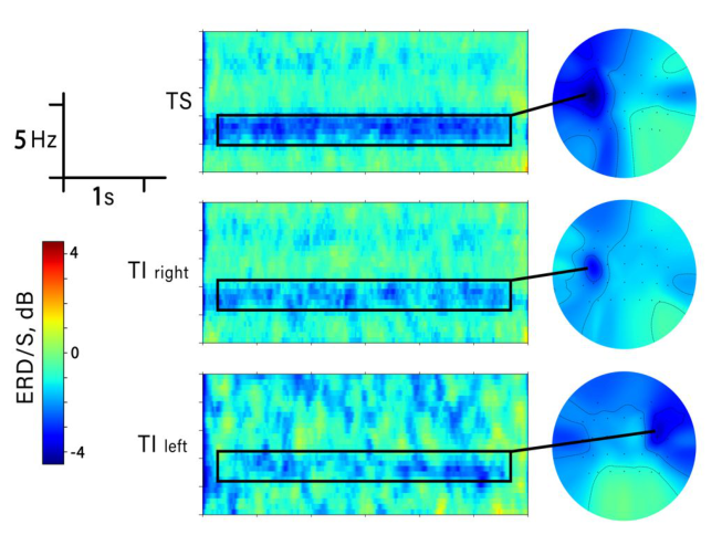
The participants consistently generated contralateral ERD patterns for the alpha (mu) activity (10-15 Hz) recorded over sensorimotor areas. These pat- terns were genetated during right-hand tactile stimulation and tactile imagery of each hand. The spatial-frequency characteristics of the ERD imagery pat- terns were quite typical for the effects of tactile stimulation (Fig. 1). Offline classification of the 3 condition pairs showed good performance in terms of accuracy, that was above the chance level (50%) and exceeded 70% for all classified conditions.
Event Related Desyncronization During Tactile Stimulation and Imagery We observed contralateral ERD in the mu range (10-15 Hz) during both tactile stimulation and imagery. ERD appeared from the start of the task
Figure 1: Left. Time-frequency dynamics of the contralateral desynchronization for tactile stimulation of the right hand (C3), tactile imagery of the same hand being stimu- lated (C3) and tactile imagery of the left hand (C4). Right. Topographic maps of ERD/S. Blue colors correspond to event related desynchronization (ERD), red designates synchronization (ERS). Green color corresponds to an absence of EEG modulations.
performance and lasted throughout the 6-s trial. Both right-hand TS and TI caused a contralaterally centered ERD with the maximum on the C3 channel. Tactile stimulation was associated with a slightly higher level of desynchroni- zation compared to the TI of the same hand (Fig. 1). Yet these differences were small, and statistical comparison at the group level did not reveal significant differences between TS and TI (Wilcoxon signed rank test). We concluded that right-hand TI led to consistent contralateral ERD, and this effect was of the same magnitude as the one observed during the actual vibrotactile stimulation.
During tactile imagery of the left hand, we observed a contralateral ERD pattern, with the maximum desynchronization intensity on the C4 chan- nel (Fig. 1). It is important to note that subjects did not practice with the actual vibrotactile stimulation of the left hand, which points to their ability to generalize this sensation to different parts of the body.
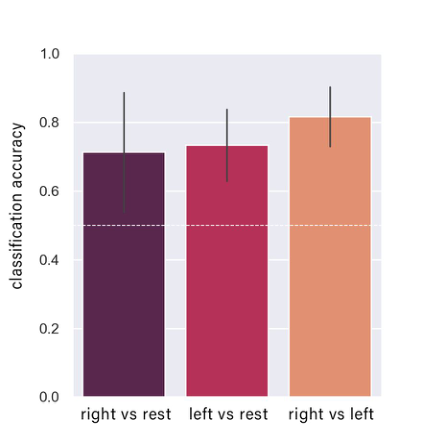
Classification Accuracy During offline classification, we calculated classification scores on the tri- als of two classes: right/left-hand TI and resting state. Average classification accuracy for right -hand TI vs rest was 0.71±0.19 with individual values ranging from 0.43 to 0.94. For the classification of the left-hand TI vs rest, average classification accuracy was 0.73±0.1 (individual ranging from 0.57 to 0.93). For the classification of the left-hand TI vs right-hand TI, ave- rage classification accuracy was 0.82±0.09 (individual ranging from 0.68 to
The Effects of Tactile Stimulation and Its Imagery on Sensorimotor EEG Rhythms 465
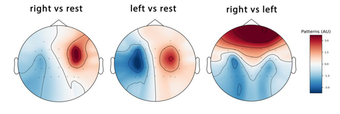
Figure 2: Mean values for classification accuracy for the explored mental states. White dashed line corresponds to the chance level.Figure 3: An example of CSP-features that differed the most across the two considered mental states. Data from subj#2.
0.97). The obtained results were statistically different from the chance level of 0.5 and comparable to the motor imagery-based BCI performance (Guger et al., 2003; Vasilyev et al., 2017). Only one participant had accuracy lower than the chance level during TI of the right hand (subj#11).
The example of CSP patterns used for classification in one subject are shown in Figure 3. For most of the subjects, CSP patterns were localized con- tralaterally over the central electrodes (Fig. 3). The results obtained showed that tactile imagery can be successfully decoded using such basic classification methods as linear discriminant analysis. The classification could be further improved using artificial neural networks.
CONCLUSION
We found that when human subjects imagine their hands receiving tactile stimulation their sensorimotor spectral characteristics exhibit consistent
changes, which could be reliably decoded with a discrete classifier. Based on these observations, we suggest that motor-imagery BCIs could be enriched by adding a tactile-imagery component. Therefore, tactile imagery-based BCIs could be especially useful for neuroprosthetic approaches intended for people suffering from somatosensory disabilities and phantom-limb pain.
ACKNOWLEDGMENT
The study was supported by the Russian Science Foundation grant 21-75- 30024.
REFERENCES
De Borst, A. W., & de Gelder, B. (2017). fMRI-based multivariate pattern analyses reveal imagery modality and imagery content specific representations in primary somatosensory, motor and auditory cortices. Cerebral cortex, 27(8), 3994–4009. Fernández-Rodríguez, Á., Velasco-Álvarez, F., & Ron-Angevin, R. (2016). Review of
real brain-controlled wheelchairs. Journal of neural engineering, 13(6), 061001. Fourkas, A. D., Ionta, S., & Aglioti, S. M. (2006). Influence of imagined posture and imagery modality on corticospinal excitability. Behavioural brain research, 168(2), 190–196. Frolov, A. A., Kozlovskaya, I. B., Biryukova, E. V., & Bobrov, P. D. (2018). Use
of robotic devices in post-stroke rehabilitation. Neuroscience and behavioral physiology, 48(9), 1053–1066. Frolov, A., Husek, D., Bobrov, P., Korshakov, A., Chernikova, L., Konovalov, R., & Mokienko, O. (2012). Sources of EEG activity most relevant to performance of brain-computer interface based on motor imagery. Neural Network World, 22(1), 21. Gramfort, A., Luessi, M., Larson, E., Engemann, D. A., Strohmeier, D., Brodbeck, C.,
... & Hämäläinen, M. (2013). MEG and EEG data analysis with MNE-Python. Frontiers in neuroscience, 267. Guger, C., Edlinger, G., Harkam, W., Niedermayer, I., & Pfurtscheller, G. (2003).
How many people are able to operate an EEG-based brain-computer interface (BCI)?. IEEE transactions on neural systems and rehabilitation engineering, 11(2), 145–147. Hari, R., & Salmelin, R. (1997). Human cortical oscillations: a neuromagnetic view through the skull. Trends in neurosciences, 20(1), 44–49. LaFleur, K., Cassady, K., Doud, A., Shades, K., Rogin, E., & He, B. (2013). Quadco-
pter control in three-dimensional space using a noninvasive motor imagery-based brain–computer interface. Journal of neural engineering, 10(4), 046003. Meng, J., Zhang, S., Bekyo, A., Olsoe, J., Baxter, B., & He, B. (2016). Noninvasive electroencephalogram based control of a robotic arm for reach and grasp tasks. Scientific Reports, 6(1), 1–15. Mizuguchi, N., Sakamoto, M., Muraoka, T., Moriyama, N., Nakagawa, K., Nakata,
H., & Kanosue, K. (2012). Influence of somatosensory input on corticospinal excitability during motor imagery. Neuroscience letters, 514(1), 127–130. Mizuguchi, N., Sakamoto, M., Muraoka, T., & Kanosue, K. (2009). Influence of touching an object on corticospinal excitability during motor imagery. Experimental brain research, 196(4), 529–535.
The Effects of Tactile Stimulation and Its Imagery on Sensorimotor EEG Rhythms 467
Naito, E., Kochiyama, T., Kitada, R., Nakamura, S., Matsumura, M., Yonekura, Y., & Sadato, N. (2002). Internally simulated movement sensations during motor imagery activate cortical motor areas and the cerebellum. Journal of Neuroscience, 22(9), 3683–3691. Neuper, C., Wörtz, M., & Pfurtscheller, G. (2006). ERD/ERS patterns refle-
cting sensorimotor activation and deactivation. Progress in brain research, 159, 211–222. Pfurtscheller, G., & Da Silva, F. L. (1999). Event-related EEG/MEG synchronization and desynchronization: basic principles. Clinical neurophysiology, 110(11), 1842–1857. Pfurtscheller, G., & Neuper, C. (2001). Motor imagery and direct brain-computer
communication. Proceedings of the IEEE, 89(7), 1123–1134. Pichiorri, F., Mrachacz-Kersting, N., Molinari, M., Kleih, S., Kübler, A., & Mattia, D.
(2017). Brain-computer interface based motor and cognitive rehabilitation after stroke–state of the art, opportunity, and barriers: summary of the BCI Meeting 2016 in Asilomar. Brain-Computer Interfaces, 4(1-2), 53–59. Ramoser, H., Muller-Gerking, J., & Pfurtscheller, G. (2000). Optimal spatial filtering of single trial EEG during imagined hand movement. IEEE transactions on rehabilitation engineering, 8(4), 441–446. Schmidt, T. T., Ostwald, D., & Blankenburg, F. (2014). Imaging tactile imagery:
changes in brain connectivity support perceptual grounding of mental images in primary sensory cortices. Neuroimage, 98, 216–224. Schmidt, T. T., & Blankenburg, F. (2019). The somatotopy of mental tactile imagery.
Frontiers in Human Neuroscience, 13, 10. Silvoni, S., Ramos-Murguialday, A., Cavinato, M., Volpato, C., Cisotto, G., Turolla, A., ... & Birbaumer, N. (2011). Brain-computer interface in stroke: a review of progress. Clinical EEG and neuroscience, 42(4), 245–252. Vasilyev, A., Liburkina, S., Yakovlev, L., Perepelkina, O., & Kaplan, A. (2017).
Assessing motor imagery in brain-computer interface training: psychological and neurophysiological correlates. Neuropsychologia, 97, 56–65. Wolpaw, J. R., Birbaumer, N., McFarland, D. J., Pfurtscheller, G., & Vaughan, T.
M. (2002). Brain–computer interfaces for communication and control. Clinical neurophysiology, 113(6), 767–791.
Read this individual research paper free through the original publisher, or explore the full digital proceedings book.
ISBN 978-1-958651-44-5
Explore this ISBN in the AHFE Bookstore ↗Listed in the July 2026 bookstore catalogue; search by ISBN to confirm current availability · Digital book purchase optional