Muhua Xu, Weiwei Ding, Haowei Deng, Qianxiang Zhou, Zhongqi Liu (2026). Regulatory Effects of Transcutaneous Electrical Acupoint Stimulation on EEG Power in 36-Hour Sleep Deprivation-Induced Cognitive Decline. In: Neuroergonomics and Cognitive Engineering. AHFE Open Access, vol. 207, pp. 58–65. https://doi.org/10.54941/ahfe1007395
Transcutaneous Electrical Acupoint Stimulation (TEAS), as a non-invasive peripheral neuromodulation technique, has been shown to improve cognitive function. However, its neurophysiological mechanisms underlying cognitive regulation remain unclear. This study aims to investigate the intervention effects of TEAS on sleep deprivation-induced cognitive brain activity and its associated neural mechanisms based on electroencephalographic (EEG) frequency domain analysis. Twenty-five healthy male volunteers were recruited as subjects, and a 36-hour sleep deprivation protocol was used to establish a cognitive decline model. Following sleep deprivation, the subjects received TEAS intervention targeting the acupoints of Neiguan, Waiguan, and Shenmen. Subjects completed the improved Go/NoGo task before and after intervention, with simultaneous EEG recording. The study focused on analyzing power spectral density (PSD) changes across five frequency bands in different electrodes. Results indicate that PSD changes following TEAS intervention exhibit state-frequency-region specificity. Alpha band activity was significantly increased in frontal and centroparietal regions in Go conditions. Meanwhile, theta band power showed widespread activation across frontal, central, centroparietal, and parietal regions in NoGo conditions. This finding suggests that TEAS may partially reverse sleep deprivation-induced cognitive impairment at the neurophysiological level by selectively activating alpha rhythms associated with response execution and theta rhythms involved in inhibitory control. The study provides electrophysiological evidence at the EEG frequency domain level for non-invasive interventions targeting sleep deprivation-related cognitive decline, demonstrating the regulatory effectiveness of peripheral electrical neuromodulation.
Sleep is a fundamental physiological process that maintains the homeostasis of the human central nervous system and supports higher-order cognitive processing (Frank and Heller, 2019). Chronic or acute sleep deprivation induces widespread and persistent negative effects at the neurobehavioral Received February 4, 2026; Revised April 1, 2026; Accepted April 18, 2026; Available online July 20, 2026
Regulatory Effects of Transcutaneous Electrical Acupoint Stimulation 59 level, impacting multiple cognitive domains including alertness, attention, working memory, decision-making, and executive function (Killgore, 2010; Griggs et al., 2022). Research indicates that even brief periods of total sleep deprivation (TSD) significantly impair cognitive speed and accuracy, with these deficits notably worsening as wakefulness duration increases (Basner et al., 2013). In modern work scenarios such as military duty and spaceflight missions, sleep deprivation often coexists with high workloads and circadian rhythm disruption, amplifying the risk of human error and safety threats (Miller et al., 2018). Therefore, developing a safe, non-invasive intervention strategy to alleviate sleep-related cognitive decline is crucial for enhancing work performance and safeguarding lives.
Among the cognitive impairments caused by sleep deprivation, inhibitory control serves as a pivotal component of the cognitive control system, governing impulse response inhibition and error detection. When inhibitory control fails, individuals tend to exhibit increased impulsive responses and difficulty in rule-keeping, leading to errors in complex tasks (Lian et al., 2023). Research indicates that post-sleep-deprivation activation in tasks related to inhibitory efficiency decreases, suggesting constrained availability of higher- level control resources (Chuah et al., 2006). Event-related potential (ERP) studies also reveal reduced amplitude and increased latency in components associated with inhibitory processing following sleep deprivation, with partial reversal observed after sleep recovery (Jin et al., 2015).
Electroencephalographic (EEG) signals are widely used to characterize brain functional states and cognitive processes due to their high temporal resolution. Sleep deprivation is typically accompanied by changes in the EEG spectrum, with theta and alpha bands often considered closely related to attention allocation and conflict detection (Haciahmet et al., 2023; Limbach and Corballis, 2017). Increased theta activity during tasks with heightened inhibitory demands often reflects stronger recruitment of cognitive control and cross-regional communication (Tan et al., 2024), while alpha oscillations correlate with attentional gating and modulation of cortical excitability (Klimesch, 2012). Under sleep-deprived conditions, research has reported decreased alpha activity and weakened network connectivity (Wu et al., 2021), alongside associations between theta activity and behavioral errors or slowed responses. Thus, tracking neural rhythm changes within a task- state framework, primarily focusing on theta and alpha frequency bands, can provide interpretable EEG evidence for intervention mechanisms.
In classical intervention strategies, caffeine and stimulant drugs are limited by adverse effects and tolerance, making them difficult to utilize long-term. In contrast, non-invasive peripheral nerve modulation techniques may offer a more suitable solution. Transcutaneous electrical acupoint stimulation (TEAS) applies electrical currents to specific acupoint regions on the skin, potentially influencing central nervous activity and network plasticity via peripheral sensory afferent pathways. Recent clinical research has reported the potential of TEAS to prevent postoperative cognitive decline, suggesting its possible positive effects on cognitive enhancement (Chen et al., 2022; Wang et al., 2023). In healthy individuals, TEAS at different frequencies can induce EEG oscillatory changes and demonstrate selective modulatory
effects on distinct brain regions and frequency bands (Lopes Alves et al., 2025). However, there remains a lack of systematic evidence regarding whether TEAS can effectively mitigate the decline in inhibition-control- related cognitive functions under acute sleep deprivation conditions, as well as its modulatory patterns across key brain regions and frequency bands.
On this basis, this study established an acute cognitive decline model through 36-hour sleep deprivation. EEG signals were synchronously recorded during the modified Go/Nogo task before and after TEAS intervention, with power spectral density (PSD) as the core metric. This method clarified the neurophysiological regulatory effects of TEAS under inhibitory control and response execution conditions. This study aims to provide EEG-level empirical evidence for non-invasive interventions targeting sleep-deprivation-related cognitive decline and to offer reference for cognitive maintenance strategies in practical scenarios.
METHOD
Participants This experiment enrolled 25 healthy male participants, aged 20 to 30 years. All participants had no cognitive impairments or psychological disorders. All participants signed informed consent forms prior to the experiment and ensured they did not take any medications that could potentially affect cognitive function during the study.
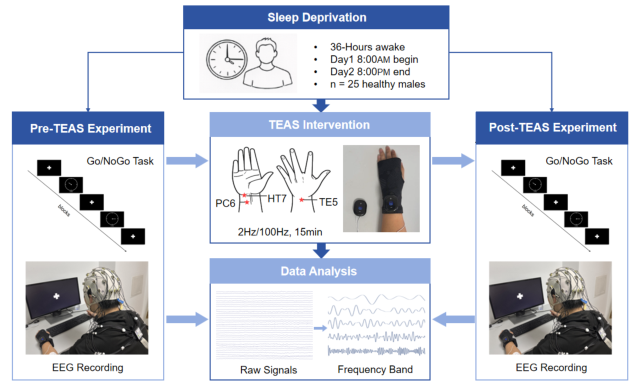
Experiment Procedure This study employed 36-hour sleep deprivation to establish a cognitive decline model. The entire process spanned 36 hours, with each experiment formally commencing at 8:00 AM on the designated day and concluding at 8:00 PM the following evening. Participants were required to remain fully awake throughout the sleep deprivation period, under continuous monitoring and supervision by the experimenter to ensure consistent and stable experimental conditions.
During the experiment, subjects completed a modified Go/NoGo task to assess their cognitive function. In the task, a round clock frame appeared on the screen containing a single hand. Within each block, the hand randomly appeared on either the left or right side of the dial. Following its appearance, the hand rotated clockwise immediately. Subjects were required to respond to target stimuli (by pressing the spacebar) while ignoring non-target stimuli. A timer located below the dial started when the hand appeared and continued until the interval ended or the subject responded. The task design aimed to test subjects’ response inhibition abilities.
EEG signals were recorded while subjects performed tasks. This experiment employed a 32-channel Brain Product EEG recorder with electrode placement following the international 10–20 system and a sampling rate of 500 Hz. After 36 hours of sleep deprivation, subjects first completed a modified Go/NoGo task with simultaneous EEG recording, followed by 15 minutes of electrical stimulation using the TEAS device. TEAS stimulation employed a 2Hz/100Hz
Regulatory Effects of Transcutaneous Electrical Acupoint Stimulation 61 sparse-dense waveform, with intensity adjusted to each subject’s maximum tolerance within a current range of 0.1–40mA. Stimulation points included Neiguan (PC6), Waiguan (TE5), and Shenmen (HT7). Following electrical stimulation, subjects repeated the identical task and signal acquisition. The experimental workflow is shown in Figure 1.
Figure 1: Experimental workflow.
Data Analysis The preprocessing of EEG signals included baseline correction, average reference of the whole brain, band-pass filtering (0.1–70 Hz), notch filtering (50 Hz), manual interpolation of bad channels and rejection of bad epochs. All signals were processed by independent component analysis (ICA), and the artifactual components were manually removed to ensure the quality of the analytical data.
In this study, analysis of EEG signals focused on changes in power spectral density (PSD), with particular attention to the power variation characteristics across five frequency bands: delta (1–4 Hz), theta (4–8 Hz), alpha (8–13 Hz), beta (13–30 Hz), and gamma (30–70 Hz). Paired t-tests were employed to compare differences in power changes before and after the TEAS intervention, with a significance level set at p < 0.05.
RESULTS
In the Go trial, TEAS-induced significant changes in PSD were primarily observed in the alpha band (8–13Hz), with the frontal and central-parietal regions as the main brain areas, as shown in Table 1. Alpha power at the midline frontal electrode Fz significantly increased post-intervention, rising from 1.02 to 1.12 (t(24) = 3.26, p = 0.003). Similarly, lateral frontal electrodes F3 and F4 showed consistent enhancement trends: PSD at F3 increased from 0.91 to 1.03 (t(24) = 2.84, p = 0.009), and F4 increased from 0.98 to 1.08 (t(24) = 2.35, p = 0.027). This suggests that under the response execution condition, frontal- related neural oscillatory activity was enhanced following the intervention.
Besides, alpha power in the central-parietal region also showed an upward trend following the intervention: PSD at CP2 increased from 0.82 to 0.92 (t(24) = 2.50, p = 0.020), at CP5 from 0.72 to 0.82 (t(24) = 2.09, p = 0.047), and at CP6 from 0.74 to 0.85 (t(24) = 2.47, p = 0.021). This indicates that the TEAS intervention effect also involves central-parietal regions associated with sensorimotor integration. Overall, spectral changes following TEAS intervention under Go conditions were characterized primarily by enhanced alpha activity in the frontal and central-parietal regions.
Table 1: Results of alpha-band PSD under Go trial.
CP6 0.74 ± 0.48 0.85 ± 0.63 2.47 0.021 In the NoGo trials, significant changes in PSD following TEAS intervention were broadly observed, primarily characterized by the theta band (4–8Hz), as shown in Table 2. Consistent activation was observed across multiple electrodes in the frontal region, with theta power at Fz increasing from 2.72 to 3.05 (t(24) = 2.62, p = 0.015), F3 from 2.18 to 2.68 (t(24) = 3.28, p = 0.003), and F4 from 2.25 to 2.61 (t(24) = 3.29, p = 0.003). This indicates a significant upregulation of frontal-related theta activity following TEAS under inhibitory control conditions. In the central region, theta power at Cz increased from 1.38 to 1.52 (t(24) = 2.23, p = 0.035);in the parietal region, Pz power increased from 1.52 to 1.77 (t(24) = 2.85, p = 0.009). Furthermore, in the central-parietal region, CP1 increased from 1.07 to 1.23 (t(24) = 2.88, p = 0.008), and CP2 from 1.09 to 1.28 (t(24) = 3.07, p = 0.005). Overall, under NoGo conditions, theta band activity showed synchronized enhancement across key brain regions including the frontal, central, parietal, and centroparietal lobes. This further supports the generality and stability of theta band changes during NoGo conditions, suggesting that rhythmic activity associated with inhibitory control and conflict monitoring is recruited across a broader network following TEAS intervention.
Table 2: Results of theta-band PSD under NoGo trial.
Regulatory Effects of Transcutaneous Electrical Acupoint Stimulation 63 Integrating the results from both conditions reveals distinct task-related PSD changes following TEAS intervention: The significant alterations in the Go condition primarily manifest as enhanced alpha activity in the frontal and centroparietal regions, whereas the NoGo condition is characterized by widespread theta band enhancement across the frontal-central-centroparietal- parietal regions. This pattern indicates that the TEAS intervention effect may exhibit different rhythmic modulation and spatial distribution characteristics under the distinct task demands of response execution and inhibitory control.
DISCUSSION
This study systematically explores the regulatory effects of TEAS on cognitive-related brain functions following sleep deprivation using a 36-hour sleep deprivation model, combined with an adapted Go/NoGo task and EEG frequency domain analysis. Results indicate that PSD changes before and after TEAS intervention exhibit clear state-frequency-region specificity. Under the response execution (Go) state, significant effects were primarily concentrated in the 8–13Hz alpha band and focused on frontal (Fz, F3, F4) and central- parietal (CP2, CP5, CP6) regions, manifesting as a general increase in PSD. Under the inhibitory control (NoGo) condition, a more extensive modulatory effect centered on the theta band was observed, spanning multiple critical brain regions including the frontal-central-centroparietal-parietal areas. Electrodes such as Fz, Cz, Pz, and CP1/CP2 similarly exhibited power enhancement. These findings suggest that TEAS modulates brain function through selective regulation across distinct cognitive networks and neural rhythms.
Alpha band oscillations represent a key rhythm associated with attentional gating, information filtering, and cortical excitability modulation. Its enhancement signifies selective activation of task-relevant processing channels. In sleep deprivation studies, individuals typically exhibit decreased attentional maintenance capacity and reduced response efficiency. In this experiment, following TEAS intervention, significant increases in alpha power were observed at electrodes Fz, F3, F4, CP2, CP5, and CP6 during Go trials. This indicates activation between frontal and central-parietal rhythmic waves, suggesting TEAS may promote the reallocation of attentional resources and enhance preparatory efficiency during response execution by synergistically restoring the top-down control-execution pathway. Theta band oscillations are associated with conflict monitoring and inhibitory control. During inhibitory control tasks, individuals must suppress dominant responses while maintaining rule representations, typically involving regulation of motor execution by prefrontal control nodes and the parietal attention system. In this study, synchronized activation patterns in the theta band during NoGo trials emerged across multiple nodes including the frontal, central, and parietal regions. This supports the hypothesis that TEAS may promote inhibition-related processing by improving rhythmic coordination within control networks.
In this study, the Go-state primarily involved response execution and sustained attention, relying on the gating and preparatory mechanisms of the frontal-centroparietal pathway, thus exhibiting prominent alpha rhythm modulation. The NoGo-state centered on inhibitory control and conflict monitoring, requiring stronger top-down control and cross-network coordination, consequently recruiting a broader range of theta-related activity. These findings suggest that TEAS intervention effects may exhibit task specificity: under different cognitive control demands, it preferentially modulates corresponding brain rhythms and network nodes to alleviate task- induced oscillatory activity caused by sleep deprivation.
CONCLUSION
This study explored the modulatory effects of TEAS on task-related EEG activity based on a 36-hour sleep deprivation experiment. Results revealed significant changes in PSD following TEAS intervention. Alpha-band power significantly increased in the frontal and centroparietal regions during the Go condition. Theta-band power showed widespread enhancement across frontal, central, centroparietal, and parietal regions during the NoGo condition. These findings suggest that TEAS may regulate task-related EEG oscillatory activity following sleep deprivation by selectively modulating key rhythmic activities within the prefrontal-parietal control network and the central-parietal motor execution network. Overall, this study provides frequency-domain EEG evidence for non-invasive intervention against sleep- deprivation-related cognitive decline, supporting the potential efficacy of peripheral acupoint electrical stimulation in modulating cognitive control- related brain activity. Future work will incorporate sham-stimulation controls, expand sample sizes, establish behavioral-EEG coupling validation, and further elucidate the mechanisms underlying TEAS’s cognitive regulation.
REFERENCES
Basner, M., Rao, H., Goel, N. and Dinges, D.F. (2013). Sleep deprivation and neurobehavioral dynamics. Current Opinion in Neurobiology, 23(5), pp. 854–863. doi: 10.1016/j.conb.2013.02.008. Chen, X., Kong, D., Du, J., Ban, Y. and Xu, H. (2022). Transcutaneous electrical
acupoint stimulation affects older adults’ cognition after general anesthesia: A meta-analysis. Geriatric Nursing, 46, pp. 144–156. Chuah, Y.L., Venkatraman, V., Dinges, D.F. and Chee, M.W. (2006). The neural basis of interindividual variability in inhibitory efficiency after sleep deprivation. Journal of Neuroscience, 26(27), pp. 7156–7162. Frank, M.G. and Heller, H.C. (2019). The function(s) of sleep. In: Sleep-wake
neurobiology and pharmacology. Cham: Springer International Publishing, pp. 3–34. Griggs, S., Harper, A. and Hickman Jr, R.L. (2022). A systematic review of sleep deprivation and neurobehavioral function in young adults. Applied Nursing Research, 63, 151552. Haciahmet, C.C., Frings, C., Beste, C., Münchau, A. and Pastötter, B. (2023).
Posterior delta/theta EEG activity as an early signal of Stroop conflict detection. Psychophysiology, 60(3), e14195.
Regulatory Effects of Transcutaneous Electrical Acupoint Stimulation 65 Jin, X., Ye, E., Qi, J., Wang, L., Lei, Y., Chen, P. and Yang, Z. (2015). Recovery sleep
reverses impaired response inhibition due to sleep restriction: Evidence from a visual event-related potentials study. PLOS ONE, 10(12), e0142361. Killgore, W.D. (2010). Effects of sleep deprivation on cognition. Progress in Brain Research, 185, pp. 105–129. doi: 10.1016/B978-0-444-53702-7.00007-5. Klimesch, W. (2012). Alpha-band oscillations, attention, and controlled access to
stored information. Trends in Cognitive Sciences, 16(12), pp. 606–617. Lian, J., Xu, L., Song, T., Peng, Z., Gong, X., Chen, J., Zhong, X., An, X., Chen, S. and Shao, Y. (2023). Decreased functional connectivity of brain networks in the alpha band after sleep deprivation is associated with decreased inhibitory control in young male adults. International Journal of Environmental Research and Public Health, 20(5), 4663. doi: 10.3390/ijerph20054663. Limbach, K. and Corballis, P.M. (2017). Alpha-power modulation reflects the
balancing of task requirements in a selective attention task. Psychophysiology, 54(2), pp. 224–234. Lopes Alves, R., Zortea, M., Mayor, D., Watson, T. and Steffert, T. (2025). Effect of different frequencies of transcutaneous electrical acupoint stimulation (TEAS) on EEG source localization in healthy volunteers: A semi-randomized, placebo- controlled, crossover study. Brain Sciences, 15(3), 270. Miller, N.L., Matsangas, P. and Shattuck, L.G. (2018). Fatigue and its effect on
performance in military environments. In: Performance under stress. Boca Raton: CRC Press, pp. 247–266. Tan, E., Troller-Renfree, S.V., Morales, S., Buzzell, G.A., McSweeney, M., Antúnez, M.
and Fox, N.A. (2024). Theta activity and cognitive functioning: Integrating evidence from resting-state and task-related developmental electroencephalography (EEG) research. Developmental Cognitive Neuroscience, 67, 101404. Wang, J., Lu, F.F., Ge, M.M., Wang, L.W., Wang, G., Gong, G.W. and Jiang, Z.W.
(2023). Transcutaneous electrical acupoint stimulation improves postoperative sleep quality in patients undergoing laparoscopic gastrointestinal tumor surgery: A prospective, randomized controlled trial. Pain and Therapy, 12(3), pp. 707–722. Wu, J., Zhou, Q., Li, J., Chen, Y., Shao, S. and Xiao, Y. (2021). Decreased restingstate alpha-band activation and functional connectivity after sleep deprivation. Scientific Reports, 11(1), 484.
LISTED IN THE AHFE DIGITAL BOOKSTORE CATALOGUE
Explore the complete digital volume
Neuroergonomics and Cognitive Engineering
Read this individual research paper free through the original publisher, or explore the full digital proceedings book.
ISBN 978-1-964867-83-0
Explore this ISBN in the AHFE Bookstore ↗Listed in the July 2026 bookstore catalogue; search by ISBN to confirm current availability · Digital book purchase optional