M. Mazzolaa, L. Forzonib, S. D’Onofriob, T. Marlerc, S. Becksd (2021). Evaluation of Professional Ultrasound Probes with Santos DHM. Handling Comfort Map Generation and Ergonomic Assessment of Different Grasps. In: Advances in Human Aspects of Healthcare. AHFE Open Access, vol. 13. https://doi.org/10.54941/ahfe100500
Sonographers’ work-related musculoskeletal disorder is a critical but often neglected problem, with reports indicating that approximately 80% of them experience pain from the extended use of sonography probes. Also transducer design can be a common cause of hand-wrist complaints. Moreover, there are many opportunities to minimize the risk of musculoskeletal disorders. A possible solution was proposed by Esaote, whereby the probe-handle shape is redesigned with a more ergonomically effective profile, appleprobe Design, that can be used with two or more different handling positions. This paper presents: details of this new design, where 6 different probes were analyzed experimentally for ergonomic purposes; virtual analyses of the evaluation of the contact surfaces (hand/probes) for each probe and each grasp, using an advanced Digital Human Model (DHM), Santos, and results of experiments to evaluate actual user discomfort. Results indicate the advantages of the proposed design and an acceptable correlation between the virtual and the experimental analyses. This work represents the first step towards the use of Santos DHM not only for comfort/ergonomics evaluation/validation after the production of the probe, but also for the simulation of new design concepts before prototypes are even manufactured, thus reducing production costs and improving quality.
The attention to sonographers’ work-related musculoskeletal disorders (WRMSD) is really crucial. Statistics report that approximately 80% of them scan in pain (Carmel and Murphy, 2000).
The origins of discomfort within a real clinical environment are different, with various effects on diverse areas of the sonographer’s body. Sonographers’ higher experienced discomfort anatomical sites are in the shoulder (76%), neck (74%), back (58%), wrist (59%) and hand/fingers (55%) (Murphy and Russo, 2000). As a consequence, there are different areas of intervention to minimize the risk of WRMSD . Among these, Ultrasound (US) transducer design seems to be the best predictor of hand-wrist complaints and muscular efforts (such as gripping the transducer, applying sustained pressure and scanning with a flexed or hyper-extended wrist) and it is significantly related to the increasing severity of symptoms in the hand, wrist and forearm area (Magnavita, 1999).
A possible solution was proposed by Esaote, who redesigned the conventional probe handle shape, proposing more ergonomically effective designed probes, appleprobe Design transducers, that offer the possibility to be grasped with two or more different positions in order to reduce the sonographer’s hand/wrist stress. As a consequence, the efficacy of the new design can be tested and validated, in order to verify if the Ergonomics requirements defined in the early stage of the Design process were achieved. Nevertheless, in the field of Professional US Probes, there is a lack of information and methods for the evaluation of the Design of a single Probe, in relation to the induced grasp.
There are many factors that concur in the Ergonomics Assessment of this Interaction. One is related to the sonographers’ subjective perception, based on the experience and/or on the first impression when testing a new product. Another factor, that can assume a great relevance, could be the level of muscular activity of the hand during a specific grasping, that can be index of fatigue. As an example, in a recent study (Vannetti et al., 2014) a Superficial Electromyography technique and a Motion Analysis Technologies were applied to US System User Interface and Probe Ergonomics Evaluation, with promising results. One additional factor to be considered in the Ergonomics assessment of a Professional Probe can be the distribution of the effort at the Hand Joints Torques (HJT) produced by the reaction forces of the probe pushing on the Patient during an exam. As proposed by Chaffin (Chaffin 2009), in Physical Ergonomics, once the design specifications were defined, and the Ergonomics requirements established, there are three different ways to proceed: the first one is to consult traditional Human Factors resources; the second one is to build and test prototypes with final users; the third one is to Test Virtual CAD prototype with Digital Human Models (DHM).
The aim of the proposed paper is to evaluate how the use of an advanced DHM, i.e. Santos Digital Human, can be reliable to simulate and to investigate the relation between the geometry of the conventional-designed model and the new-designed model of transducers in a virtual environment, to produce a hands-joint torque distribution map and to compare it with the user’s subjective discomfort perception.
The presented work consists of three different phases: the first one is related to the definition of the different grasps adopted by professional sonographers, and to the determination of the perceived contact points and the effort at the hand level; the second phase is the simulation of the observed grasps inside the Santos Virtual Environment. In the third and final phase, information about the reaction forces acting on the avatars hand were entered in the HJT Visualizer, an innovative capability of the Santos Software, to determine the distribution of the HJT at the Hand Level.
MATERIAL AND METHODS Sonography can be performed in different applications. As patient size plays a major role in the gripping and force application of the transducer, the experiment was performed in two parts, Abdominal and Vascular applications, to meet different conditions to simulate a scan. In addition, various probes are used with different consoles and dissimilar grasps are applied for diverse applications.
Experimental Set-Up and Simulation Conditions Grasps
The orientation of the transducer within the hand, which must be rotated to obtain different images, determined the type of grip used. There are 3 different types of grasp, depending on the analyzed probe application: Longitudinal Pincer Grip, Transversal Pincer Grip and Palmar Grip. An example of each grasp is shown in Figure 1.
Figure 1: from left to right, the longitudinal pincher grip, the transversal pincher grip and the palmar grip.
Figure available in the original PDF paper.
In the first step of the analysis, these grasps were observed for a Male and a Female expert sonographer, for each probe used in the presented work.
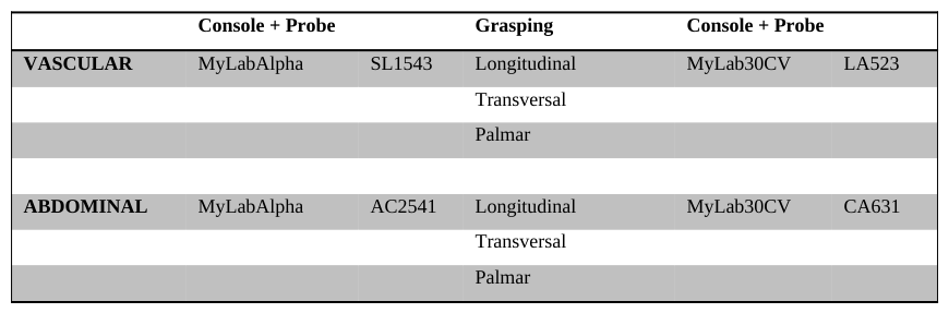
Probes 4 different US probes (Convex array probes CA631 and AC2541; Linear array probes LA523 and SL1543; Esaote S.p.A., Firenze, Italy; see Figure 2) were used for ergonomic analysis; each probe was used with a specific console and with three different grasps, as a consequence of the specific application (Abdominal/Vascular). AC2541 and SL1543 are ergonomically designed probes, appleprobe Design transducers. Each probe can be grabbed with the three different grasps described above, while each of them is associated with a single application and a specific console. In table 1, all the simulation conditions were reported.
Figure 2. From left to right: probe CA631, AC2541,LA523, SL1543Table 1: Experimental conditions for Simulations Console + Probe Grasping Console + Probe
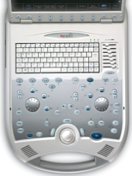
Consoles Two different Esaote US systems (Esaote S.p.A., Firenze, Italy) were used for the investigation: MyLabAlpha and MyLab30CV.
Figure 3: Left: MyLab30CV Console, presented as a simplified .obj model imported in the Santos Environment, and a picture of the real interface. In red are shown the different areas of investigations for the simulation conditions of the Left Hand. Right: MyLabAlpha Console presented as a simplified .obj model imported in the Santos Environment, and a picture of the real interface. In this case, the investigated zones for the simulations are shown in blue.
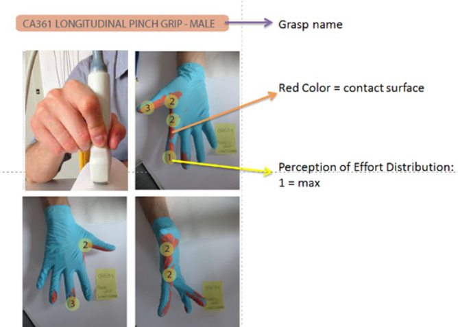
Contact Points and Efforts Evaluation The evaluation of the contact surfaces (hand/probes) for each probe and each grasp was conducted by two professional sonographer, one male and one female. Each sonographer was asked to grab all the probes with the three grasps, with no specific restrictions. For each condition, they were asked to identify the points of major effort perception and, whenever they were more than one, to order them from the highest (1 in the scale) to the lowest level of effort.
This experimental phase was planned with three main objectives: the first one was to analyze all the possible grasps for each probe, as a model for the simulations; the second one was to define the contact surfaces between the hand and the probe. To do that, each probe was painted, in order to visualize these surfaces on the hand (Figure 4). The third objective was to evaluate the subjective perception of the reaction forces and the final result is the determination of the contact points and the distribution of the force values according to the user’s rating. (Figure 4).
Figure 4. Contact Points and Effort distribution
To quantify the force acting at each contact point, a Mecmesin Compact Force Gauge 500N was used for both sonographers and the force produced was estimated in 25 N. This value of the force, higher than the maximum force applied to a probe, was considered in order to maximize the strength effects. Table 2 presents the force distribution used in this study, according to the number of contact points and the rates perceived by the subjects.
Table 2. Force distribution
RESULTS
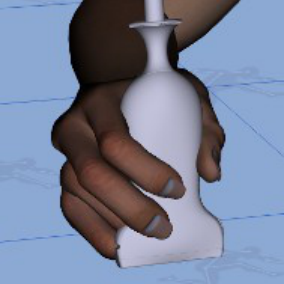
Figure 5 presents the observed grasps, the respective simulation inside Santos DHM virtual environment and the contact points and efforts, according to the sonographers’ subjective evaluation. In particular, Figure 5 shows respectively pictures of the palmar, the longitudinal and the transversal pinch for probes AC2541 and CA631, for the male subject and the palmar, the longitudinal and the transversal pinch for probes LA523, SL1543 for the female subject. This is just as a representation of the simulated grasps, the observed grasp and the contact surface and effort distribution for the 4 probes; the same information was evaluated for all the probes, for both sonographers.
Figure 5. From left to right, DHM simulated grasps, observed grasp, contact surface and effort distribution for different probes and for the two sonographers
Results demonstrate that the Palmar grip presents a limited number of stressed points (contact points) for both the male and the female sonographers. This result is promising, considering the hypothesis that a limited number of contact points, if the joints torques values are comparable, produces a lower level of hand stress. This results was confirmed by the subjective perception of the sonographers.
About the simulated grasp, results demonstrate that there are two main limitations in reproducing the correct grasps in the digital environment. The first one is related to the hand dimensions of both male and female avatars, that present different anthropometrical measures and types from the sonographers’ hands. In particular, the avatar male hand is shorter, while the avatar female hand is thinner than the corresponding sonographer’s one. The second evidence is that the Biomechanical model provided by Santos DHM does not permit a complete grasp manipulation, i.e. some rotational degrees of freedom, that could be useful in the reproduction of the different grasps. As an example, the biomechanical limitation of the avatar’s hand does not permit to completely wrap the probes in the different grasps, while in the real environment this is allowed. This limitation is also related to the fact that the human body has a variable and natural joint flexibility that, when appropriate, allows some movements like the hyper flexion of the middle phalanx.
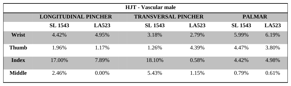
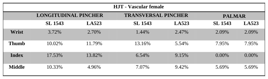
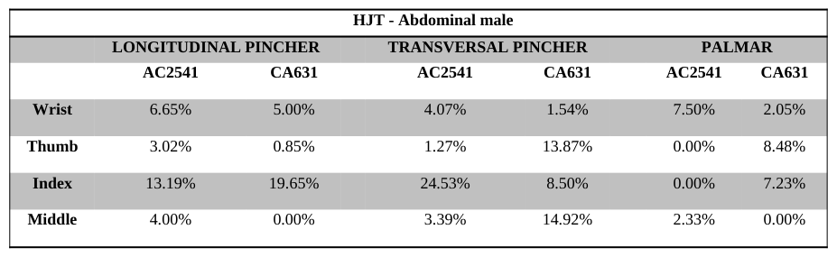
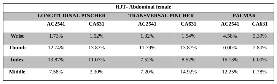
Hand Joint Torques Distribution Results of the HJT distribution are presented in Tables 3, 4, 5 and 6. Despite the biomechanical model provided by Santos DHM considers from 1 to 3 different joints for each finger, in this study each finger was considered with a global torques value and results are presented considering the mean value of the single joint torque computed by the simulation software. The reason for this choice is related to the fact that in this study what is investigated is the effort distribution among the single hand fingers. According to the HJT Visualizer capability of Santos DHM, results are presented in percentage, where 100% represents the maximum Joint torques value allowed.
Results demonstrate that, especially for the Abdominal application, there is an improvement in the torques values and distribution, due to the new appleprobe Design of the AC2541 transducer. In particular, the thumb and the index finger are completely not stressed and the numerical values of the torques at the wrist and middle finger level are commensurate with the values produced by the CA631 probe. On the contrary, these results are not confirmed for the female avatar.
Considering the experimental results presented in the recent studies (Vannetti et al., 2014), the probes evaluation provided by the Santos DHM is not completely coherent.
CONCLUSIONS
In this study, the ergonomic Evaluation of Professional US Probes with Santos DHM was proposed. The analysis were conducted considering two professional sonographers, and integrating an observational analysis of specific grasps, with a subjective comfort perception evaluation and quantitative data about HJT distribution in a virtual environment, using Santos DHM. This study is well promising, considering the attention of the development and the use of innovative ergonomic tools for a pro-active approach to ergonomic evaluation. Santos DHM, in fact, allows the evaluation of physical parameters, such as the HJT distribution, that is difficult to estimate with direct measures.
Results of this study demonstrate the needs of an integrated methodology (qualitative and quantitative data) to produce reliable data of the ergonomics of a professional US device. The first reason is that the information from real users, in terms of grasps, contact points and effort perception, is necessary to correctly prepare the simulation environment and the simulation conditions. In addition, these data can represent a validation of the simulation.
Results of the handling comfort distribution (torques map) are not completely coherent with results of other studies about the ergonomic assessment of professional probes. Nevertheless, this work is the first one using tools such DHM for this kind of evaluation, and future development can provide additional information about the reliability of these results.
Future development should consider advances in the Hand Modeling, especially for the female avatar, in term of link lengths (and consequently of the biomechanical response to the grasp simulation and the HJT generation), but also considering additional degrees of freedom, and an augmented flexibility of the fingers, that is more natural, even if very variable.
In conclusion, DHM seems to be promising as an early design tool for the ergonomic assessment of medical devices, offering innovative capabilities that are difficult to develop with traditional approaches. Results of the study demonstrate to be reliable, even if not completely coherent with other studies with traditional methods, but a refinement in specific software capabilities, together with a more strengthened methodological approach, can overcome these limitations and guarantee more reliable data, which are useful for the design process.
REFERENCES
Abdel-Malek, K., Arora, J., Yang, J., Marler, T., Beck, S., Swan, S., Frey-law, L., Mathai, A., Rahmatalla, S., and Patrick, A.
(2006), “Santos: A Physics-Based Digital Human Simulation Environment”, Paper presented at the 50th Annual Meeting of the Human Factors and Ergonomics Society, San Francisco, CA. Abdel-Malek, K., Yang, J., Kim, J. K., Marler, T., Beck, S., Swan, C., Frey-Law, L., Mathai, A., Murphy, C., Rahmatalla, S., and Arora, J. (2007), “Development of the Virtual-Human Santos, in Digital Human Modeling”, First International Conference on Digital Human Modeling, Beijing, China, Lecture Notes in Computer Science, 4561, Springer-Verlag, Berlin, 490-499. Abdel-Malek, K., Yang, J., Kim, J. K., Marler, T., Beck, S., Swan, C., Frey-Law, L., Mathai, A., Murphy, C., Rahmatalla, S., and
Arora, J. (2007, July), “Development of the virtual-human Santos”, Paper presented at the 12th International Conference on Human-Computer Interaction, Beijing, China. Abdel-Malek, K., Yang, J., Marler, T., Beck, S., Mathai, A., Zhou, X., Patrick, A., and Arora, J. (2006), “Towards a New Generation of Virtual Humans, in International Journal of Human Factors Modeling and Simulation”, 1(1), 2-39. Andreoni G., Costa F., Mazzola M., Fusca M., Romero M., Carniglia E., Zambarbieri D., Santambrogio G.C. (2013), “A
multifactorial approach and Method for Assessing Ergonomic Characteristics in Biomedical Technologies”, Advances in Human Aspects of Healthcare, edited by Duffy V., Chapter 1, 1- 12.
Brown, G., Baker, J., (2004), “Work-related musculoskeletal disorders in sonographers”, JDMS 20, 85–93. Gregory, V. (1998), “Musculoskeletal Injuries: Occupational Health and Safety Issues in Sonography”, Sound Effects 30. International Standard ISO 3411 (2007), “Earth-moving machinery – Physical dimensions of operators and minimum operator
space envelope”, Fourth edition. Magnavita, N., Bevilacqua, L., Mirk, P., Fileni, A, and Castellino, N. (1999), “Work-related Musculoskeletal Complaints in Sonologists”, J Occup Environ Med, Nov; 41(11):981-8. Mirk, P., Magnavita, N., Masini., L., Bazzocchi., M ., and Fileni., A. (1999), “Frequency of Musculoskeletal Symptoms in
Diagnostic Medical Sonographers. Results of a Pilot Survey”, Radiol Med, Oct; 98 (4):236-41. Murphy, C. and Russo, A. (2000), “Report: An Update on Ergonomic Issues in Sonography”, Healthcare Benefit Trust, EHS - Employee Health and Safety Services. NIOSH Technical Report, HETA 99-0093-2749 Smith, A.C., Wolf, J. G., Xie, G.Y, and Smith, M.D. (1997), “Musculoskeletal
Pain in Cardiac Ultrasonographers: Results of a Random Survey”, J. Am. Soc Echocardiogr, May; 10(4):357-62. Smith and Sainfort (1989) in Kuorinka et al. (1995), “Work-Related Musculoskeletal Disorders: A Reference for Prevention”, Philadelphia: Taylor & Francis. Society of Diagnostic Medical Sonography (2003), “Industry Standards for the Prevention of Work-Related Musculoskeletal
Disorders in Sonography”. Vanderpool, J.E., Friis, E.A., Smith, B.S., Harma, K.L. (1993), “Prevalence of carpal tunnel syndrome and other work-related musculoskeletal problems in cardiac sonographers”, JOM 36 (6), 604–610. Vannetti et al (2014), “Superficial Electromyography and Motion Analysis Technologies applied to Ultrasound System User
Interface and Probe Ergonomics Evaluation”, AHFE 2014. In press Village J., Trask C. (2007), “Ergonomic analysis of postural and muscular loads to diagnostic sonographers”, Int. Jour. Ind.
Ergonomics 37, 781-789. Yang, J., Marler, T., Beck, S., Abdel-Malek, K., and Kim, H.-J. (2006), “Real-Time Optimal-Reach Posture Prediction in a New Interactive Virtual Environment”, Journal of Computer Science and Technology, 21(2), 189-198. Yang, J., Marler, R. T., Kim, H., Arora, J. S., and Abdel-Malek, K. (2004), “Multi-Objective Optimization for Upper Body
Posture Prediction”, 10th AIAA/ISSMO Multidisciplinary Analysis and Optimization Conference, Albany, NY. Yang, J., Verma, U., Penmatsa, R., Marler, T., Beck, S., Rahmatalla, S., Abdel-Malek, K., and Harrison, C. (2008), “Development of a Zone Differentiation Tool for Visualization of Postural Comfort”, SAE 2008 World Congress, Detroit, MI.
LISTED IN THE AHFE DIGITAL BOOKSTORE CATALOGUE
Explore the complete digital volume
Advances in Human Aspects of Healthcare
Read this individual research paper free through the original publisher, or explore the full digital proceedings book.
ISBN 978-1-4951-2093-0
Explore this ISBN in the AHFE Bookstore ↗Listed in the July 2026 bookstore catalogue; search by ISBN to confirm current availability · Digital book purchase optional