F. Vannettia, T. Atzoria, G. Pasquinia, L. Forzonib, L. Modib, R. Molino-Lovaa (2021). Superficial Electromyography and Motion Analysis Technologies applied to Ultrasound System User Interface and Probe Ergonomics Evaluation. In: Advances in Human Aspects of Healthcare. AHFE Open Access, vol. 13. https://doi.org/10.54941/ahfe100493
The present work describes the ergonomics and usability preliminary tests regarding the human upper limb kinematics and strength evaluation of two diagnostic portable ultrasound scanners. The tests were performed by one expert sonographer in vivo on one subject in abdominal and vascular clinical applications and in vitro using a dynamometer. The two portable systems had different user interfaces and probe design: one system had traditionally-designed probes, a classic Soft keys interactive Menu, placed in the lower part of the screen, with toggles and buttons for the activation and adjustment of the functions, then a physical qwerty keyboard and a control panel. The other system had appleprobe-designed transducers, a touch screen integrated in the control panel with an interactive user interface and reduced number of physical buttons. Comparisons between the two systems considering the sonographer’s use are provided. Motion Analysis and Superficial Electromyography results will be presented and discussed on both systems regarding tests in vivo and in vitro only concerning Superficial Electromyography.
Diagnostic Ultrasound (US) systems are characterized by real-time examination, which forces to manage the US Probe with one hand (usually the right one in most clinical applications) and the US system User Interface (UI) with the other hand.
The concepts of UI usability and workflow, Probe design and ergonomics are familiar to US system designers and users (sonographers, physicians, nurses, paramedics, etc..) since many years. The issue of US systems and Probe ergonomics has been described and treated in many Standard and Guidance Documents from Regulatory Organizations, Healthcare Institutions and Sonographers Associations (Craig, 1985; Atjak and Gattinella, 1989; Pike et al. 1997; Smith et al., 1997; Gregory, 1998; Schoenfeld, 1998; Gregory, 1999; Magnavita et al., 1999; Evans et al., 2009; Sommerich et al., 2011). However, today no Industrial Standards dedicated to the design of US systems (control panels, UI and probes) are available. The different design results provided by the diverse US system producers are usually implemented through the company own experience and in most of the cases continuing to prefer interface solutions easy to be used by their loyal customers, even if not ergonomically optimized in general terms. Sub-optimally designed US systems UI and Probe ergonomics led to a high presence of work related musculoskeletal disorders (WRMSD) among US system users (Habes and Baron, 1999; Society of Diagnostic Medical Sonography, 2003; DHHS (NIOSH), 2006; The Society of Radiographers, 2007). The problem of WRMSD is nowadays even more crucial, due to the increased number of US systems sold per year and consequently the enlarged number of users, the growth of the total annual amount of exams worldwide performed, the increased number of scanned patients, working stress and working hours. Recently, also Regulatory bodies, such as FDA, have increased their attention to the ergonomics and usability aspects of US systems (U.S. Department of Health and Human Services, Food and Drug Administration, 2011; European Standard EN 62366, Medical devices, 2008).
Ergonomics improvements related to US system UI and US Probes are characterized by a complex and varied group of aspects, depending on the clinical application, the kind of US system and Probe used and the level of US system user expertise. Therefore, it’s a complex topic which has to consider multiple aspects, whose characterization and analysis approach is difficult to be defined and targeted.
The present preliminary work describes the measurement of ergonomics and usability performances of a classic UI portable US system equipped with classically-designed US Probes, compared to a new one with an innovative UI and a design characterized also by innovatively-designed US Probes.
MATERIALS AND METHODS
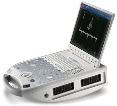
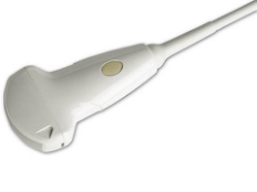
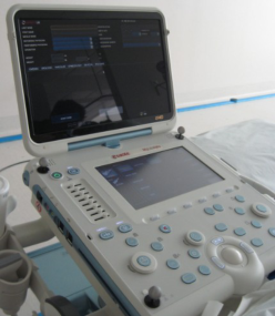
Two US systems (MyLabAlpha and MyLab30CV, Esaote S.p.A., Firenze, Italy; see Figure 1) and four probes (two Linear array transducers and two Convex array transducers: SL1543, LA523, AC2541 and CA631, Esaote S.p.A., Firenze, Italy; see Figure 2) were evaluated by one expert sonographer (with the same level of usage knowledge for both systems and for all probes) in two different clinical applications (vascular and abdominal) on one subject, following a detailed US scanning examination procedure, simulating as much as possible the real clinical practice. In vitro tests were also performed regarding Probe’s ergonomics evaluation using a Dynamometer.
A B
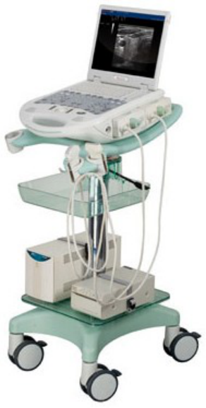
Figure 1. A) MyLab30CV US system; B) MyLabAlpha US system
A B
C D
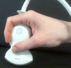
Figure 2. A) Linear array probe Sl1543 appleprobe (MyLabAlpha); B) Convex array probe AC2541 appleprobe (MyLabAlpha); C) Linear array probe LA523 (MyLab30CV); D) Convex array probe CA631
(MyLab30CV)
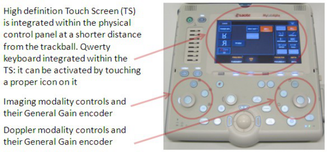
MyLabAlpha US system has a high definition touch screen (TS), that is integrated within the physical control panel at a shorter distance from the trackball. A reduced number of well-spaced physical controls (buttons, toggles, sliders and encoders) is available on the panel, in order to facilitate their detection and use by the operator. Buttons and controls which can be activated (or are already active in a certain system status) are lighted on. Some of the most frequently used controls/adjustments are physical, in order to be activated/changed while the sonographer’s eyes are focused on the main screen where the echo image is shown: Freeze, General Gain, TGC, Automatic Adjustment of imaging and Doppler traces, line/update, saving options (image/clip/print). The same concept is applied to modality buttons like B-Mode, Color Doppler (CD), Pulsed Wave (PW) Doppler, Continuous Wave (CW) Doppler, M-Mode. (Andreoni et al., 2013; Forzoni et al., 2012). See Figure 3.
Figure 3. MyLabAlpha UI
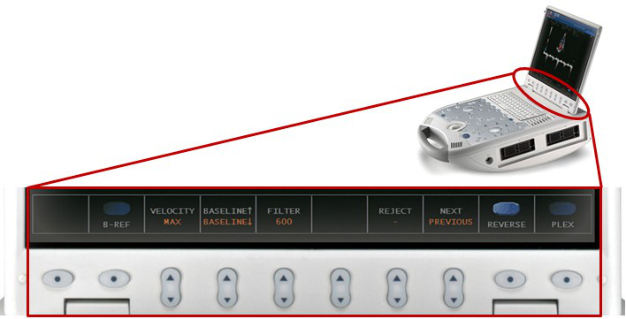
MyLab30CV has a typical UI for portable US systems: a reconfigurable, multi-level, Graphical UI (GUI) - Soft key - menu is present in the lower part of the monitor, with direct controls right under the screen. A physical control panel with encoders for General Gain and Time Gain Compensation (TGC) controls and with buttons for modality, measure, annotations, body-marks, saving options and line/update operations are placed around the trackball. Between the Soft key menu controls and the physical control panel there is a qwerty keyboard for Patient ID data entering, free text annotations and Report fulfilling and comments (Andreoni et al., 2013; Forzoni et al., 2012). See Figure 4.
A
B
Figure 4. A) MyLab30CV UI; B) ; Close up of MyLab30CV Soft key Menu UI
SL1543 and AC2541 are, respectively, a linear and a convex probe with an innovative appleprobe design which enables a dual-possibility hand grip (pinch grip and palmar grip) in order to provide a neutral wrist position (see Figure 4).
A B
Figure 4: A) palm hold; B) pincer hold
Measurements of both US system usability and workflow and US Probe ergonomics and handling were performed using Motion Analysis (MA) and Superficial Electromyography (SEMG), with in vitro and in vivo tests. In vitro tests were performed focusing only on the probe handling strength using SEMG: a dynamometer (BFG 1000N, Mecmesin, United Kingdom) was used in order to test the muscle strength with the different probes pressed directly on its measurement interface considering a maximized force (to enhance the possible strength differences between the tested probes). The examination protocols for the in vivo tests were performed in abdominal application (liver and abdominal Aorta - AA) and vascular application (Common Carotid Artery - CCA) according to the following scanning protocols.
Abdominal application:
Probe, application and user preset selection.
Start real-time examination.
B-Mode Imaging: visualization of AA, adjustment of depth, frequency, general gain and Time Gain Compensation (TGC) parameters and image parameters such as XView (speckle reduction and image quality enhancement algorithm) in order to optimize the image quality. Image freeze and generic measurement of AA width (distance); storage of imaged AA (still frame) with the performed measure.
CD: visualization of the AA CD signal, CD Region of Interest (ROI) dimensions, position and electronic steering (considering the longitudinal scanning) adjustment, optimization of the Pulse Repetition Frequency (PRF) parameter, the Doppler frequency and the Doppler general gain, in order to avoid Aliasing effect.
PW Doppler: sample of the AA Doppler signal, adjustment of PW line of sight, PW sample volume (SV) position,. Doppler angle correction (θ), velocity (scale) and PW General Gain parameters . Velocity generic measurement over the PW Doppler trace of the AA blood flow signal: measurement of maximum velocity point and storage of AA still frame of the performed measure.
Exam closure and storage in the internal local Hard Disk of the system.
Vascular application:
Probe, application and user preset selection.
Start real-time examination.
Left Side examined.
B-Mode Imaging: visualization of the Left CCA. Adjustment of depth, frequency, general gain and TGC parameters in order to optimize the image quality. Optimization of the image parameters such as XView and MView. Image freeze and generic measurement of CCA width; storage of CCA still frame of the performed measure.
CD: visualization of the CCA CD signal, CD Region of Interest (ROI) dimensions, position and electronic steering (considering the longitudinal scanning) adjustment, optimization of the Pulse Repetition Frequency (PRF) parameter, the Doppler frequency and the Doppler general gain, in order to avoid Aliasing effect.
PW Doppler: sample of the CCA Doppler signal, adjustment of PW line of sight, PW sample volume (SV) position,. Doppler angle correction (θ), velocity (scale) and PW General Gain parameters . Velocity generic measurement over the PW Doppler trace of the CCA blood flow signal: measurement of maximum velocity point and storage of CCA still frame of the performed measure.
Exam closure and storage in the internal local Hard Disk of the system. Human Aspects of Healthcare (2021)
The tests were performed in a simulated clinical environment, focusing only on UI workflow of the two compared systems and on the tested Probes. The US systems were positioned on their proper height adjustable cart (with four swiveling wheels) at the same height (see Figure 5).
A B
Figure 5: A) MyLab30CV on its height-adjustable cart with four swiveling wheels; B) MyLabAlpha on its height-adjustable cart with four swiveling wheels
The presence of a prompter was considered necessary, in order to remind the scheduled sequence of the actions as listed in the protocol. Upper-body kinematic and muscle strength of the sonographer were, respectively, recorded through a six cameras optoelectronic system (SmartDx400, BTS Spa, Milano, Italy) and a Multi-channel Superficial Electromyography (Mizar40, EB Neuro Spa, Firenze, Italy), while the operator performed the clinical US examinations. The analysis was performed on the left arm (the one used to work on the US system) and on the right arm (used to handle the Probe).
RESULTS
For in vitro test the SEMG strength measurements were performed considering a value of 15 N force impressed to the linear probes (SL1543 and LA523) and a value of 40 N force impressed to the convex probes (AC2541 and CA631): the appleprobe designed transducers were tested with pinch longitudinal and palmar grip, the conventionally designed probes were tested with pinch longitudinal and pinch transverse grip (see Figure 6).
Figure 6: In vitro SEMG tests for the probe strength mesurements
The SEMG was performed placing the electrodes on the following muscles: on the right side first dorsal interosseus (FDI) muscle, flexor carpi (FDC), extensor carpi (EDC), deltoid (DELT) and trapezius; on the left –side the electrical activity of the trapezius and the pectoralis major has been measured. The probes were pressed directly on the dynamometer measurement interface, with a proper support developed for the interaction probe/dynamometer; the sonographer was recommended to maintain the compression force direction aligned to the measurement axis of the dynamometer. The data shows an higher muscular effort during the compression with the conventionally- designed probe (AC2541appleprobe with palmar grip and CA631 conventionally-designed probe with transverse grip). See Figure 7 and Figure 8.
Figure 7: The picture shows the different SEMG traces between the in vitro test using the AC2541 convex appleprobe palmar grip (left) and the CA631 conventional convex probe with transverse pincer
Figure available in the original PDF paper.
grip (right) which induces a globally higher muscular effort
Figure 8: In vitro tests: the graph represents the mean value of the standard deviation of the rectified
SEMG signals for each considered muscle
The in vivo tests were performed on a simulated clinical environment, with the systems positioned at the cart height of 86 cm (height measured from ground to trackball position, which can be considered the center of the US system user interface). The examination bed was positioned at a height of 60 cm.
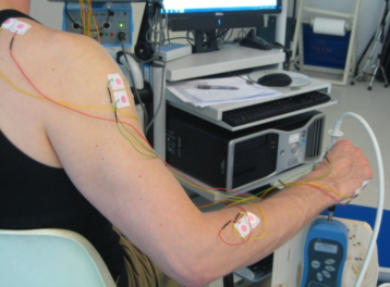
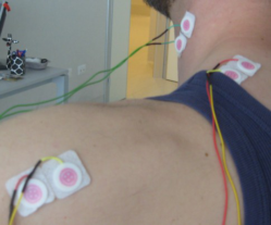
The motion analysis was performed with 3 passive reflective markers, placed on the system control panel as a reference and with two markers on the left hand (see Figure 9). The SEMG was performed placing the electrodes on the following muscles: on the right side first dorsal interosseus muscle (FDI), abductor digiti minimi (ADM), adductor brevis pollicis (ABP), flexor carpi (FDC), extensor carpi (EDC), deltoid (DELT) and trapezius (TRAP); on the left –side we measure the electrical activity of the trapezius (TRAP) and the pectoralis major (PET). See Figure 10.
A B C
Figure 9. A and B) Three refelective markers placed on the system control panels; C) Two reflective markers placed on the left handFigure 10. Example of SEMG electrodes positioning for the in vivo test
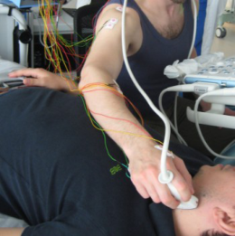
The mean duration of the test both in abdominal application (convex probe – AA examination) and in the vascular protocol (linear array probe – neck area CCA examination) was 157 ± 4 s. See Figure 11.
Figure 11. Example of in vivo test: A) Vascular application CCA; B) Abdominal application AA scanning
The left hand trajectories analysis demonstrated that the MyLab30CV UI required a higher distance to be covered to perform all the tasks for both the operators. The total distance covered by the left hand during the test on MyLabAlpha was lower than the one on MyLab30CV (see Figure 12).
Figure 12. The graph represents the total distance, in mm, covered by the left hand during both the abdominal and the carotid exams.
Figure available in the original PDF paper.
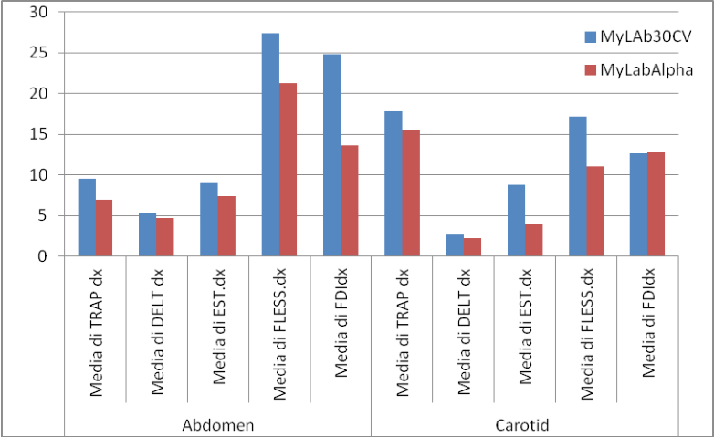
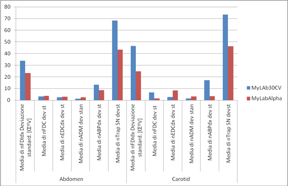
The SEMG muscle strength analysis demonstrated that the sonographer presented lower EMG values using the MyLabAlpha system and its related apple-design probes: in particular the reduction is appreciable in the left side muscles, in the first dorsal interosseus and in the adductor pollicis, especially during the vascular (in particular, carotid) examination (figure 3). These EMG values, using the setting with MyLabAlpha, achieved reductions from 31% (in the FDI) to 79% (in the ABP). See Figure 13.
These data may explain the sonographer’s perception of a more comfortable posture during the tests, using the MyLabAlpha system and its related apple-design probes.
Figure 13. In vivo tests: the graph represents the mean value of the standard deviation of the rectified
SEMG signals for each considered muscle
DISCUSSION
The new portable US system MyLabAlpha offers an easier UI with a reduced number of controls, in order to have few commands only when necessary and what is needed. Results demonstrated that there was a significant reduction of the left hand movements and muscle strength in all the performed tests. MyLabAlpha system produced an improvement of the physical usability and this was coherent with the innovative design of the physical interface.
The original appleprobe-designed transducers reduced the muscle strength in all the applications tested, in comparison to traditionally designed linear and convex probes.
CONCLUSION
MyLabAlpha has a smaller physical control panel that lowered the users’ hand/arm movements. The trackball closer to the General Gain encoders, TGC sliders and the qwerty keyboard (virtual qwerty keyboard), the reconfigurable controls (toggles) and GUI (TS) are solutions which reduced the strength and the time-of-flight of the operator’s hand and arm, reducing the upper limb stress while examining. The use of a high definition TS concentrates all the main features of the reconfigurable GUI in a well-defined area close to the trackball, reducing repetitive distant movements related to reaching and several repeated uncomfortable actions obtaining a 40% reduction during the abdominal exam and a 16% reduction in the carotid exam. The new probes SL1543 and AC2541demonstrated a reduced muscle strength regarding both the tested clinical applications, with respect to the traditionally-designed transducers.
The performed measurements represent a preliminary evaluation of the innovative UI of the MyLabAlpha and its related appleprobe-designed transducers, with respect to a traditional portable US system UI solution such as the one of the MyLab30CV. The obtained kinematic measurement indicates a reduction in the possible causes of WRMSD using an innovative portable US system UI (MyLabAlpha) with respect to the use of a traditionally designed UI. Same considerations were obtained regarding appleprobe design of the transducers and the traditionally-designed ones.
REFERENCES
Andreoni, G., Zambarbieri, D., Forzoni, L., Mazzola, M., D’Onofrio, S., Viotti, S., Santambrogio, G., Baselli, G. (2013), “Motion Analysis and Eye Tracking Technologies applied to Portable Ultrasound Systems User Interfaces Evaluation”, ICCMA 2013, International Conference on Computer Medical Applications. Atjak, A., and Gattinella, J.A, (1989). “Ergonomics in ultrasound equipment: productivity and patient throughput”, Radiol
Manage, vol. 11(4), pp. 38-40.Pike, I., et al. (1997), “The prevalence of musculoskeletal disorders among diagnostic medical sonographers”, Journal of Diagnostic Medical Sonography, vol. 13(5), pp.219-227. Craig, M. (1985), “Sonography: an occupational health hazard”, Journal of Diagnostic Medical Sonography, vol.1, pp. 121-125. DHHS (NIOSH, 2006), “Preventing Work-Related Musculoskeletal Disorders in Sonography”. Publication No. 2006–148. European Standard EN 62366, Medical devices (2008), “Application of usability engineering to medical devices”, IEC 62366:2007. Evans, K., Roll, S., Baker, J. (2009), “Work-Related Musculoskeletal Disorders (WRMSD) Among Registered Diagnostic
Medical Sonographers and Vascular Technologists. A Representative Sample”, Journal of Diagnostic Medical Sonography, vol. 25(6), pp.287-299. Forzoni, L., Guraschi, N., Fertino, C., Delpiano, M., Santambrogio, G., Baselli, G., Costa, F., Romero, M., Andreoni, G. (2012), “Case Study of Integrated Ergonomic Assessment of a Portable Ultrasound System”, AHFE 2012, Applied Human Factors and Ergonomics in Healthcare. Gregory, V. (1998), “Musculoskeletal Injuries: Occupational Health and Safety Issues in Sonography”, Sound Effects, vol. 30. Gregory, V. (1999), “Report on the results of the Australian Sonography. Survey on the prevalence of musculoskeletal disorders
among Sonographers, OHS Update”, Sound Effects, vol. 42. Habes, D.J., and Baron, S. (1999), “NIOSH Ergonomics evaluation of sonographers at St. Peter’s University Hospital”, Health Hazard Evaluation Report, HETA, 99-0093-2749. Magnavita, N., Bevilacqua, L., Mirk, P., Fileni, A., and Castellino, N. (1999), “Work-related Musculoskeletal Complaints in
Sonologists”, J. Occup. Environ. Med., vol. 41(11), pp. 981-8. Schoenfeld, A. (1998), “Ultrasonographer’s wrist – an occupational hazard”, Ultrasound Obstet Gynecol, vol. 11, pp. 313–316. Smith, A.C. (1997), Wolf, J.G., Xie, G.Y., and Smith, M.D, “Musculoskeletal Pain in Cardiac Ultrasonographers: Results of a Random Survey”, J. Am. Soc. Echocardiogr., vol.10(4), pp.357-62. Society of Diagnostic Medical Sonography (2203), “Industry Standards for the Prevention of Work-Related Musculoskeletal
Disorders in Sonography”, Plano, Texas, USA. Sommerich, C., et al. (2011), “Participatory Ergonomics Applied to Sonographers’ Work”, Proceedings of the Human Factors and Ergonomics Society Annual Meeting, vol. 55(1), pp.1067-1070. The Society of Radiographers (2007), “Prevention of Work Related Musculoskeletal Disorders in Sonography”, UK. U.S. Department of Health and Human Services, Food and Drug Administration, Center for Devices and Radiological Health,
Office of Device Evaluation (2011), “Draft Guidance for Industry and Food and Drug Administration Staff. Applying
Human Factors and Usability Engineering to Optimize Medical Device Design”.
LISTED IN THE AHFE DIGITAL BOOKSTORE CATALOGUE
Explore the complete digital volume
Advances in Human Aspects of Healthcare
Read this individual research paper free through the original publisher, or explore the full digital proceedings book.
ISBN 978-1-4951-2093-0
Explore this ISBN in the AHFE Bookstore ↗Listed in the July 2026 bookstore catalogue; search by ISBN to confirm current availability · Digital book purchase optional