Zain Tariq, Mahmood Al Zubaidi, Marco Agus, Mowafa Househ (2026). FADA: An AI-Enabled Platform for Prenatal Ultrasound Analysis, Clinical Decision Support, and Automated Reporting. In: Human Interaction and Emerging Technologies (IHIET 2026). AHFE Open Access, vol. 236, pp. 401–408. https://doi.org/10.54941/ahfe1008115
Prenatal ultrasound examination is the primary screening tool for monitoring fetal development and identifying hereditary abnormalities before birth. Despite its widespread use, accurately interpreting fetal brain ultrasound images requires specialized expertise and considerable clinical experience. Variations in operator skill, image quality, reporting practices, and access to specialist services can influence diagnostic consistency and timeliness. These challenges highlight the need for decision-support tools that can assist clinicians in image interpretation while fitting naturally into existing clinical workflows.This paper presents the Fetal Abnormality Detection Application (FADA), an AI-enabled healthcare informatics platform designed to support fetal brain ultrasound assessment through automated image analysis, clinical decision support, interactive consultation, and report generation. The platform provides an integrated environment where healthcare professionals can upload ultrasound images, receive automated measurements and abnormality assessments, interact with an intelligent clinical assistant, and obtain structured diagnostic reports through a unified interface.At the core of FADA is a curated dataset containing more than 3,000 expert-annotated fetal brain ultrasound images. The dataset focuses on clinically important anatomical structures, including the Cavum Septum Pellucidum (CSP) and Lateral Ventricles (LV), both of which serve as important indicators of fetal neurological development. The annotated dataset forms the basis for machine learning models trained to identify relevant anatomical regions, perform measurements, and detect patterns associated with developmental abnormalities. The platform supports automated assessment of these structures and produces quantitative measurements that can assist clinicians during prenatal evaluation.The system architecture follows a modular healthcare informatics design consisting of four primary functions: user interaction, clinical data processing, intelligent analysis, and results management. Healthcare professionals interact with the platform through a web-based clinical workspace that supports image submission, consultation, and review of findings. Submitted images are processed through an AI analysis pipeline that performs anatomical identification, measurement extraction, abnormality assessment, and generation of preliminary findings. In parallel, an intelligent conversational assistant enables users to ask questions regarding imaging findings, measurements, and clinical interpretation, providing contextual guidance and explanatory information. The outputs of these services are consolidated into structured clinical summaries and downloadable reports suitable for review and incorporation into existing clinical documentation workflows.A distinguishing aspect of FADA is the integration of image interpretation and clinical reporting within a single workflow. Rather than functioning solely as a diagnostic algorithm, the platform acts as a healthcare information system that connects image acquisition, automated analysis, clinician interaction, and report generation. This approach reduces repetitive administrative tasks, promotes reporting consistency, and supports traceable documentation of imaging findings.The proposed platform demonstrates how artificial intelligence can be embedded within healthcare information systems to support routine prenatal screening and fetal assessment. By combining expert-annotated imaging data, automated anomaly detection, conversational clinical assistance, measurement extraction, and report generation, FADA provides a practical model for computer-assisted prenatal diagnostics. The platform illustrates the potential of healthcare informatics to improve the efficiency, consistency, and accessibility of fetal ultrasound assessment while preserving clinician oversight in the diagnostic process.
Prenatal ultrasound is the primary imaging modality for monitoring fetal development and detecting structural abnormalities during pregnancy due to its safety, accessibility, and real-time imaging capability (Lawrie, 2017). Assessment of the fetal brain is particularly important because abnormalities
affecting structures such as the Cavum Septum Pellucidum (CSP) and Lateral Ventricles (LV) may indicate underlying neurological developmental disorders (Salomon et al., 2022).
Advances in artificial intelligence have led to significant progress in automated analysis of fetal ultrasound images. Deep learning techniques have been applied to fetal standard-plane detection, anatomical localization, segmentation, biometric measurement, and abnormality identification, demonstrating improved consistency and reduced operator dependence (Baumgartner et al., 2017; Burgos-Artizzu et al., 2020). More recent studies have reported promising results for automated fetal brain assessment and anomaly detection from ultrasound examinations (Yaqub et al., 2017; Yousefpour et al., 2023).
Despite these advances, most existing solutions remain focused on isolated analytical tasks and are typically evaluated as research prototypes. Clinical functions such as workflow integration, decision support, auditability, clinician interaction, and automated report generation are often absent, limiting their practical adoption in healthcare environments (Chen et al., 2021). Consequently, a gap persists between high-performing AI models and operational healthcare informatics platforms that support end-to-end prenatal assessment.
To address this gap, this paper presents the Fetal Abnormality Diagnostic Application (FADA), an AI-enabled healthcare informatics platform for prenatal ultrasound analysis. FADA integrates image processing, anatomical structure identification, measurement extraction, abnormality screening, clinician review, and automated report generation within a unified workflow. Supported by a repository of more than 3,000 expert-annotated fetal brain ultrasound images, the platform focuses on CSP and LV assessment while providing decision-support and documentation capabilities. By combining AI-driven image interpretation with clinical workflow support, FADA aims to improve consistency, efficiency, and traceability in prenatal ultrasound assessment.
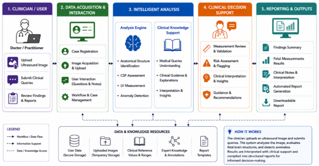
Figure 1: High-level functional architecture and workflow of the FADA platform for fetal anomaly diagnosis, clinical decision support and automated reporting in prenatal care.
FADA: An AI-Enabled Platform for Prenatal Ultrasound Analysis, Clinical Decision 403 FADA: AN AI-ENABLED PRENATAL ULTRASOUND PLATFORM
FADA (Fetal Abnormality Diagnostic Application) is a healthcare informatics platform designed to support prenatal ultrasound assessment through artificial intelligence, clinical decision support, and automated reporting. The platform combines image analysis, clinician interaction, and documentation services within a unified workflow to assist fetal brain abnormality screening and reporting.
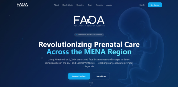
Figure 2: Web view of the FADA platform (HBKU, n.d.).
Dataset and Clinical Context Accurate AI-assisted diagnosis requires clinically representative and expertly annotated data. FADA is therefore built upon a curated fetal brain ultrasound dataset developed specifically for prenatal neurological assessment. The web view of the FADA platform is shown in Figure 2.
The platform utilizes a repository of more than 3,000 expert-annotated fetal brain ultrasound images. Dataset development focused on clinically significant anatomical structures, particularly the Cavum Septum Pellucidum (CSP) and Lateral Ventricles (LV). The CSP is a fluid-filled midline structure in fetal brain, whose absence may indicate severe neurodevelopmental abnormalities, while enlargement of the lateral ventricles beyond 10 mm is commonly associated with ventriculomegaly and abnormal brain development. Expert annotations provide the reference information required for model training, validation, and performance evaluation.
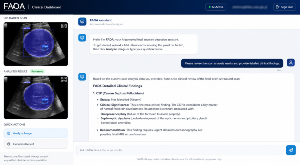
System Architecture FADA employs a modular architecture that separates user interaction, application services, AI inference, and data management functions, as shown in Figure 1. This design enables scalability, maintainability, and integration with existing healthcare workflows. Healthcare practitioners access the platform through a web-based interface that supports ultrasound image upload, result visualization, report retrieval, and clinical interaction as shown in Figure 3. The architecture consists of a presentation layer, an
application programming interface (API) layer, an AI intelligence layer, and a data management layer.
API Layer: The API layer coordinates communication between platform components, while dedicated AI services perform image analysis and provide clinical guidance. Supporting data assets include trained machine learning models, uploaded ultrasound images, reference measurement datasets, and report-generation templates.
AI Intelligence Layer: The core inference engine uses a YOLOv5s object detection model (∼7.2 M parameters, 6.7 MB checkpoint) trained to localize four anatomical targets in the transventricular plane: biparietal diameter (BPD) endpoints, occipitofrontal diameter (OFD) endpoints, cavum septi pellucidi (CSP), and lateral ventricular atria (LV). The model predicts bounding boxes and class labels; BPD/OFD are derived from skull box extents, while CSP length and LV width are measured directly from detected regions. Biometric estimation (gestational age, head circumference, fetal weight) follows established clinical formulas applied to the detected landmarks.
AI Analysis Pipeline The core functionality of FADA is provided through an automated analysis pipeline that transforms ultrasound images into clinically meaningful findings.
The workflow begins when a clinician uploads a fetal brain ultrasound image through the platform interface as shown in Figure 1. The image is forwarded to the fetal anomaly detection service, where trained machine learning models identify relevant anatomical structures and extract diagnostic measurements. The system evaluates the presence or absence of the CSP, measures ventricular dimensions, and assesses findings against established clinical thresholds. Detected abnormalities and measurement deviations
FADA: An AI-Enabled Platform for Prenatal Ultrasound Analysis, Clinical Decision 405 are automatically flagged, producing quantitative outputs that support subsequent clinical assessment and documentation.
Clinical Decision Support and Automated Reporting Beyond image interpretation, FADA provides decision-support capabilities that assist clinicians in translating analytical outputs into actionable clinical information.
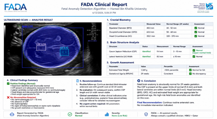
Figure 4: Clinical report generated by the FADA platform. (HBKU, n.d.).
Analysis results are presented through an interactive dashboard that summarizes detected findings, anatomical measurements, and clinical interpretations, as shown in Figure 3. These outputs are incorporated into automatically generated clinical reports that can be reviewed, downloaded, and integrated into routine documentation processes as shown in Figure 4. The reporting framework standardizes presentation of diagnostic findings while reducing administrative workload. By combining AI-driven image analysis, clinician interaction, decision support, and report generation within a single workflow, FADA functions as a complete prenatal ultrasound assessment platform rather than a standalone image-processing tool.
EVALUATION AND DISCUSSION This section examines the operational capabilities of FADA, its integration within prenatal ultrasound workflows, and the current limitations that motivate future development. The discussion focuses on platform-level evaluation and practical applicability in clinical environments.
Performance Evaluation The FADA platform was evaluated using a repository of more than 3,000 expert-annotated fetal brain ultrasound images. Assessment focused on the
platform’s ability to identify clinically relevant anatomical structures, extract measurements, detect abnormalities associated with CSP and LV assessment, and generate structured outputs for clinician review.
Experimental observations demonstrated that the integrated AI pipeline successfully performed automated image analysis, measurement extraction, abnormality flagging, and report generation within a single workflow. The system could produce consistent outputs across uploaded ultrasound images while reducing the manual effort associated with measurement interpretation and documentation. The evaluation confirmed the feasibility of combining AI-driven fetal brain assessment with healthcare informatics functions in a unified platform environment.
Clinical Workflow Integration A key objective of FADA is to bridge the gap between AI-based image analysis and routine clinical practice. Unlike conventional research prototypes that terminate at prediction generation, FADA incorporates image upload, automated analysis, clinician interaction, decision support, and report generation within a single operational workflow.
The platform enables healthcare practitioners to upload fetal ultrasound scans, review automated findings, obtain contextual guidance through the integrated clinical assistance module, and generate structured reports without requiring multiple disconnected systems. This workflow reduces administrative overhead and improves consistency in documentation. The modular architecture also supports future integration with electronic health record (EHR) systems, hospital information systems, and telemedicine platforms.
Limitations and Future Directions Although FADA demonstrates the potential of AI-assisted prenatal ultrasound assessment, several limitations remain. The current implementation focuses primarily on fetal brain analysis, with emphasis on CSP and LV assessment. Additional anatomical structures and fetal abnormality categories remain outside the present scope of the platform.
Furthermore, evaluation has been conducted using curated and expert- annotated datasets. Broader validation across multiple healthcare institutions, imaging devices, patient populations, and clinical settings is required to establish generalizability and robustness. Regulatory, interoperability, privacy, and clinical governance considerations will also require further investigation before large-scale deployment.
Future development will focus on expanding the annotated dataset, incorporating additional fetal anatomical structures, enhancing anomaly detection capabilities, and conducting multi-center clinical validation studies. Planned enhancements also include integration with healthcare information systems, explainable AI mechanisms for improved transparency, and advanced decision-support functionalities to further assist clinicians during prenatal assessment and reporting.
CONCLUSION
FADA is an AI-enabled healthcare informatics platform for prenatal ultrasound assessment that integrates fetal brain image analysis, clinical decision support, and automated report generation within a single workflow. Built upon a repository of more than 3,000 expert-annotated fetal brain ultrasound images, the platform supports assessment of clinically significant structures such as the Cavum Septum Pellucidum (CSP) and Lateral Ventricles (LV). The paper presented the clinical rationale, system architecture, AI analysis pipeline, and workflow integration mechanisms that transform ultrasound images into measurements, abnormality indicators, clinical interpretations, and structured reports. By combining automated image interpretation with clinician-facing decision-support and documentation capabilities, FADA extends beyond conventional algorithm-centric solutions toward practical deployment in prenatal care settings. The platform demonstrates the feasibility of unifying AI-assisted diagnosis and healthcare informatics functions, while future development will focus on broader anatomical coverage, larger multi-institutional datasets, and clinical validation across diverse healthcare environments.
ACKNOWLEDGMENT
This work has been funded by the Canadian International Development Research Centre (IDRC) under the Grant Agreement 110060-001, managed by the Global Health Institute at the American University of Beirut through the Global Health and Artificial Intelligence Network in MENA (GHAIN MENA) (GHI, n.d.).
REFERENCES
Baumgartner, C.F., Kamnitsas, K., Matthew, J., Fletcher, T.P., Smith, S., Koch, L.M., Kainz, B. and Rueckert, D. (2017) ‘SonoNet: Real-time detection and localisation of fetal standard scan planes in freehand ultrasound’, IEEE Transactions on Medical Imaging, 36(11), pp. 2204–2215. Burgos-Artizzu, X.P., Coronado-Gutiérrez, D., Valenzuela-Alcaraz, B., Bonet-
Carne, E., Eixarch, E., Crispi, F. and Gratacós, E. (2020) ‘Evaluation of deep convolutional neural networks for automatic classification of common maternal fetal ultrasound planes’, Scientific Reports, 10(1), p. 10200. Chen, Z., Liu, Z., Du, M. and Wang, Z. (2021) ‘Artificial intelligence in obstetric ultrasound: an update and future applications’, *Frontiers in Medicine, 8, p. 733468. GHI (n.d.) ‘Fetal Abnormality Detection Algorithm (FADA)’, American University of
Beirut. Available at: https://ghi.aub.edu.lb/fetal-abnormality-detection-algorithm/ (Accessed: 9 June 2026). HBKU (n.d.) ‘FADA: Fetal Abnormality Diagnostic Application’, HikmaTech.
Available at: https://hikma.hbku.edu.qa/fada/ (Accessed: 9 June 2026). Lawrie, T. (2017) ‘WHO recommendations on antenatal care for a positive pregnancy experience: going beyond survival’, BJOG: An International Journal of Obstetrics and Gynaecology, 124(6), pp. 860–862.
Salomon, L.J., Alfirevic, Z., Berghella, V., Bilardo, C.M., Chalouhi, G.E., Costa, F.D.S., Hernandez-Andrade, E., Malinger, G., Munoz, H., Paladini, D. and Prefumo, F. (2022) ‘ISUOG Practice Guidelines (updated): performance of the routine mid- trimester fetal ultrasound scan’, Ultrasound in Obstetrics and Gynecology, 59(6), pp. 840–856. Yaqub, M., Kelly, B., Papageorghiou, A.T. and Noble, J.A. (2017) ‘A deep learning
solution for automatic fetal neurosonographic diagnostic plane verification using clinical standard constraints’, Ultrasound in Medicine & Biology, 43(12), pp. 2925–2933. Yousefpour Shahrivar, R., Karami, F. and Karami, E. (2023) ‘Enhancing fetal anomaly detection in ultrasonography images: a review of machine learning- based approaches’, Biomimetics, 8(7), p. 519.
SEARCH THE AHFE DIGITAL BOOKSTORE BY ISBN
Explore the complete digital volume
Human Interaction and Emerging Technologies (IHIET 2026)
Read this individual research paper free through the original publisher, or explore the full digital proceedings book.