Xinyi Pan, Wenjing Yang (2026). Application of 3D Neck Modeling in Ergonomic Product Design. In: Human Factors in Simulation, Software and Systems Engineering. AHFE Open Access, vol. 220, pp. 112–122. https://doi.org/10.54941/ahfe1007699
Neck pain is a common health problem and has had a significant impact worldwide. The products designed for the neck aim to provide healthcare functions. To fully realize their potential, they typically need to fit snugly against the body, and user comfort must be considered to ensure ergonomic design. Therefore, obtaining accurate anthropometric data is crucial for ergonomic product design. Traditional anthropometric methods are time consuming, labor intensive, and prone to error. With the continuous development of image processing and computer aided design and modeling technologies, people have been able to construct high precision and reliable 3D neck models. 3D modeling has become a key technology for developing human centered designs for the neck. As a versatile and essential domain, 3D neck modeling provides the fundamental basis for innovation in ergonomic design, healthcare, and safety engineering. This review follows the Preferred Reporting Items for Systematic Reviews and Meta-Analyses (PRISMA) framework for systematic reviews and meta-analyses, analyzing research on 3D neck modeling published up to August 2025. This paper presents a systematic review of existing approaches to developing 3D neck models. It also examines the techniques used for processing and analyzing 3D data, including their limitations, and discusses the applications of these models in ergonomic product design. The results show that 3D neck modeling is increasingly used in product design, and its applications extend far beyond ergonomic product design. These models have become essential tools in many other professional fields. However, no single technique can fully capture the complexity of the neck; therefore, future work should combine multiple modeling techniques.
The human neck, or cervical spine, is a marvel of biomechanical engineering, tasked with the paradoxical roles of providing both stable support for the head and a vast range of motion. This inherent duality of requiring both significant mobility and robust protective stability renders the neck biomechanically complex and a critical subject of investigation (Bland & Boushey, 1990). The high global prevalence of neck pain as a musculoskeletal disorder underscores the urgent need for improved preventive solutions in product design (Hoy et al., 2010). Most neck related products, including support devices, cervical orthoses, and pillows, are developed for healthcare applications. Since these products typically require prolonged contact with the body, comfort and proper fit are crucial to their effectiveness. Therefore,
Application of 3D Neck Modeling in Ergonomic Product Design 113 incorporating human centered and ergonomic design principles into the development process is essential.
Anthropometry is valuable for understanding the morphological characteristics of neck disorders (Ameer et al., 2024; Weerakoon et al., 2021) and can further aid in designing customized, ergonomic orthoses. Traditionally, anthropometry data is obtained manually using tools such as a weight scale, camera, measuring tape, anthropometer, spreading caliper, sliding compass, and head spanner (Norton & Olds, 1996). These methods are time consuming and prone to human error due to soft tissue deformation during measurement. With recent advances in technology, 3D light-based body scanners can now capture a wide range of anthropometric data with high precision. This enables faster acquisition of anthropometric measurements for design purposes, reduces the time required for processing and manual data entry, and lowers the risk of errors associated with traditional measurement methods and instruments. With the development of 3D modelling technologies, product designers can more readily utilise 3D anthropometric data to create and refine ergonomic products that offer enhanced comfort and improved fit. Many researchers have used 3D anthropometric data in the design of various products because this method can accurately represent the shape and contour (Niu & Li, 2012). Effectively achieving human centered and ergonomic design requires a deep understanding of cervical spine anthropometry to create products that optimize user comfort and support long- term health.
This paper aims to provide a systematic review of the field of 3D neck modeling. This paper assesses the advantages and limitations of the technologies currently used to develop 3D neck models for product design, along with the various processing and analysis techniques involved. Furthermore, it summarizes the applications of 3D neck modeling in ergonomic product design and other fields.
METHODOLOGY
A literature search for relevant papers indexed in the PubMed, Scopus, Web of Science, and IEEE Xplore databases was conducted, with no publication date restriction, and included all published articles until August 2025. The following conjugated search terms were used: (“neck” OR “cervical spine” OR “human neck”) AND (“3D OR three-dimensional” OR “digital” OR “virtual”) AND (“modeling” OR “scanning” OR “anthropometry” OR “reconstruction”) AND (“ergonomic” OR “design” OR “human factors”). No language restrictions were applied during the initial search.
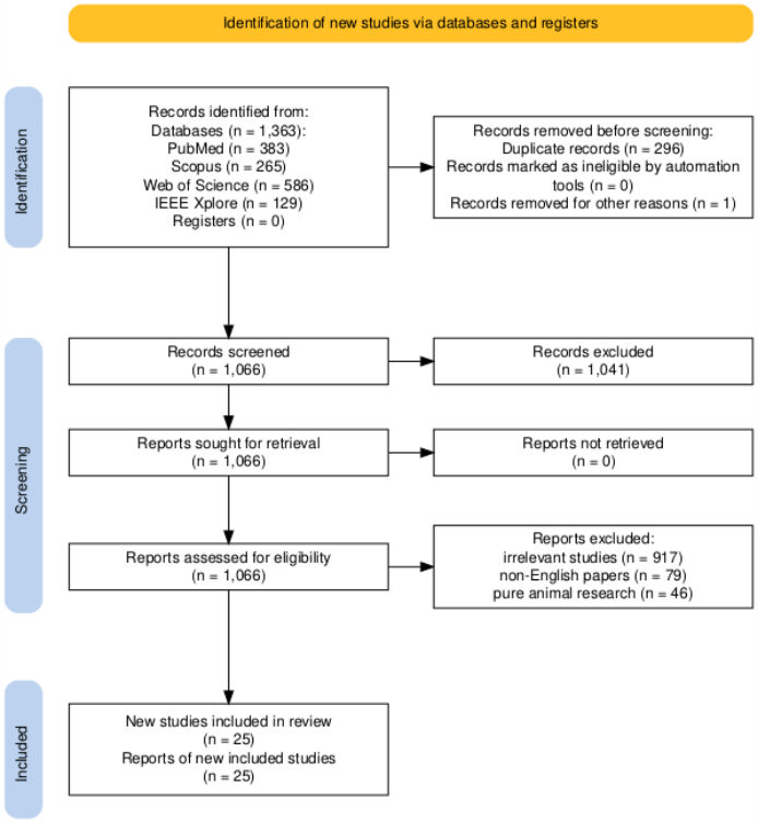
The database developed for this study was first screened for duplicate records, which were subsequently removed. A language filter was then applied to exclude all articles not published in English. As the present study focused on the neck, only neck related articles were retained. In the first stage of screening, the titles and abstracts of the retrieved articles were evaluated. The inclusion criterion at this stage was that the study addressed 3D modelling of the human neck and its applications. The remaining articles were then examined in detail. The inclusion criteria at the second stage required
114 Pan and Yang that the study be relevant to at least one of the following key areas: 3D data acquisition and processing, parametric and geometric models, digital human models, and statistical shape models. At this stage, exclusion criteria comprised non-3D research and purely animal-based studies.
A total of twenty-five articles met the inclusion criteria for the study. Figure 1 depicts the flow diagram of the systematic review process. All the articles included in the study focused on one of the decided key areas.
Figure 1: PRISMA (Preferred reporting items for systematic reviews and meta- analyses) flow diagram of the study selection process.
Among the selected articles, five investigated a range of 3D modeling techniques, including laser scanning, structured light scanning, photogrammetry, Computed Tomography (CT) and Magnetic Resonance Imaging (MRI). Four articles examined the most commonly used alignment techniques. Two of these specifically addressed anatomical landmark-based alignment, while the other two focused on 3D data processing. One of the latter highlighted Principal Component Analysis (PCA) as the most frequently employed method in the
Application of 3D Neck Modeling in Ergonomic Product Design 115 included studies, whereas the other applied numerical optimization to register a subject specific model to a reference model. In addition, four studies discussed the use of 3D neck models in Statistical Shape Models (SSMs) to advance anatomical analysis. Finally, five articles explored the use of 3D models in the design of ergonomic products to improve personalization and comfort.
RESULTS
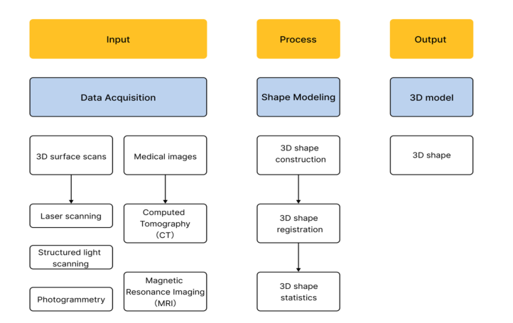
We summarized the 3D shape modeling workflow from the included studies into three phases: data acquisition, shape modeling, and 3D model (see Figure 2).
Figure 2: Flow chart of the 3D neck modeling process.
Data Acquisition for 3D Neck Modelling Creating a complete 3D model of the human neck is a complex task. The neck contains numerous distinct anatomical structures, including the skin and internal components such as muscles, ligaments, and vertebrae. Consequently, to build a comprehensive model, researchers must employ a range of data acquisition technologies.
Surface Acquisition In constructing a 3D neck model, the first step is typically to capture its external morphology. Three main non-contact technologies are commonly used for this purpose: laser scanning, structured scanning, and photogrammetry. Laser scanners are widely used for capturing surface morphology. These devices operate by emitting laser beams and measuring the reflected light to determine the 3D coordinates of points on a surface, thereby generating a dense point cloud that represents the object’s shape. For example, in the development of a full body model, Gayzik et al. (2011) used a laser scanner
116 Pan and Yang arm to acquire a detailed point cloud of a subject’s skin surface while the subject was seated. This method is rapid, non-contact, and well suited for collecting anthropometric data (Lu & Wang, 2008). Structured light scanners function in a similar manner. Instead of a single laser point, they project a known pattern of light, such as stripes or grids, onto an object. A camera records how the pattern deforms over the object’s surface, and dedicated software then calculates the 3D shape from this deformation (Remondino, 2011). This technique is particularly effective for capturing the shape of smaller, geometrically complex objects. Laser scanning and structured light scanning can achieve high geometric accuracy, but they are very sensitive to surface material, ambient light, and occlusion. As a result, they often fail on reflective, dark, transparent, or cluttered scenes, and they struggle with fine details and complete coverage in complex environments.
To improve scan accuracy, various researchers have employed stereophotogrammetry in their studies. Photogrammetry is another important technology that uses images to create 3D models. This method relies on multiple photographs of an object taken from different viewpoints. Specialized software identifies corresponding points in these images and uses them to compute the 3D geometry (Remondino, 2011). Photogrammetry is highly flexible, requiring cameras and is considered a reliable technique for producing accurate and detailed 3D information, depending on camera resolution, calibration, and surface properties. The limitations of this technique primarily relate to its setup and environmental sensitivity. The equipment arrangement is complex, as multiple cameras and supporting systems result in substantial wiring that typically requires a controlled laboratory environment. The system is also highly sensitive to physical disturbances: even minor changes in camera position can introduce artifacts into the 3D model. Moreover, its accuracy depends strongly on environmental factors, particularly ambient lighting, which can adversely affect the results.
Internal Acquisition To model the internal structures of the neck, researchers rely on medical imaging technologies. The two most essential methods for 3D neck modelling are CT and MRI. CT scans are the preferred method for modelling hard tissues such as bone. A CT scanner acquires a series of X-ray images from different angles to create cross-sectional slices of the body. These slices can then be stacked to reconstruct a highly detailed 3D model of the skeleton. For instance, Meyer et al. (2004) have used CT scans of a human volunteer to generate the detailed geometry of each cervical vertebra for a finite element model of the neck. This technique is also employed to create high resolution reference models of bones, which can then be adapted to fit data from individual patients (Delorme et al., 2003).
For modelling soft tissues, MRI is the method of choice. MRI provides excellent contrast between different types of soft tissues, such as muscles, ligaments, and intervertebral discs (Gayzik et al., 2011). This makes it ideal for capturing the geometry of these non-bony structures. Often, researchers build a complete model by combining CT and MRI data, using CT for the
Application of 3D Neck Modeling in Ergonomic Product Design 117 vertebrae and MRI for the surrounding soft tissues. This multi-modality approach enables the creation of a comprehensive, detailed model of the entire neck structure.
The following table summarizes the comparison of these technologies based on the information provided above.
Table 1: Comparison of technologies for 3D neck modeling.
Table available in the original PDF paper.
Technology Principle Primary Use Strengths Laser Scanning Measures reflected laser light to create a point cloud.
Capturing external body shape.
Fast and contact- free.
Capturing smaller, complex surfaces.
Effective for complex geometry.
Structured Light Calculates shape from the deformation of a projected light pattern.
Flexible and reliable.
Creating 3D models from images.
Photogrammetry Calculates 3D geometry from multiple 2D photographs.
Computed Tomography
Uses X-rays to create cross- sectional images.
Modeling hard tissues.
Provides high resolution models of the skeleton.
Uses magnetic fields to image the body.
Modeling soft tissues.
Magnetic Resonance Imaging
Excellent contrast between different soft tissues.
Construction Methods and Analysis Techniques for 3D Neck Modeling A common approach to constructing 3D neck models is to use geometric and parametric methods. These methods rely on mathematical formulations to describe neck morphology, allowing models to be generated or modified by adjusting a limited set of key parameters. Typically, parametric modeling begins by representing complex anatomical structures using simple geometric primitives to approximate cervical vertebrae (Laville et al., 2009; Maurel et al., 1997), with lengths and angles defining these primitives. Other studies focus on the external contour of the neck. By analyzing large datasets of 3D scans, they identify key anthropometric variables that capture overall size and shape (Jiang et al., 2008). Wang and Gu (2021) further demonstrate that these measurements can be estimated from simple 2D photographs, greatly reducing the cost and complexity of data collection. The central objective is to establish robust mappings between these key measurements and the final 3D surface geometry. Once such mappings are defined, an individualized neck model can be generated from a small set of body measurements (Jiang et al., 2008; Wang, 2005), which is particularly valuable for applications requiring customized models, such as the design of specialized clothing collars. Across the reviewed studies, there is broad consensus that models must be parameterized, geometry is the key link between form and function, low-dimensional shape descriptors are essential, and automated pipelines are necessary, motivating the development of new, fully parameterized, shape-driven methods. SSMs are statistical tools for quantifying and analyzing
118 Pan and Yang anatomical shape. An SSM is trained on a set of CT-derived bone models of the cervical spine, and captures both the average shape and the dominant modes of variation within the population (Sarkalkan et al., 2014). SSMs offer substantial advantages for anatomical analysis because they describe shape variability across the entire 3D structure rather than relying solely on simple scalar measures such as lengths or angles (Gaffney et al., 2019). For example, Harris et al. (2013) used an SSM to identify detailed 3D differences in bone shape associated with a specific pathology, providing a more comprehensive understanding of disease related morphological changes. SSMs are also highly effective for patient specific modeling. Because an SSM encodes the statistically plausible variability of normal anatomy, it can reconstruct a 3D model for a particular patient from limited imaging data (Sarkalkan et al., 2014; Zheng & Yu, 2017). This capability supports more accurate surgical planning and facilitates quantitative comparison between an individual patient and a healthy reference population.
Generating an accurate 3D neck model from scan data also requires careful spatial alignment. One common strategy is anatomical landmark–based alignment: researchers select reliable landmarks on the 3D model and use them to define axes and reference planes (Lee et al., 2006), after which the model is transformed into the corresponding coordinate frame (Jager, 1996). Computational methods provide alternative alignment strategies. PCA can be used to derive three orthogonal axes from the distribution of points on the surface and to adopt these as the model’s coordinate system. However, several studies indicate that PCA based alignment is not always sufficiently accurate and may yield orientations that are not anatomically intuitive (Chaouch & Verroust-Blondet, 2009). Other methods exploit geometric symmetry to obtain more meaningful alignments. Another class of techniques relies on numerical optimization to register a subject specific model to a reference model, which is particularly useful for assessing the fidelity of physical prototypes relative to their digital designs (Taft et al., 2011). Therefore, constructing a high quality 3D neck model requires selecting alignment and modeling strategies that are appropriate for the specific objective, whether the focus is on external surface shape, bony structures, or soft tissues.
In summary, early approaches often used geometric and parametric methods. These methods use a few key measurements, like neck girth, to build a model. This is useful for applications such as custom clothing design because the models are flexible and easy to modify. However, these simple models often cannot capture the full, complexity of neck anatomy. They provide a general shape but miss the subtle variations between different people. To address this, researchers developed more advanced techniques. SSMs learn from a large collection of 3D scans to understand both the average neck shape and its common variations. This data driven approach creates much more realistic and detailed models. It is especially useful for medical applications, where accuracy is critical. While SSMs are powerful, a gap still exists. Previous work has focused either on simple parametric models for general shape or on complex statistical models for specific anatomy, such as bones. There is a clear need for a method that combines the simple control
Application of 3D Neck Modeling in Ergonomic Product Design 119 of parametric systems with the detailed accuracy of statistical models for the external neck surface.
Applications in Ergonomic Product Design 3D neck models play a crucial role in contemporary ergonomic design. They enable designers to develop products that better accommodate the complex morphology and motion of the human body, with an emphasis on personalization, comfort, and safety. One of the primary applications of 3D neck models is improving fit and comfort in wearable products. By starting with precise anatomical data, designers can create devices that closely conform to the user’s body, thereby reducing discomfort and improving overall functionality.
Custom neck orthoses, or braces, illustrate this approach clearly. Researchers now employ 3D scanning systems to capture a patient’s specific neck and shoulder anatomy. This process is rapid and yields a detailed digital representation of the user. The resulting model then serves as the basis for designing a custom-fit orthosis. Advanced software can further utilize this model to generate optimized ventilation patterns, making the final device lighter and more breathable and thereby substantially enhancing user comfort during long-term wear (Ambu et al., 2024). Beyond static fit, 3D models are also used in the design of wearable robotic and assistive devices, such as cervical exoskeleton systems. For example, kinematic models describing the neck’s range of motion are used to develop robotic neck braces for therapeutic traction. These models ensure that the device can guide the user’s head through prescribed flexion and extension movements while applying controlled forces, rendering the therapy both effective and comfortable (Kulkarni & Agrawal, 2024). The same kinematic modeling principles are also used in the design of assistive technologies, such as robotic arms controlled by head movements for individuals with quadriplegia, where 3D models of neck motion help optimize the device’s workspace to match the user’s physical capabilities (Ouerfelli et al., 1999).
Although the referenced studies primarily address medical devices, the underlying principles are broadly applicable to consumer products. The general practice of using 3D human body models to assess fit is well established in domains such as apparel design (Li, 2023). The same methodology can be extended to the design of helmets, virtual reality (VR) headsets, and other wearable devices to ensure they fit securely and comfortably around the neck, without generating pressure points or limiting movement.
The application of 3D neck models extends well beyond ergonomic product design. These models have become valuable tools in several other professional fields, particularly clinical medicine and professional training. In clinical practice, 3D models have transformed surgical planning. Surgeons use Computer Aided Design (CAD) and Computer Aided Manufacturing (CAM) systems based on patient CT data to construct detailed 3D models of the head and neck (Copelli et al., 2024). These models are then used to plan complex reconstructive procedures, such as those required following tumor resection. Surgeons can virtually simulate the procedure and 3D print patient-specific
120 Pan and Yang cutting guides and implants. This workflow improves reconstruction accuracy, reduces operative time, and leads to better functional outcomes for patients.
CONCLUSION
Over the past few decades, 3D scanning tools and data handling methods have made advanced significantly, enabling designers to use 3D neck models for ergonomic products with better comfort. This paper provided an ordered review of recent work, examining primary methods and data steps currently used. While identifying the strengths and weakness of various modeling techniques, this study acknowledges that the current scope is limited by a relatively small number of included papers. No single technique can fully capture neck complexity; therefore, a combination of methods is necessary. With improvements in methods and continuous advances in CAD technology, the research space in ergonomic product design is expected to grow significantly. Future work should combine multiple modeling techniques to better capture the complex morphology of the neck.
ACKNOWLEDGMENT
This work was supported by the National Natural Science Foundation of China (Grant No. 52508338), the Guangdong Philosophy and Social Science Foundation (Grant No. GD25YYJ54), the Guangdong Provincial Education Science Project (Grant No. 2025GXJK0600), and the Shenzhen Education Science Research Project (Grant No. yb24003).
REFERENCES
Ambu, R., Oliveri, S. M., & Calì, M. (2024). Neck orthosis design for 3D printing with user enhanced comfort features. International Journal on Interactive Design and Manufacturing (IJIDeM), 18(8), 6055–6068. https://doi.org/10.1007/s12008- 023-01507-1 Ameer, M. K., Mehboob, F., Ahmad, N., Moeed, K., Ahmed, H., & Irshad, F. (2024).
Comparative Evaluation of Neck Length, Relative Neck Length and Total Body Height in Cervical Spondylosis Affected and Non-Affected Individual: Neck Length and Height in Cervical Spondylosis. Pakistan Journal of Health Sciences, 156–160. https://doi.org/10.54393/pjhs.v5i06.1665 Bland J. H., & Boushey D. R. (1990). Anatomy and physiology of the cervical spine.
Seminars in Arthritis and Rheumatism, 20(1), 1–20. https://doi.org/10.1016/0049- 0172(90)90090-3 Chaouch, M., & Verroust-Blondet, A. (2009). Alignment of 3D models. Graphical Models, 71(2), 63–76. https://doi.org/10.1016/j.gmod.2008.12.006 Copelli, C., Cacciatore, F., Cocis, S., Maglitto, F., Barbara, F., Iocca, O., & Manfuso,
A. (2024). Bone reconstruction using CAD/CAM technology in head and neck surgical oncology. A narrative review of state of the art and aesthetic-functional outcomes. Acta Otorhinolaryngologica Italica, 44(2 Suppl 1), S58–S66. https:// doi.org/10.14639/0392-100X-suppl.1-44-2024-N2819 Delorme, S., Petit, Y., de Guise, J. A., Labelle, H., Aubin, C.-E., & Dansereau, J.
(2003). Assessment of the 3-D reconstruction and high-resolution geometrical modeling of the human skeletal trunk from 2-D radiographic images. IEEE Transactions on Biomedical Engineering, 50(8), 989–998. https://doi.org/10.1109/ TBME.2003.814525
Application of 3D Neck Modeling in Ergonomic Product Design 121 Gaffney, B. M. M., Hillen, T. J., Nepple, J. J., Clohisy, J. C., & Harris, M. D. (2019).
Statistical shape modeling of femur shape variability in female patients with hip dysplasia. Journal of Orthopaedic Research, 37(3), 665–673. https://doi. org/10.1002/jor.24214 Gayzik, F. S., Moreno, D. P., Geer, C. P., Wuertzer, S. D., Martin, R. S., & Stitzel, J. D.
(2011). Development of a full body CAD dataset for computational modeling: A multi-modality approach. Annals of Biomedical Engineering, 39(10), 2568–2583. https://doi.org/10.1007/s10439-011-0359-5 Harris, M. D., Datar, M., Whitaker, R. T., Jurrus, E. R., Peters, C. L., & Anderson, A. E. (2013). Statistical shape modeling of cam femoroacetabular impingement. Journal of Orthopaedic Research, 31(10), 1620–1626. https://doi.org/10.1002/ jor.22389 Hoy, D. G., Protani, M., De, R., & Buchbinder, R. (2010). The epidemiology of neck
pain. Best Practice & Research Clinical Rheumatology, 24(6), 783–792. https:// doi.org/10.1016/j.berh.2011.01.019 Jager, M. M. D. (1996). Mathematical head-neck models for acceleration impacts.
Eindhoven University of Technology. https://doi.org/10.6100/IR460661 Jiang, L., Zhang, W., Xia, M., & Hou, X. (2008). Neck characteristic parameters mathematical model. 2008 9th International Conference on Computer-Aided Industrial Design and Conceptual Design, 188–192. https://doi.org/10.1109/ CAIDCD.2008.4730548 Kulkarni, P., & Agrawal, S. K. (2024). Design and validation of a novel robotic neck
brace for cervical traction. IEEE/ASME Transactions on Mechatronics, 29(4), 3092–3099. https://doi.org/10.1109/TMECH.2024.3402614 Laville, A., Laporte, S., & Skalli, W. (2009). Parametric and subject-specific finite element modelling of the lower cervical spine. Influence of geometrical parameters on the motion patterns. Journal of Biomechanics, 42(10), 1409–1415. https://doi. org/10.1016/j.jbiomech.2009.04.007 Lee, Y. S., Oh, S. H., Seon, J. K., Song, E. K., & Yoon, T. R. (2006). 3D femoral neck
anteversion measurements based on the posterior femoral plane in ORTHODOC® system. Medical and Biological Engineering and Computing, 44(10), 895–906. https://doi.org/10.1007/s11517-006-0104-7 Li, Y. (2023). Application of computer virtual reality technology in clothing 3D cutting design. 2023 2nd International Conference on Data Analytics, Computing and Artificial Intelligence (ICDACAI), 124–129. https://doi.org/10.1109/ ICDACAI59742.2023.00029 Lu, J.-M., & Wang, M.-J. J. (2008). Automated anthropometric data collection using
3D whole body scanners. Expert Systems with Applications, 35(1), 407–414. https://doi.org/10.1016/j.eswa.2007.07.008 Maurel, N., Lavaste, F., & Skalli, W. (1997). A three-dimensional parameterized finite element model of the lower cervical spine, study of the influence of the posterior articular facets. Journal of Biomechanics, 30(9), 921–931. https://doi. org/10.1016/S0021-9290(97)00056-0 Meyer, F., Bourdet, N., Deck, C., Willinger, R., & Raul, J. S. (2004). Human neck
finite element model development and validation against original experimental data. 2004-22–0008. https://doi.org/10.4271/2004-22-0008 Niu, J., & Li, Z. (2012). Using Three-Dimensional (3D) Anthropometric Data in Design. In V. R. Preedy (Ed.), Handbook of Anthropometry: Physical Measures of Human Form in Health and Disease (pp. 3001–3013). Springer. https://doi. org/10.1007/978-1-4419-1788-1_185
(1996). Measurement techniques in anthropometry. Anthropometrica, 1, 25-75. Ouerfelli, M., Kumar, V., & Harwin, W. S. (1999). Kinematic modeling of headneck movements. IEEE Transactions on Systems, Man, and Cybernetics - Part A: Systems and Humans, 29(6), 604–615. https://doi.org/10.1109/3468.798064 Remondino, F. (2011). Heritage recording and 3D modeling with photogrammetry
and 3D scanning. Remote Sensing, 3(6), 1104–1138. https://doi.org/10.3390/ rs3061104 Sarkalkan, N., Weinans, H., & Zadpoor, A. A. (2014). Statistical shape and appearance models of bones. Bone, 60, 129–140. https://doi.org/10.1016/j.bone.2013.12.006 Taft, R. M., Kondor, S., & Grant, G. T. (2011). Accuracy of rapid prototype models
for head and neck reconstruction. The Journal of Prosthetic Dentistry, 106(6), 399–408. https://doi.org/10.1016/S0022-3913(11)60154-6 Wang, C. C. L. (2005). Parameterization and parametric design of mannequins.
Computer-Aided Design, 37(1), 83–98. https://doi.org/10.1016/j.cad.2004.05.001 Wang, T., & Gu, B. (2021). Automatic recognition and 3D modeling of the neck-shoulder human shape based on 2D images. International Journal of Clothing Science and Technology, 33(5), 796–810. https://doi.org/10.1108/IJCST-05-2020-0079 Weerakoon, T. C. S., Dissanayake, P. H., Jayakody, S., Weerasekera, M. M., &
Yasawardene, S. G. (2021). Intensity of neck pain and its association with anthropometric measurements | Sri Lanka Anatomy Journal. https://doi.org/10.4038/ slaj.v5i1.130 Zheng G., & Yu W. (2017). Statistical shape and deformation models based 2D–3D reconstruction. In Statistical Shape and Deformation Analysis (pp. 329–349). Academic Press. https://doi.org/10.1016/B978-0-12-810493-4.00015-8
LISTED IN THE AHFE DIGITAL BOOKSTORE CATALOGUE
Explore the complete digital volume
Human Factors in Simulation, Software and Systems Engineering
Read this individual research paper free through the original publisher, or explore the full digital proceedings book.
ISBN 978-1-964867-96-0
Explore this ISBN in the AHFE Bookstore ↗Listed in the July 2026 bookstore catalogue; search by ISBN to confirm current availability · Digital book purchase optional