Pramath Kakodkar, Nooshin Shekari, Jay Kalra (2025). Organ-Specific Biomarkers of Aging: An Innovative Framework for Biological Age Assessment. In: Human Factors in Design, Engineering, and Computing. AHFE Open Access, vol. 199, pp. 1562–1576. https://doi.org/10.54941/ahfe1006967
As population ages, there is an immense need to identify reliable biomarkers that reflect biological age, which is representative of the cumulative burden of physiological decline across all organ systems. The current model for estimating the systemic assessment of biological age relies on epigenetic and multiomic signatures, but there remains a gap in the literature regarding the modular assessment of organ-specific aging. We describe a conceptual and evidence-based framework for evaluating organ-specific aging biomarkers across major physiological systems and integrating them with systemic aging metrics to construct a holistic assessment of biological age. We reviewed and critically appraised emerging ageing biomarkers for the cardiovascular (e.g., VO₂ max, pulse wave velocity, ApoB), hepatic (e.g., ALT, GGT, elastography), renal (e.g., eGFR, cystatin C), pulmonary (e.g., FEV1), immune (e.g., hs-CRP, CD8:CD4 ratio), musculoskeletal (e.g., grip strength, DEXA-derived lean mass), neurocognitive (e.g., processing speed, MRI volumetrics), endocrine (e.g., DHEA-S, IGF-1, cortisol rhythm), and integumentary (e.g., dermal elasticity, wrinkle depth) systems. We evaluated these biomarkers and their relationship to the trajectory of age-related decline, response to interventions, and prognostic ability for morbidity, frailty, and mortality. The overall ageing trajectory can be estimated using a tiered model that integrates organ-level biomarkers with systemic DNA methylation indices (Horvath, GrimAge, DunedinPACE), blood-based aging calculators (PhenoAge, inflammaging indices), and functional aging metrics (e.g., gait speed, reaction time, sleep architecture). This work advocates for a modular yet integrated approach to biological age assessment that captures both organ-level and systemic aging signals. As longevity medicine and preventive geriatrics advance, such frameworks may support the development of personalized interventions to extend health span, improve clinical risk stratification, and facilitate early detection of organ-specific decline before the onset of overt disease. We emphasize the importance of validated outcome measures and caution against overreliance on unverified surrogate endpoints.
Organ-Specific Biomarkers of Aging: An Innovative Framework 1563
INTRODUCTION
Aging remains a fundamental process indicating the linear progression of our physical world. At an organism level, aging can be conceptually explained as the cumulative burden acquired by a gradual decline in physiological function across organ systems (Rutledge et al., 2022). While most people are familiar with chronological age, defined as the numerical age from birth, this metric fails to encapsulate the wide variability in how individuals age biologically. Contrastingly, biological age is a nuanced reflection of cumulative molecular and functional deterioration within an organism (Moqri et al., 2023). This comprehensive metric provides a better prognostication for functional capacity, morbidity, and mortality compared to the conventional chronological age.
There is an avid interest in understanding and modulating biological ageing. This fascination extends far beyond the scientific community. In popular culture and media, the discordance between chronological and biological aging is revered in high-performance athletes such as Cristiano Ronaldo and Lebron James, who maintain elite physical metrics well into their late 30s. Notably, tech entrepreneur Bryan Johnson had made significant publicly declared efforts to achieve the biological profile of an 18-year-old across multiple organs with his Blueprint project. This project has drawn widespread attention for its granular monitoring of biological age using DNA methylation clocks, imaging, and a battery of clinical biomarkers. These high- profile cases fostered both public enthusiasm and skepticism. It is important to note that while these individual case studies underscore the aspirational goals of longevity science, they also illustrate the gaps in the standardization, validation, and interpretation of aging biomarkers.
On the opposite end of the spectrum, there exist rare genetic disorders such as progeria (Hutchinson-Gilford Progeria Syndrome), which offer a stark reminder of what an accelerated biological aging phenotype looks like (Bejaoui et al., 2022). Pediatric cases with progeria display early- onset cardiovascular disease, growth failure, and shortened lifespan, which illustrates the systemic consequence of accelerated biological aging (Bejaoui et al., 2022). These cases underscore the profound biological underpinnings of aging and highlight the urgency of understanding how aging manifests differently across tissues and individuals.
In this review, we present a conceptual and evidence-based framework for assessing biological age across major physiological systems; cardiovascular, hepatic, renal, pulmonary, immune, musculoskeletal, neurocognitive, endocrine, and integumentary domains. We critically appraise candidate biomarkers based on their mechanistic validity, responsiveness to intervention, feasibility, and prognostic value. We also explore how these modular assessments can be synthesized with whole-body aging metrics and functional performance indicators (e.g., gait speed, grip strength, sleep architecture) to create a comprehensive biological age profile.
By bridging scientific evidence with practical considerations, this review aims to identify candidate biomarkers that span all major organ systems and utilize them to create an organ-specific aging model. This model will
assess the current composite biological age and subsequently predict the rate of aging. We also caution against the overuse of surrogate markers in the absence of clinical outcome data, particularly in commercial or self- experimentation contexts. As personalized medicine and preventive geriatrics evolve, robust frameworks for biological age assessment will be essential for risk stratification, early intervention, and extending healthspan.
FROM MOLECULES TO MOBILITY: INTERLINK BETWEEN mTOR, AMPK, AND SIRTUINS WITH HUMAN PERFORMANCE AND ERGONOMICS The molecular mechanistic explanation for ageing is the concept of ex- differentiation, wherein there is a loss in the fidelity of the epigenetic signatures, which is inherently responsible for maintaining the specialized differentiated state of a mature cell type. The dogma here is that cells now become generalized instead of maintaining their carefully epigenetically curated specialization state, and this alteration is heralded as the central molecular event leading to ageing. From a human factors perspective, such molecular drift is expressed at the system level as reduced cognitive performance, slowed reaction times, and diminished physicality. All these directly impact an individual’s ability to interact with their environment, workplace performance, independence in daily living.
At a molecular level, longevity-related pathways are governed by mTOR (mechanistic target of rapamycin), AMPK (AMP-activated protein kinase), and the sirtuin family of NAD+-dependent enzymes. These 3 genes networks work in consort to orchestrate the balance between growth, repair, and stress resistance by regulating cellular energy expenditure and supporting the epigenetic fidelity of cells (Sadria and Layton, 2021).
mTOR integrates nutrient and growth signals, a gene that gets activated when it senses amino acids, and induces protein synthesis. A low activity of mTOR is a trigger for the cell to induce survival mode and optimize energy efficiency. mTOR hyperactivity promotes vascular fibrosis and inflammation, resulting in increased arterial stiffness (Pulse Wave Velocity (PWV)). Additionally, aerobic functional capacity (VO2 max) decreases with mTOR-mediated mitochondrial dysfunction. These changes manifest not just as abstract molecular phenomena, but as measurable decrements in our cardiovascular performance and fatigue resistance, which are critical ergonomic determinants in both occupational and aging populations.
Contrastingly, AMPK activation supports metabolic efficiency during fasting and increases insulin sensitivity and mitochondrial efficiency (Salminen and Kaarniranta, 2012). Interestingly, AMPK activation by lifestyle modifications (caloric restriction, exercise) and pharmacological interventions (metformin) demonstrate the direct human-factor relevance in modulating these pathways. People experience can improve their endurance, attain stringent glycemic control, and develop stress tolerance. AMPK also downregulates mTOR and upregulates other longevity-promoting genes.
Lastly, sirtuin family genes are the central regulators of longevity, activated by fasting and exercise. Sirtuin gene products produce proteins that help
Organ-Specific Biomarkers of Aging: An Innovative Framework 1565
reset the epigenetic machinery, which essentially reverse ex-differentiation (Ji et al., 2022). Functionally, this translates to improved neurocognitive performance, preserved musculoskeletal function, and greater resilience to environmental stressors. All of these are human-centered outcomes that align biological mechanisms with ergonomics and applied aging research.
METHODOLOGY
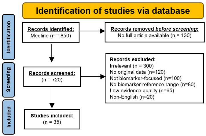
We conducted a comprehensive review (Figure 1) on Medline to identify biomarkers of organ-specific ageing. The search query utilized a combination of terms: “biomarkers”OR “marker”AND “aging”. The query was targeted to studies from 2015 to 2025, English language, and human studies. A total of 35 studies were included after screening for clinical relevance, normalized data (median, 95% percentiles), and age-specific ranges in healthy cohorts. Studies were also critically appraised based on their study design and clinical utility for inclusion.
Figure 1: Flowchart showing literature review process for evaluation of candidate biomarkers for aging.
RESULTS
To construct a comprehensive model for holistic evaluation of biological age, we identified 17 biomarkers across all major organ systems that had established associations with ageing and a predominantly non-invasive methodology for testing. Table 1 shows a summary of each biomarker, including the level of evidence available based on the presence of a longitudinal cohort study or meta-analysis for clinical validation.
Table 1: Summary of organ system specific biomarkers of ageing: Evidence, biological implication and age-matched metrics.
Table available in the original PDF paper.
Biomarker [System]
Biological Implication
Reference [Evidence Level]
Example of Age-Related Metrics
Pulse wave Velocity (PWV) [CV]
(Díaz et al., 2014; Vieira-da-Silva et al., 2025) [High]
Measures arterial stiffness an indicator for vascular aging. This reflects cumulative oxidative and inflammatory damage. Increased risk of HTN, CVA, MI. Higher velocities indicate less vessel elasticity.
(Hawkins and Wiswell, 2003; Kaminsky et al., 2015; Mandsager et al., 2018) [High]
VO2 Max [CV] Maximal rate at which a person consumes O2 during a bout of intense exercise. It declines 0.5–1% per year after age 30. Predicts functional capacity, CV fitness, and mortality risk.
MRI based Brain Volume [Neuro]
(Bethlehem et al., 2022; Cumplido-Mayoral et al., 2025; Fujita et al., 2023) [High]
T1-weighted voxel-based morphometry indicating cortical atrophy by measuring GMV. GMV is more sensitive to aging and correlate with cognitive decline, dementia risk, and neurodegenerative processes such as AD.
Continued
Organ-Specific Biomarkers of Aging: An Innovative Framework 1567
Table 1: Continued Biomarker [System] Biological Implication
Table available in the original PDF paper.
Reference [Evidence Level]
Example of Age-Related Metrics Plasma upper 95th
Neurofilament Light Chain (NfL) [Neuro]
(Simrén et al., 2022; Sukhonpanich et al., 2025) [High]
percentile (pg/mL) 5-17y: 7 18-50y: 10 >70y: 35
NfL is a structural protein of the neuronal cytoskeleton that becomes detectable in blood when neurons undergo damage or degeneration. Blood-based NfL measurement can serves as a sensitive aging biomarker in healthy individuals.
Digit Symbol Substitution Test (DSST) [Neuro]
(Erdodi et al., 2017; Shaaban et al., 2023) [Moderate]
DSST score is a numerical value of correct symbol-digit pairings completed in 90 seconds from validated Wechsler Adult Intelligence Scale. DSST score declines with age due to slower processing speed and cognitive aging.
(Quanjer et al., 2012) [High]
Median (upper 95th percentile) L 20-29y: 3.8 (4.5) 40-49y: 3.3 (4.0) >70y: 2.2 (2.9)
FEV1 [Resp] Measures lung capacity decline and is sensitive to lung aging. FEV1 predicts COPD, respiratory infection risk, mortality.
Cystatin C [Renal] Assesses kidney filtration decline; clinically utilized for predicting chronic kidney disease, CV risk, and mortality. Cystatin C is less affected by muscle mass than creatinine.
(Le Couteur et al., 2010; Najmy et al., 2019) [Moderate]
ALT is a hepatocyte injury marker. Increases with inflammation and decreases with advanced age, correlating with metabolic disorders, liver disease, and mortality
Gamma-Glutamyl Transferase (GGT) [Hepatic]
(Long et al., 2014; Praetorius Björk and Johansson, 2018) [Moderate]
GGT is an enzyme with oxidative stress and biliary function marker. GGT increases with age due to metabolic changes, correlating with hepatocyte fatty deposition, diabetes, and CV mortality
FibroScan [Hepatic]
(Colombo et al., 2011; Selman and Pardo, 2021; Sharma et al., 2023) [Moderate]
This transient elastography assesses hepatic fibrosis via parenchyma stiffness. Stiffness correlated with age-related subtle collagen buildup, degree of steatosis, and mortality.
Continued
Organ-Specific Biomarkers of Aging: An Innovative Framework 1569
Table 1: Continued Biomarker [System] Biological Implication
Table available in the original PDF paper.
Reference [Evidence Level]
Example of Age-Related Metrics
Grip strength [MSK]
(Roman-Liu et al., 2024; Wang et al., 2018) [High]
This reflects sarcopenia and predicts ageing, frailty, falls, and mortality.
Median (upper 95th percentile) Kg 20-29y: 36 (45) 40-49y: 35 (43) >70y: 24 (32)
(Kirk et al., 2021) [High]
DEXA lean mass [MSK]
This measures sarcopenia and predicts physical decline.
Median (upper 95th percentile) Kg 20-29y: 36 (45) 40-49y: 35 (43) >70y: 24 (32)
(Gabin et al., 2018; Wang et al., 2016) [Moderate]
hsCRP [Immune] Serum marker of low-grade systemic inflammation; predicts frailty, and mortality.
IGF-1 [Endocrine] IGF-1 is somatotropic hormone. It declines with ageing due to reduction in GH secretion. Serum IGF-1 correlates with frailty, muscle loss, cognitive decline, and mortality.
Continued
Table 1: Continued Biomarker [System] Biological Implication
Table available in the original PDF paper.
Reference [Evidence Level]
Example of Age-Related Metrics
Dermal Elasticity [Derm]
(Everett and Sommers, 2013; Ryu et al., 2008) [Moderate]
Cutometer is a non-invasive suction device that measures the skin deformation to evaluate the degree of collagen degradation.
Cardiovascular (CV), Hypertension (HTN), Cerebrovascular attack (CVA), Myocardial Infarction (MI), Confidence Interval (CI), Volume of Oxygen (VO2), Magnetic Resonance Imaging (MRI), Alzheimer’s Disease (AD), total brain volume (TBV), gray matter volume (GMV), Forced Expiratory Volume in 1 Second (FEV1), Respiratory (Resp), chronic obstructive pulmonary disease (COPD), musculoskeletal (MSK), dual-energy X-ray absorptiometry (DEXA), high sensitivity C reactive protein (hsCRP), Insulin-Like Growth Factor-1 (IGF-1) It is important to note that these biomarkers in Table 1 can have numerous pathophysiological conditions and preanalytical variables such as metabolic disorders, infections, medications, and assay variability (sample collection or equipment calibration). One notable example is that an elevated hsCRP assay could indicate acute inflammation rather than underlying inflammaging. Similarly, a reduced FEV1 could also indicate undiagnosed COPD instead of age-related decline. To assess biological aging accurately with these biomarkers, the patients must be disease-free, medication-naïve, and devoid of confounding factors, which must be ruled out via comprehensive clinical evaluation. It is recommended that there is oversight by corresponding subspecialty physician (e.g. Neurologist for NfL or hematologist for T- Cell Senescence Markers) or a clinician well-versed in aging biomarker assessment that can exclude the corresponding pathophysiological causes and contextualize the results.
SYSTEMIC BIOLOGICAL AGING ASSESSMENT There are many different blood-based methylation arrays, such as Horvath, GrimAge, and DunedinPACE. The Horvath assay is a first-generation model that evaluates the methylation status at 353 CpG sites across multiple tissues to estimate the biological age (r = 0.85 with chronological age) by reflecting on the cumulative epigenetic dysregulation (Lu et al., 2023). The GrimAge assay is a second-generation clock that evaluates the methylation status at 1030 CpG sites and incorporates surrogate plasma protein (PAI-1, TIMP-1) as well as extrinsic factors such as smoking status to provide an estimated mortality risk (r= 0.9 with chronological age) (Lu et al., 2019). Contrastingly, the DunedinPACE assay evaluates the methylation status at 173 CpG sites
Organ-Specific Biomarkers of Aging: An Innovative Framework 1571
along with 19 additional physiological biomarkers, which quantifies the rate of aging (e.g. 0.5 is aging at half the rate of a normal person) (Belsky et al., 2022).
DISCUSSION
In this paper, we present a framework that estimates overall biological aging by integrating the age from validated organ system-specific biomarkers and utilizing a modulating factor based on the systemic epigenetic metrics to provide a multidimensional assessment for biological age. The selected candidates from our Table 1 span across multiple organ systems such as cardiovascular (Pulse Wave Velocity, VO2 Max), neurological (MRI-based brain volume, NfL, DSST), pulmonary (FEV1), renal (eGFR or Cystatin C), Hepatic (ALT, GGT, FibroScan), MSK (Grip strength, DEXA-derived lean mass), immune (hs-CRP, T-cell senescence markers), endocrine (IGF-1), and integumentary (dermal elasticity) systems. These non-invasive biomarkers were included in our model as they predominantly endorsed a strong level of evidence for predicting frailty, morbidity, and mortality in our literature review.
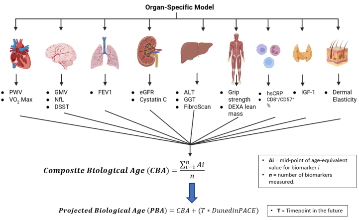
A composite biological Age (CBA) metric is derived by averaging the midpoint values obtained from the age-matched estimates from each biomarker. This CBA reflects the current biological age of the patient, independent of their chronological age. Additionally, the cumulative rate of biological aging across the organs over a shorter timeframe (approximately 2 years) can then be supplemented with the DunedinPACE value as a modulating factor to yield an integrated age projection. This modulation adjusts for disproportionate aging across organ systems while providing a prediction that highlights the discordance between chronological and biological age. Figure 2 summarizes how the biomarkers are utilized to compute the CBA and how the DunedinPACE is used to predict the biological age.
One current limitation in our model is that we assume that each of the biomarkers has an equal contribution towards the CBA. There is a need for an evidence-based weightage model that appropriately incorporates the weight of each biomarker into the CBA formula. Further longitudinal studies are required to categorize which biomarkers are considered fast-aging (hepatic (ALT, GGT, FibroScan)) versus slow-aging (Lungs (FEV1)) organs (Le Couteur et al, 2010; Najmy et al., 24; Quanjer et al., 2012; Sharma et al., 2023). This would allow refinement of our unweighted model into an outcome-driven composite model. Another limitation in our current model is the partial overlap between biomarkers (GGT, FEV1, hsCRP, and grip strength) included in our proposed CBA and those already incorporated in the proprietary DunedinPACE epigenetic clock. This redundancy could introduce multicollinearity and potentially introduce more bias towards the contribution from these biomarkers. Future iterations of this model can utilize alternative epigenetic assays such as Horvath or GrimAge, which have less biomarker overlap.
Figure 2: Summary of organ-specific biological aging model. The mid-point value from each of the biomarkers is averaged to obtain the composite biological age. Abbreviations: Pulse wave Velocity (PWV), Maximum Volume of Oxygen (VO2 Max), Gray Matter Volume (GMV), Neurofilament Light Chain (NfL), Digit Symbol Substitution Test (DSST), Forced Expiratory Volume in 1 Second (FEV1), estimated Glomerular filteration Rate (eGFR), Alanine Aminotransferase (ALT), Gamma-Glutamyl Transferase (GGT), dual-energy X-ray absorptiometry (DEXA), high sensitivity C-reactive protein (hsCRP), Insulin-Like Growth Factor-1 (IGF-1).
Compared to the other blood-based aging calculators, such as PhenoAge or inflammaging indices, our organ-based model offers a higher resolution at the tissue level. PhenoAge utilizes nine biomarkers (such as glucose, albumin, hsCRP, and lymphocyte %) from routine blood tests to estimate the biological age and a mortality prediction (Cribb et al., 2022). This makes PhenoAge more economical than our model, but it does dilute the assessment by not covering organ-level insights from specialized tests like (MRI cortical volume, and FibroScan). Contrastingly, the inflammaging indices (such as interleukin-6 (IL-6), and Tumour necrosis factor alpha (TNF-α)) quantify the longstanding low-grade inflammation and correlate it with the frailty and multi-organ decline prediction (Cribb et al., 2022).
Similarly, functional aging metrics such as gait speed (4-meter walk test), reaction time (choice reaction tasks), and sleep architecture (Rapid eye movement (REM) fragmentation) capture real-world functional decline beyond molecular signals (Deatsch et al., 2025). Our organ-based model offers an alternative to these functional markers, such as grip strength instead of the gait speed test, DSST instead of the reaction time test, and IGF-1, which is a more accessible blood test instead of the polysomnography.
CONCLUSION
Despite advances in systemic biological age estimation through epigenetic clocks like Horvath, GrimAge, and DunedinPACE, there is a critical gap in the
Organ-Specific Biomarkers of Aging: An Innovative Framework 1573
literature regarding modular organ-specific assessment for aging. Given that different organs may age at different rates and have unique vulnerabilities, our integrated approach that combines both organ-level aging biomarkers using CBA score and future rate of aging projection with DunedinPACE. This model bridges molecular and epigenetic insights and offers a practical and comprehensive review for personalized aging interventions as well as biomarkers for monitoring the impact of the intervention. Future studies can validate its specificity against these alternatives.
REFERENCES
Astley, M. E., Chesnaye, N. C., Hallan, S., Gambaro, G., Ortiz, A., Carrero, et al.
2025. Age- and sex-specific reference values of estimated glomerular filtration rate for European adults. Kidney Int. 107, 1076–1087. https://doi.org/10.1016/ j.kint.2025.02.025. Bejaoui, Y., Razzaq, A., Yousri, N. A., Oshima, J., Megarbane, A., Qannan, et al.
2022. DNA methylation signatures in Blood DNA of Hutchinson-Gilford Progeria syndrome. Aging Cell 21, e13555. https://doi.org/10.1111/acel.13555. Belsky, D. W., Caspi, A., Corcoran, D. L., Sugden, K., Poulton, R., Arseneault, L.
et al. 2022. DunedinPACE, a DNA methylation biomarker of the pace of aging. eLife 11. https://doi.org/10.7554/eLife.73420. Bethlehem, R. A. I., Seidlitz, J., White, S. R., Vogel, J. W., Anderson, K. M., Adamson, C., et al. 2022. Brain charts for the human lifespan. Nature 604, 525–533. https:// doi.org/10.1038/s41586–022-04554-y. Chang, S.-T., Chuang, Y.-F., Li, A.-H., Fan, Y.-T., Liao, M.-R., Chen, I.-Y., et al.
2024. Age-dependent immune profile in healthy individuals: an original study, systematic review and meta-analysis. Immun. Ageing 21, 75. https://doi.org/ 10.1186/s12979–024-00480-x. Colombo, S., Belloli, L., Zaccanelli, M., Badia, E., Jamoletti, C., Buonocore, M., Del Poggio, P., 2011. Normal liver stiffness and its determinants in healthy blood donors. Dig. Liver Dis. 43, 231–236. https://doi.org/10.1016/j.dld.2010.07.008. Conover, C. A., Oxvig, C., 2025. The IGF System and Aging. Endocr. Rev. 46,
214–223. https://doi.org/10.1210/endrev/bnae029. Cribb, L., Hodge, A. M., Yu, C., Li, S. X., English, D. R., Makalic, E., et al.
2022. Inflammation and Epigenetic Aging Are Largely Independent Markers of Biological Aging and Mortality. J. Gerontol. A. Biol. Sci. Med. Sci. 77, 2378–2386. https://doi.org/10.1093/gerona/glac147. Cumplido-Mayoral, I., Sánchez-Benavides, G., Vilor-Tejedor, N., López-Martos, D., Brugulat-Serrat, A., Milà-Alomà, M., et al., 2025. Neuroimaging-derived biological brain age and its associations with glial reactivity and synaptic dysfunction cerebrospinal fluid biomarkers. Mol. Psychiatry 30, 3718–3728. https://doi.org/10.1038/s41380–025-02961-x. Díaz, A., Galli, C., Tringler, M., Ramírez, A., Cabrera Fischer, E. I., 2014.
Reference values of pulse wave velocity in healthy people from an urban and rural argentinean population. Int. J. Hypertens. 2014, 653239. https://doi.org/10.1155/ 2014/653239. Deatsch, A., McKenna, M., Palumbo, J., Tian, Q., Simonsick, E., Ferrucci, L., et al.
2025. Prediction of future aging-related slow gait and its determinants with deep learning and logistic regression. PloS One 20, e0325172. https://doi.org/10.1371/ journal.pone.0325172.
Erdodi, L. A., Abeare, C. A., Lichtenstein, J. D., Tyson, B. T., Kucharski, B., Zuccato, B. G., Roth, R. M., 2017. Wechsler Adult Intelligence Scale-Fourth Edition (WAIS- IV) processing speed scores as measures of noncredible responding: The third generation of embedded performance validity indicators. Psychol. Assess. 29, 148–157. https://doi.org/10.1037/pas0000319. Everett, J. S., Sommers, M. S., 2013. Skin viscoelasticity: physiologic mechanisms,
measurement issues, and application to nursing science. Biol. Res. Nurs. 15, 338–346. https://doi.org/10.1177/1099800411434151. Fujita, S., Mori, S., Onda, K., Hanaoka, S., Nomura, Y., Nakao, T., et al. 2023.
Characterization of Brain Volume Changes in Aging Individuals With Normal Cognition Using Serial Magnetic Resonance Imaging. JAMA Netw. Open 6, e2318153. https://doi.org/10.1001/jamanetworkopen.2023.18153. Gabin, J. M., Saltvedt, I., Tambs, K., Holmen, J., 2018. The association of high sensitivity C-reactive protein and incident Alzheimer disease in patients 60 years and older: The HUNT study, Norway. Immun. Ageing A 15, 4. https://doi.org/ 10.1186/s12979–017-0106–3. Groesbeck, D., Köttgen, A., Parekh, R., Selvin, E., Schwartz, G. J., Coresh,
J., Furth, S., 2008. Age, gender, and race effects on cystatin C levels in US adolescents. Clin. J. Am. Soc. Nephrol. CJASN 3, 1777–1785. https://doi.org/ 10.2215/CJN.00840208. Hawkins, S., Wiswell, R., 2003. Rate and mechanism of maximal oxygen consumption decline with aging: implications for exercise training. Sports Med. Auckl. NZ 33, 877–888. https://doi.org/10.2165/00007256–200333120-00002. Ji, Z., Liu, G.-H., Qu, J., 2022. Mitochondrial sirtuins, metabolism, and aging. Spec.
Issue Metab. 49, 287–298. https://doi.org/10.1016/j.jgg.2021.11.005. Kaminsky, L. A., Arena, R., Myers, J., 2015. Reference Standards for Cardiorespiratory Fitness Measured With Cardiopulmonary Exercise Testing: Data From the Fitness Registry and the Importance of Exercise National Database. Mayo Clin. Proc. 90, 1515–1523. https://doi.org/10.1016/j.mayocp.2015.07.026. Kirk, B., Bani Hassan, E., Brennan-Olsen, S., Vogrin, S., Bird, S., Zanker, J., et al. 2021. Body composition reference ranges in community-dwelling adults using dual-energy X-ray absorptiometry: the Australian Body Composition (ABC) Study. J. Cachexia Sarcopenia Muscle 12, 880–890. https://doi.org/10.1002/ jcsm.12712. Le Couteur, D. G., Blyth, F. M., Creasey, H. M., Handelsman, D. J., Naganathan, V.,
Sambrook, P. N., et al. 2010. The association of alanine transaminase with aging, frailty, and mortality. J. Gerontol. A. Biol. Sci. Med. Sci. 65, 712–717. https:// doi.org/10.1093/gerona/glq082. Long, Y., Zeng, F., Shi, J., Tian, H., Chen, T., 2014. Gamma-glutamyltransferase predicts increased risk of mortality: a systematic review and meta-analysis of prospective observational studies. Free Radic. Res. 48, 716–728. https://doi.org/ 10.3109/10715762.2014.902055. Lu, A. T., Fei, Z., Haghani, A., Robeck, T. R., Zoller, J. A., Li, C. Z., et al.
2023. Universal DNA methylation age across mammalian tissues. Nat. Aging 3, 1144–1166. https://doi.org/10.1038/s43587–023-00462–6. Lu, A. T., Quach, A., Wilson, J. G., Reiner, A. P., Aviv, A., Raj, K., et al. 2019.
DNA methylation GrimAge strongly predicts lifespan and healthspan. Aging 11, 303–327. https://doi.org/10.18632/aging.101684. Mandsager, K., Harb, S., Cremer, P., Phelan, D., Nissen, S. E., Jaber, W.,
2018. Association of Cardiorespiratory Fitness With Long-term Mortality Among Adults Undergoing Exercise Treadmill Testing. JAMA Netw. Open 1, e183605. https://doi.org/10.1001/jamanetworkopen.2018.3605.
Organ-Specific Biomarkers of Aging: An Innovative Framework 1575
Moqri, M., Herzog, C., Poganik, J. R., Justice, J., Belsky, D. W., Higgins-Chen, A., et al. 2023. Biomarkers of aging for the identification and evaluation of longevity interventions. Cell 186, 3758–3775. https://doi.org/10.1016/j.cell.2023.08.003. Najmy, S., Duseja, A., Pal, A., Sachdev, S., Sharma, R. R., Marwah, N., Chawla,
Y., 2019. Redefining the Normal Values of Serum Aminotransferases in Healthy Indian Males. J. Clin. Exp. Hepatol. 9, 191–199. https://doi.org/10.1016/ j.jceh.2018.06.003. Praetorius Björk, M., Johansson, B., 2018. Gamma-Glutamyltransferase (GGT) as a biomarker of cognitive decline at the end of life: contrasting age and time to death trajectories. Int. Psychogeriatr. 30, 981–990. https://doi.org/10.1017/ S1041610217002393. Quanjer, P. H., Stanojevic, S., Cole, T. J., Baur, X., Hall, G. L., Culver, B. H., et al.
2012. Multi-ethnic reference values for spirometry for the 3–95-yr age range: the global lung function 2012 equations. Eur. Respir. J. 40, 1324–1343. https:// doi.org/10.1183/09031936.00080312. Roman-Liu, D., Kami´nska, J., Tokarski, T. M., 2024. Population-specific equations of age-related maximum handgrip force: a comprehensive review. PeerJ 12, e17703. https://doi.org/10.7717/peerj.17703. Rutledge, J., Oh, H., Wyss-Coray, T., 2022. Measuring biological age using
omics data. Nat. Rev. Genet. 23, 715–727. https://doi.org/10.1038/s41576–022- 00511–7. Ryu, H. S., Joo, Y. H., Kim, S. O., Park, K. C., Youn, S. W., 2008. Influence of age and regional differences on skin elasticity as measured by the Cutometer. Skin Res. Technol. Off. J. Int. Soc. Bioeng. Skin ISBS Int. Soc. Digit. Imaging Skin ISDIS Int. Soc. Skin Imaging ISSI 14, 354–358. https://doi.org/10.1111/ j.1600–0846.2008.00302.x. Sadria, M., Layton, A. T., 2021. Interactions among mTORC, AMPK and SIRT:
a computational model for cell energy balance and metabolism. Cell Commun. Signal. 19, 57. https://doi.org/10.1186/s12964–021-00706–1. Salminen, A., Kaarniranta, K., 2012. AMP-activated protein kinase (AMPK) controls the aging process via an integrated signaling network. Ageing Res. Rev. 11, 230–241. https://doi.org/10.1016/j.arr.2011.12.005. Selman, M., Pardo, A., 2021. Fibroageing: An ageing pathological feature driven
by dysregulated extracellular matrix-cell mechanobiology. Ageing Res. Rev. 70, 101393. https://doi.org/10.1016/j.arr.2021.101393. Shaaban, C. E., Rosano, C., Zhu, X., Rutherford, B. R., Witonsky, K. R., Rosso, A. L., et al. 2023. Discordant Biological and Chronological Age: Implications for Cognitive Decline and Frailty. J. Gerontol. A. Biol. Sci. Med. Sci. 78, 2152–2161. https://doi.org/10.1093/gerona/glad174. Sharma, D., Choudhary, N. S., Dhampalwar, S., Saraf, N., Duseja, A., Gautam, D.,
et al. 2023. Liver Stiffness Values in Persons with Normal Histology. J. Clin. Exp. Hepatol. 13, 10–14. https://doi.org/10.1016/j.jceh.2022.10.008. Simrén, J., Andreasson, U., Gobom, J., Suarez Calvet, M., Borroni, B., Gillberg, C., et al. 2022. Establishment of reference values for plasma neurofilament light based on healthy individuals aged 5–90 years. Brain Commun. 4, fcac174. https:// doi.org/10.1093/braincomms/fcac174. Stojanovic, M., Popevic, M., Pekic, S., Doknic, M., Miljic, D., Medic-Stojanoska, M.,
et al. 2021. Serum Insulin-Like Growth Factor-1 (IGF-1) Age-Specific Reference Values for Healthy Adult Population of Serbia. Acta Endocrinol. Buchar. Rom. 2005 17, 462–471. https://doi.org/10.4183/aeb.2021.462. Sukhonpanich, N., Ongphichetmetha, T., Uawithya, E., Jitprapaikulsan, J., Rattanathamsakul, N., Prayoonwiwat, N., Siritho, S., 2025. Reference range for serum neurofilament light chain: findings from healthy Thai adults. Brain Commun. 7, fcaf166. https://doi.org/10.1093/braincomms/fcaf166.
Terekhova, M., Swain, A., Bohacova, P., Aladyeva, E., Arthur, L., Laha, A., et al. 2023. Single-cell atlas of healthy human blood unveils age-related loss of NKG2C+GZMB-CD8+ memory T cells and accumulation of type 2 memory T cells. Immunity 56, 2836–2854.e9. https://doi.org/10.1016/ j.immuni.2023.10.013. Vieira-da-Silva, M. A., Bauab Filho, A. B., Imanichi, F., Lessa Silva, R. C., Marchiori
Vieira, L., Roma Uyemura, J., et al. 2025. The correlation between age, blood pressure variability and estimated pulse wave velocity. Sci. Rep. 15, 6990. https:// doi.org/10.1038/s41598–025-91023-x. Waas, T., Schulz, A., Lotz, J., Rossmann, H., Pfeiffer, N., Beutel, et al. 2021.
Distribution of estimated glomerular filtration rate and determinants of its age dependent loss in a German population-based study. Sci. Rep. 11, 10165. https:// doi.org/10.1038/s41598–021-89442–7. Wang, Y.-C., Bohannon, R. W., Li, X., Sindhu, B., Kapellusch, J., 2018. Hand-Grip Strength: Normative Reference Values and Equations for Individuals 18 to 85 Years of Age Residing in the United States. J. Orthop. Sports Phys. Ther. 48, 685–693. https://doi.org/10.2519/jospt.2018.7851. Wang, Z., Wang, X., Chen, Z., Zhang, L., Zhu, M., 2016. Distribution of High-
Sensitivity C-Reactive Protein and Its Relationship with Other Cardiovascular Risk Factors in the Middle-Aged Chinese Population. Int. J. Environ. Res. Public. Health 13. https://doi.org/10.3390/ijerph13090872.
LISTED IN THE AHFE DIGITAL BOOKSTORE CATALOGUE
Explore the complete digital volume
Human Factors in Design, Engineering, and Computing
Read this individual research paper free through the original publisher, or explore the full digital proceedings book.
ISBN 978-1-964867-75-5
Explore this ISBN in the AHFE Bookstore ↗Listed in the July 2026 bookstore catalogue; search by ISBN to confirm current availability · Digital book purchase optional