Sodai Kondo, Hisaya Tanaka (2024). Comparison of Alpha Waves and SSVEP Based on Ear-EEG Using Conductive Paste and Gel Sheet. In: Neuroergonomics and Cognitive Engineering. AHFE Open Access, vol. 126, pp. 231–241. https://doi.org/10.54941/ahfe1004756
The ear-electroencephalogram (ear-EEG) method is used to measure brain activity from regions around ear. It is becoming a popular EEG measurement method due to its characteristics such as wearability, simplicity, and long term measurability. In our previous study, we developed and evaluated a brain-computer interface (BCI) by means of steady-state visual evoked potential (SSVEP) measured via ear-EEG. Some ear-EEG-based SSVEP-BCIs have demonstrated practical performance in assisting people with disabilities in various ways. However, the EEG measurement electrodes used in these devices establish contact with skin through conductive paste that stains subject’s hair and skin. This impairs the simplicity of measurement, impacting the advantages of using ear-EEG. Hence, in this study, we measured and evaluated SSVEP and alpha waves by using electrodes coated with conductive gel sheets. A total of 20 channels of electrodes were installed around the subject’s left and right ears, of which EEG components of 10 channels each were made of conductive paste and gel sheet. While the SSVEP and alpha waves were detected by using both conductive paste and gel sheet as the electrode-skin interface, the electrodes attached to the skin with conductive paste showed better detection performance. This indicates that an ear-EEG-based BCI system can be constructed by using conductive gel sheets instead of conductive paste as the electrode-skin interface. In future studies, we aim to improve the signal detection performance of electrodes by using conductive gel sheets as the electrode-skin interface, subsequently developing SSVEP-BCI systems that are implemented for the progress of society.
Wearable devices that collect biometric information, such as Apple watch, improve the quality of life through health monitoring. Ear- electroencephalogram (EEG) is a noninvasive method of obtaining brain wave signals from regions around ears; hence, the applications of ear-EEG for designing wearable devices have been investigated in previous studies in recent years (Kidmose et al., 2013; Guermandi et al., 2018; Sun et al., 2022; Goverdovsky et al., 2016; Ahn et al., 2018). Ear-EEG is superior to conventional EEG measurement methods that include the electrodes being
attached to the scalp; hence, ear-EEG shows improved wearability, sim- plicity, and long-term measurement capability. In our previous study, we developed an ear-EEG-based steady-state visual evoked potential (SSVEP) brain-computer interface (BCI) system. When a subject is gazing at a flashing stimulus, SSVEP is predominantly evoked in the primary visual cortex region in their occipital lobe, being subsequently observed in the EEG signal (Lotte et al., 2018). BCI is an interface that connects people and computers (Vidal, 1973) and is expected to have numerous applications to support people with disabilities. Some studies have improved the performance of ear-EEG-based SSVEP-BCI systems by the following means: combining regular EEG and ear-EEG (Kondo and Tanaka, 2023), employing ear-EEG instrumentation equipment that uses ear-hook attachments (Kondo and Tanaka, 2023), and employing a four-level classification type ear-EEG-based SSVEP-BCI (Kondo and Tanaka, 2023). Considering previous studies, it can be inferred that ear- EEG-based BCI has a high potential to support people with disabilities (Sun et al., 2022; Zhu et al., 2021; Kwak et al., 2019; Ahn et al., 2018). However, the electrodes used in our previous study all utilize conductive paste. The conductive paste is used to bring the electrodes into close contact with the skin, and while it provides a high-quality EEG signal, it also stains the hair and skin. Conductive paste impairs the comfort during measurement, which is an advantage of ear-EEG. Some study on ear-EEG electrodes has proposed electrodes that are more comfortable by using dry electrodes or gel sheet electrodes (Ahn, 2018; Goverdovsky, 2015; Guermandi, 2018; Mikkelsen, 2015). These previous studies have shown that it is not sufficient to sim- ply detect the desired components from ear-EEG as in our previous study, and that it is necessary to change to dry electrodes or gel sheet electrodes. Therefore, in this study, we investigated the use of conductive gel sheets as an alternative to that of conductive paste. The measurement of SSVEP and alpha wave components was conducted by using electrodes attached to skin with conductive paste and conductive gel sheet; the results were subsequently evaluated. Further, this study aimed to evaluate whether the conductive gel sheet functioned correctly as the electrode-skin interface in the ear hook type electrode. Finally, this study also explored technical issues related to the inte- gration of conductive gel sheets as electron-skin interface into BCI, mainly considering SSVEP-BCI system.
THEORY Detection of Alpha Waves via ear-EEG
Spontaneous brain waves are brain waves that are emitted regardless of sen- sory input, such as external stimuli or recollection of motor imagery. Alpha waves are a type of spontaneous brain waves and were discovered by Berger in 1929. These waves are predominantly expressed around the range of 8–13Hz when the eyes are closed or at rest. They are dominant in the pari- etal and occipital areas, showing a significant increase in the occipital area (Adrian et al., 1934). Alpha waves are dominant in EEG signals than evoked potentials such as SSVEP; therefore, they are highly resistant to noise and
Comparison of Alpha Waves and SSVEP Based on Ear-EEG Using Conductive Paste 233
easily detectable. Hence, when evaluating ear-EEG electrodes, their perfor- mance is confirmed by comparing the alpha wave components when the eyes are open (hereinafter referred to as “eyes-open condition”) and closed (hereinafter referred to as “eyes-closed condition”) (Mikkelsen 2015).
In this study, ear-EEG performance was measured with both eyes-open and eyes-closed conditions, and the alpha wave component in both conditions was analyzed and compared. Our previous study showed that alpha wave components can be detected by using ear-EEG attached to skin with conduc- tive paste (Kondo and Tanaka, 2023); therefore, in this study, we conducted measurements by using both conductive paste and conductive gel sheets as the electrode skin interface, subsequently comparing the obtained results.
In this study, fast Fourier transform (FFT) was used to detect the frequency components of alpha wave activity. Although it shows lower noise resistance as compared to that of canonical correlation analysis (Chen et al., 2014), it is widely used for alpha wave detection because of its suitability for calculating power values in a certain band. In this study, a bandpass filter of 8–13 Hz was applied to the data measured at a sampling rate of 1k Hz. Subsequently, FFT was performed and the converted one-sided frequency information was used to obtain the alpha wave component. The alpha wave components in the eyes-open and eyes-closed conditions were averaged over multiple trials, and the sum of the alpha wave band power values was considered as the alpha wave power. The electrode performance with the use of conductive paste and conductive gel sheet as electron-skin interface was conducted for both eyes-opened and eyes-closed conditions.
Detection of SSVEP via ear-EEG SSVEP is a potential that increases in the same frequency component as that of the blinking stimulus in the primary visual cortex of the occipital lobe when the subject repeatedly gazes at a blinking stimulus for a fixed period (Lotte et al., 2018). In SSVEP, in addition to the stimulus frequency component, harmonic components such as 2 and 3 times increase (Zhu et al., 2010). Generally, the frequency range in which SSVEP can be observed is 1–75 Hz (Herrmann, 2001). The peak amplitude is approximately 15 Hz (Kus et al., 2013; Pastor et al., 2003). The SSVEP component gradually increases from 1 Hz to approximately 15 Hz; after 15 Hz, it decreases as the frequency increases (Herrmann, 2001). One of this study’s aims was to detect the basic SSVEP response via ear-EEG.
Liquid crystal displays (LCDs) are often used as a flashing stimulus to induce SSVEP in subject’s brain due to the following reasons: there exists a rich lineup of LCDs, they are widely used, they are inexpensive, and they are easy to control with software. Hence, in this study, we used an LCD to implement flashing stimuli. It is important to note that when configur- ing blinking stimuli by using an LCD there remain some restrictions due to the refresh rate of the LCD. When only blinking stimuli consisting of simple ON/OFF stimuli are used, the types of blinking stimulus frequencies that can be expressed by the LCD can only be divisors of the LCD refresh rate. This problem has been solved by sinusoidally modulating the brightness of the
234 Kondo and Tanaka
flashing stimulus (Chen et al., 2014). Hence, based on previous studies, we designed a blinking stimulus in this study according to Eq. (1) as follows:
n
io
Stim
Equations and mathematical formulas are available in the original paper PDF.
1 + sin h
n, fi
R
When fi is the stimulation frequency, R is the LCD refresh rate, and n is the frame number. In this study, stimulation frequencies of 10–60 Hz were used in 2 Hz steps by implementing Eq. (1). When a subject looked at a flashing stimulus, one of the frequency components between 10 and 60 Hz increased, and we detected this increase by using ear-EEG. We also compared the EEG measurement results obtained by using conductive paste and conductive gel sheet as the electrode-skin interface.
Methods for detecting SSVEP include FFT, canonical correlation analy- sis (CCA), and task-related component analysis (TRCA). CCA shows better performance than FFT in terms of noise tolerance (Chen et al., 2014), and TRCA shows better performance than CCA (Nakanishi et al., 2018). There- fore, TRCA is most suitable for SSVEP-BCI system; however, TRCA requires measurement of training data for analysis. In this study, we only clarify the SSVEP response; therefore, we used CCA to detect and evaluate SSVEP. CCA is used to extract common information contained in multiple observational data. It comprises determining linear transformation parameters that maxi- mize the correlation coefficient of values obtained by linearly transforming multiple data sets (Akaho, 2013). In SSVEP detection using CCA, if the EEG signal obtained via ear-EEG is data x and the reference frequency signal is data y, data y is defined by Eq. (2) (Chen et al., 2014) as follows:
Where i is the frequency number, fi is the stimulation frequency, j is the variable that controls the stimulation frequency, fs is the sampling rate of the ear-EEG, and T is the number of sample points. When the time series of data x, y mentioned above is t = 1, 2, . . . , T, the combination of data is
x1, y1
x2, y2
xT, yT
. When the sample average of x, y is set to 0, the values u(x), v(y) obtained by linearly transforming x, y, respectively, are expressed by Eqs. (3) and (4) as follows: u (x) =aTx (3)
v (y) =bTy (4) Where a, b in Eq. (3) and Eq. (4) are the parameters to be found in CCA. The correlation coefficient of these values is drived by the covariance divided by the respective standard deviation. Furthermore, when the average of x, y is set to zero in advance, Eq. (5) based on Akaho (2013) is used.
xyT
ρ
a, b
b q
h
h
TE
aTE
xxT
a
b
yyT
b
E
E
u (x)2ir
Comparison of Alpha Waves and SSVEP Based on Ear-EEG Using Conductive Paste 235
Where E[f (x)] represents the sample mean. In this study, the correlation for each column of u,v is defined as the sample canonical correlation vec- tor r, and the sum of r is defined as the SSVEP component C. Since C is calculated for each stimulation frequency, the sum C of sample canonical correlation vectors r for each stimulation frequency is expressed as Ci in the same way as the stimulation frequency fi. In this study, one stimulus object was used to induce SSVEP; hence, SSVEP component C is classified into two types. The SSVEP component Ctarget is the same as the stimulation frequency presented by the LCD, and the SSVEP component Cnon–target is different from the stimulation frequency presented by the LCD. Under the two measurement conditions of conductive paste and conductive gel sheet as electron-skin inter- face, we define an index Ratio as Ratio that indicates to what extent Ctarget has increased or not as compared to Cnon–target. Ratio is derived as follows:
EXPERIMENT Electrode Attached to Skin With Conductive Paste and Gel Sheet
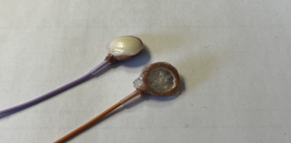
In this study, silver dish electrodes were used; the electrodes were categorized based on their coating: the first type of coating was conductive paste and the second type of coating was conductive gel sheet. In Figure 1, the pur- ple cable indicates the silver electrode coated with conductive paste, and the orange cable indicates the silver electrode coated with a transparent conduc- tive gel sheet. In this study, we investigated whether alpha waves and SSVEP components can be detected by using these two types of electrodes.
Figure 1: A purple cable electrode coated with conductive paste and an orange cable electrode coated with a transparent conductive gel sheet.
Alpha Wave Measurement in the Eyes-Open and Eyes-Closed Conditions Alpha waves decrease in the eyes-open condition and increase when in the eyes-closed condition (Adrian et al., 1934). In this study, we used this charac- teristic to calculate the alpha wave component obtained via ear-EEG. Subjects wore electrodes at the regions around their ears described below, and ear-EEG
236 Kondo and Tanaka
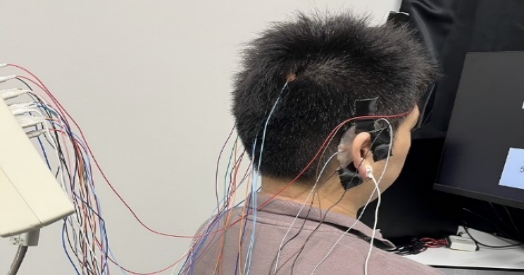
measured brain activity while they were at rest, in the eyes-open condition and eyes-closed condition. Figure 2 shows the EEG measurement process. While an overhead light is perceived in the room in which the experiment was conducted, it was switched on for ease of viewing of the reader of this paper; however, it was turned off during the experiment. Measurements were taken every 10 s; measurement trails were taken five times for the eyes-open condition and five times for the eyes-closed condition to average the alpha wave components for each trial during analysis. During these 10 measure- ments, subjects were instructed in advance to not do anything that would cause noise, such as grinding their teeth or moving their bodies. Additionally, they were instructed to relax and not to focus too much on their ears, as that might result in their neck muscles becoming strained, causing noise.
Figure 2: Measurement scene from the subject’s right ear position.
Frequency Response Measurement of SSVEP In this study, SSVEP was measured by presenting a single stimulus in the range of 10–60 Hz, presented in 2 Hz increments. In other words, the mea- sured SSVEP stimulation frequencies were the following 26 frequencies: 10, 12, 58, 60 Hz. The EEG measurement site was the same as the alpha wave measurement task. To maintain a constant distance and visual angle from the blinking stimulus for SSVEP induction, the subject’s position was adjusted so that the distance between them and the LCD was 50 cm and their line of sight was near the center of the display. The subjects gazed at the flashing stim- ulus screen shown in Figure 3. The start and end of the measurement were controlled by MATLAB (R2022b) and signaled by sound. The measurement period per measurement was 3 s, and the interval between measurements was 3 s. Four measurements were taken for each stimulation frequency, resulting in 104 measurements across the 26 stimulation frequencies. Stim- ulation frequencies were presented in an ascending order from 10 Hz in the or descending order from 60 Hz. This was performed to reduce the effects of subject fatigue over time and the order of measurements on the SSVEP com- ponent. In this study, a Nihon Koden EEG-1000 (1 kHz) was used. A 27-inch Z-EDGE LCD monitor was used. The LCD resolution was 1920 × 1080
Comparison of Alpha Waves and SSVEP Based on Ear-EEG Using Conductive Paste 237
pixels and the refresh rate was 240 Hz. The blinking stimulus shown in Figure 3 was implemented according to Eq. (1) by using the Unity game engine (2021.3.11f1).
Figure 3: Flickering stimuli display for SSVEP induction.
Measurement Area and Subjects
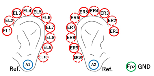
Figure 4 shows the positions of the EEG electrodes attached to the sub- ject. Electrodes were placed at 20 locations, 10 on each side of the head, surrounding the left and right ears. The GND and reference electrodes were Fpz (GND), A1, and A2 (Ref.) based on the extended 10–20 method recommended by the International Federation of Clinical Neurophysiology Societies (Nuwer et al., 1998).
Figure 4: Measurement area (the solid line indicates the conductive paste-coated electrode, and the dotted line indicates the conductive gel sheet-coated electrode).
A total of nine male and female students of Kogakuin University between the ages of 20 and 24 years participated in this study. This experiment adhered to Kogakuin University’s study ethics review for humans, “Psychobi- ological measurements for the development of new interfaces 2022-A-31.”
RESULTS
The alpha wave measurement results obtained by using conductive paste- coated electrodes, conductive gel sheet-coated electrodes, in the eyes-open
238 Kondo and Tanaka
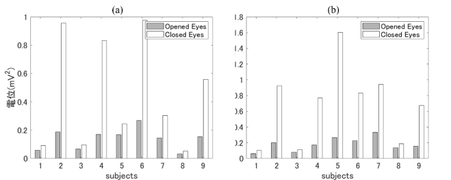
condition, and in the eyes-closed condition are presented below. Figure 5 (a) and (b) show the alpha wave power values for all subjects in the eyes-open and eyes-closed conditions. Figure 5 (a) is the measurement result obtained by using conductive paste-coated electrodes, and Figure 5 (b) is the measurement result obtained by using conductive gel sheet-coated electrodes.
Figure 5: Alpha wave power in the eyes-open and eyes-closed (a: paste, b: gel sheet).
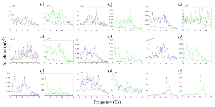
Figure 6 shows the alpha wave spectra of each subject. The results were divided by four factors: conductive paste-coated electrodes, conductive gel sheet-coated electrodes, eyes-open condition, and eyes-closed condition. The left side of each subject’s results is the result obtained by using the conductive paste-coated condition, and that on the right side is the result obtained by using the conductive gel sheet-coated condition. For the alpha wave spectrum, the solid line is the value for the eyes-open condition, and the solid line with circles is the value for the eyes-closed condition.
Figure 6: Alpha wave power spectrum for the eyes-open and eyes-closed (left: paste, right: gel sheet, solid line: when eyes are open, solid line with circles: when eyes are closed).
Comparison of Alpha Waves and SSVEP Based on Ear-EEG Using Conductive Paste 239
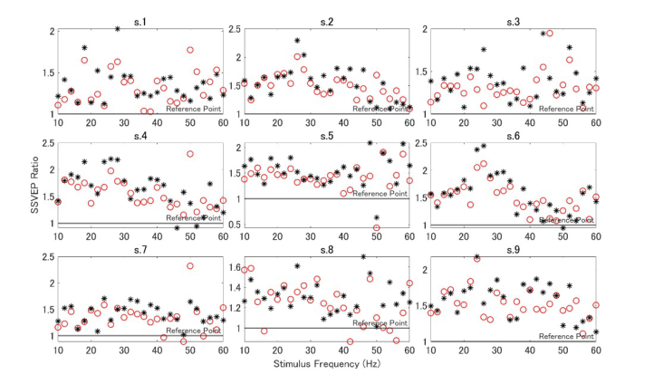
Figure 7 shows the SSVEP measurement results obtained by using the con- ductive paste-coated electrodes and conductive gel sheet-coated electrodes. The horizontal axis is the stimulation frequency, and the vertical axis is the SSVEP increase rate Ratio shown in Eq. (7). Since Ratio is an index of how much Ctarget has increased as compared to Cnon - target, the reference point was set to Ratio = 1.
Figure 7: Frequency response of SSVEP obtained by using conductive paste-coated electrodes and gel sheet-coated electrodes (red o mark: paste, black * mark: gel sheet).
DISCUSSION
As can be seen from Figures 5 and 6, the alpha wave power for all subjects was greater for the eyes-closed condition as compared to the eyes-open condi- tion. This is consistent with the general properties of alpha waves; therefore, it can be said that the alpha wave component during the experimental task was correctly detected via ear-EEG equipment designed in this study. It can also be seen that both the conductive paste-coated electrodes and gel sheet- coated electrodes are functioning appropriately. This shows that alpha waves can be measured even if the conductive paste-coated electrode is replaced by a conductive gel sheet-coated electrode. As conductive gel sheet-coated elec- trodes are worn in the ear, they are compatible with ear-EEG, that has the advantages of comfort and wearability. In future studies, we aim to investi- gate mechanisms to prevent the in-ear-EEG electrodes from falling out when conducting measurements for long periods.
The gel sheet-coated electrode showed good results in the alpha wave mea- surements; however, the results regarding the SSVEP measurements were slightly inferior as compared to that of the paste-coated electrodes. As shown in Figure 7, the SSVEP component measured with the conductive paste- coated electrode was generally larger than that measured with the conductive
240 Kondo and Tanaka
gel sheet-coated electrodes. This tendency was particularly strong for s. 2, s. 4, and s. 6. When using ear-EEG as a signal source for SSVEP-BCI system, it is desirable that the Ratio in equation (7) exceeds 1.5. This is because the BCI will not function appropriately if the SSVEP component induced while gazing at a blinking stimulus is buried in the SSVEP component during normal times. Although we succeeded in detecting SSVEP components from gel sheet-coated electrodes in all subjects, improvements in analysis methods and electrodes are required to operate SSVEP-BCI system by using this method. Among the subjects, s. 9 recorded the best SSVEP component. This subject stably recorded a Ratio of approximately 1.5 by using both conductive paste-coated electrodes and gel sheet-coated electrodes. This tendency was particularly strong when the frequency was less than 50 Hz; hence, improvements must be made in future studies so that this result can be reproduced with other subjects. We aim to continue to investigate whether the frequency character- istics of the SSVEP component depend on the individual or whether they can be improved by changing the shape of the electrode or analysis algorithm.
CONCLUSION
The advantages of ear-EEG are its wearability and simplicity. Since conduc- tive paste-coated electrodes negate the advantages of ear-EEG by staining the skin and hair of subjects, we tested the performance of conductive gel sheet-coated electrodes, and compared their performance to that of paste- coated electrodes. In this study, we measured alpha waves for the eyes-open and eyes-closed conditions and measured the frequency response of SSVEP to stimulation frequencies of 1–60 Hz. Consequently, we succeeded in clearly detecting the alpha wave power for both conductive paste-coated electrodes and gel sheet-coated electrodes. However, although the SSVEP component clearly increased in subjects when exposed to blinking stimulus, it was smaller in the results obtained via gel sheet-coated electrodes as compared to that obtained via paste-coated electrodes. Hence, in future studies, we aim to investigate the effectiveness of using gel sheet-coated electrodes to con- struct SSVEP-BCI systems, and verify whether any additional processing or electrode improvements are necessary to achieve 90% BCI accuracy.
ACKNOWLEDGMENT
This work was supported by JSPS KAKENHI Grant Number JP23K11202.
REFERENCES
Adrian, E. D. et al. (1934), The Berger Rhythm: Potential Changes From the Occipital Lobes in Man, Brain, 54(7), pp. 413–414. Ahn, J. W. et al. (2018), Wearable in-the-ear EEG system for SSVEP-based brain-
computer interface, Electronics Letters, 54(7), pp. 413–414. Akaho, S. (2013), Introduction to Canonical Correlation Analysis -Mutual Information Extraction from Multimodal Observations, Societas Neurologica Japonica, 20(2), pp. 62–72. Chen, X. et al. (2014), A high-ITR SSVEP-based BCI speller, Brain-Computer
Interfaces, 1(3–4), pp. 181–191.
Comparison of Alpha Waves and SSVEP Based on Ear-EEG Using Conductive Paste 241
Chen, X. et al. (2014), Hybrid frequency and phase coding for a high-speed SSVEPbased BCI speller, in 2014 36th Annual International Conference of the IEEE Engineering in Medicine and Biology Society, pp. 3993–3996. Goverdovsky, V. et al. (2015), In-ear EEG from viscoelastic generic earpieces: Robust
and unobtrusive 24/7 monitoring, IEEE Sensors Journal, 16(1), pp. 271–277. Guermandi, M. et al. (2018), A wearable device for minimally-invasive behind-theear EEG and evoked potentials, in 2018 IEEE Biomedical Circuits and Systems Conference (BioCAS), pp. 1–4. Herrmann, S. C. (2001), Human EEG responses to 1–100 Hz flicker: Resonance phe-
nomena in visual cortex and their potential correlation to cognitive phenomena, Experimental Brain Research, 137, pp. 346–353. Kidmose, P. et al. (2013), A study of evoked potentials from ear-EEG, IEEE Transactions on Biomedical Engineering, 60(10), pp. 2824–2830. Kondo, S. and Tanaka, H. (2023), Improvement of Accuracy of SSVEP-BCI with
In-Ear EEG Using Multiple Regression Analysis, Neuroergonomics and Cognitive Engineering, 102, pp. 264–273. Kondo, S. and Tanaka, H. (2023), Evaluation of Ear EEG Measurement Electrode Assuming SSVEP-BCI, Proceeding of Human Interface Symposium 2023, 6T-D18, pp. 116–123 (in Japanese). Kondo, S. and Tanaka, H. (2023), Ear EEG Measurement Method and SSVEP-BCI
Application, Proceeding of IEEE IM Society, Tokyo/Section Joint Chapter, IM09 Student Research Presentation, S23–023, pp. 13–14 (in Japanese). Ku´s, R., et al. (2013), On the quantification of SSVEP frequency responses in human EEG in realistic BCI conditions, PloS one, 8(10), pp. e77536. Kwak, N. S., & Lee, S. W. (2020), Error correction regression framework for
enhancing the decoding accuracies of ear-EEG brain–computer interfaces, IEEE transactions on cybernetics, 50(8), pp. 3654–3667. Lotte, F. et al. (2018), A review of classification algorithms for EEG-based brain– computer interfaces: a 10 year update, Journal of neural engineering, 15(3), 031005. Mikkelsen, K. B., et al. (2015), EEG recorded from the ear: characterizing the ear-
EEG method, Frontiers in neuroscience, 9(438), pp. 1–8. Nakanishi, M., et al. (2018), Enhancing detection of SSVEPs for a high-speed brain speller using task-related component analysis, IEEE Transactions on Biomedical Engineering, 65(1), pp. 104–112. Nuwer, M. R., et al. (1998), IFCN standards for digital recording of clinical EEG,
Electroencephalography and clinical Neurophysiology, 106(3), pp. 259–261. Pastor, M. A., et al. (2003), Human cerebral activation during steady-state visualevoked responses, Journal of neuroscience, 23(37), pp. 11621–11627. Sun, Y. et al. (2022), Cross-subject fusion based on time-weighting canonical
correlation analysis in SSVEP-BCIs, Measurement, 199, 111524. Vidal, J. J. (1973), Toward direct brain-computer communication, Annual review of Biophysics and Bioengineering, 2(1), pp. 157–180. Zhu, D. et al. (2010), A survey of stimulation methods used in SSVEP-based BCIs,
Computational intelligence and neuroscience, 2010, pp. 1–12. Zhu, Y. et al. (2021), EEGNet with ensemble learning to improve the crosssession classification of SSVEP based BCI from ear-EEG, IEEE Access, 9, pp. 15295–15303.
LISTED IN THE AHFE DIGITAL BOOKSTORE CATALOGUE
Explore the complete digital volume
Neuroergonomics and Cognitive Engineering
Read this individual research paper free through the original publisher, or explore the full digital proceedings book.
ISBN 978-1-964867-02-1
Explore this ISBN in the AHFE Bookstore ↗Listed in the July 2026 bookstore catalogue; search by ISBN to confirm current availability · Digital book purchase optional