Yuqian Wang, Junjian Chen, Yan Luximon (2024). A Pilot Study for Understanding the Lying Neck Posture with Flexion and Extension. In: Creativity, Innovation and Entrepreneurship. AHFE Open Access, vol. 125, pp. 106–111. https://doi.org/10.54941/ahfe1004726
1 School of Design, The Hong Kong Polytechnic University, Hong Kong SAR, China; Laboratory for Artificial Intelligence in Design, Hong Kong Science Park, Hong Kong SAR, China ↗
2 School of Design, The Hong Kong Polytechnic University, Hong Kong SAR, China ↗
Due to the prolonged focus on mobile phones or computers, people are exposed to a higher risk of neck issues. To avoid the radiation exposure of CT (Computed Tomography) scans, the MRI (magnetic resonance imaging) facilities with a lying posture were explored in this pilot study with 5 participants and three head postures (flexion, neutral, and extension). The vertebra angles from a sagittal view are measured, and the contribution of each pair of successive vertebras is compared with other literature. The results of this study show that the mean flexion and extension contributions fluctuated much more than others. This may be because the passive lying posture based on physical support is adopted rather than active flexion and extension maximum with a sitting posture. This suggests that active and passive head rotation postures have distinct mechanisms and should be studied separately. The lying posture with an MRI scan could be used for vertebra postures with different supports like pillows.
Nowadays, due to the prolonged focus on mobile phones or computers and adopting problematic postures throughout daily life, many people experi- ence issues related to their necks. These issues can manifest in various ways, including neck pain, neck stiffness, cervical radiculopathy, text neck, and so on (AlAbdulwahab et al., 2017).
Disruptions in neck movement can cause clinical symptoms that need to be diagnosed and treated properly. Studying the kinematics of the cervical spine is essential for understanding spinal diseases, improving motion-related prod- ucts, and differentiating between normal and abnormal motion (Lindenmann et al., 2022). This knowledge can also aid surgeons in preoperative planning. Hence, spine kinematics is quite important.
However, it is pretty difficult to figure out spine kinematics. Most work is based on CT (Computed Tomography) scans (Anderst, 2015; Anderst et al., 2011; Wan et al., 2021). However, CT scans come with potential risks and harms, like any medical procedure involving radiation exposure. Hence, this pilot study would like to use MRI to figure out the positions of each neck
A Pilot Study for Understanding the Lying Neck Posture With Flexion and Extension 107
bone and calculate related angles, as there is no radiation exposure in MRI scans, which is safer than CT (Parizel et al., 2001).
Also, as participants needed to keep static and lie on the MRI platform during the scan, we used pillows to mimic the head flexion and extension and compare them with related works.
METHOD
Participant Five young adult participants, with an average age of 28.9 years, were recruited for this pilot study. None of the participants had neck pain or clinical neurological or psychiatric disorders. Prior to the study, ethical approval was obtained from the Hong Kong Polytechnic University Insti- tutional Review Board, which operates under the Research Committee. All participants were given a thorough introduction to the study’s objec- tives and procedures, and they provided signed consent forms before their participation.
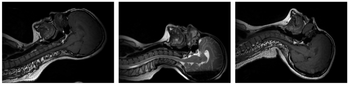
Facilities For each participant, he or she was required to have a small pillow, which is rolled by a towel under the neck to have a neutral position scan. Then, with another designed pillow to hold their head and shoulder to perform a flex- ion and extension posture. The examples of flexion, neutral, and extension postures are shown in Figure 2 (a, b, c), respectively.
For the scan setting, the participants will be able to undergo three T1 scanning (TR = 450ms; TE = 23ms; FoV = 382x382x272mm; voxel size = 0.6*0.6*0.8mm; total time 2:33min).
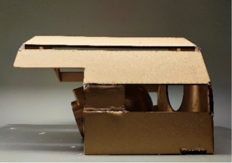
Figure 1: The wooden coil holder used in the MRI scan to hold the body coil with a head model with a brown color inside it with a neutral posture.Figure 2: The MRI scan results with a sagittal view of (a) flexion posture with physical support under the head; (b) neutral posture with a small pillow under the neck position; (c) extension posture with physical support under the shoulder and neck position.
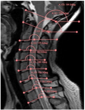
Measurement of Vertebrae Angles To measure the vertebra angle accurately from a sagittal view, it is essential to establish reference lines. For C0 (occipital bone), the reference line is deter- mined by the tangent to the cross sections of the basion and the opisthion (Botelho & Ferreira, 2013). In the case of C1 (Atlas), the reference line is tangent to the cross sections of the posterior and anterior arch (Ishii, Mukai, Hosono, Sakaura, Nakajima et al., 2004). Similarly, for the remaining cervi- cal vertebrae (C2 to C7), the reference line is established by the tangent to the cross-section of the vertebral body (Ishii, Mukai, Hosono, Sakaura, Fujii et al., 2004). Additionally, the reference line for the first thoracic vertebra (T1) will be designated as the origin line. An example measurement is shown in Figure 3.
Figure 3: An example measurement of vertebra angles in a sagittal view.
In the software Slicer (version 5.22), the absolute angles of all lines in the world coordinate system are measured relative to the horizontal line. Subse- quently, the relative angles between C0-C1, C1-C2, C2-C3, C3-C4, C5-C6, C6-C7, C7-T1, and C0-T1 are calculated using the measured data. The rel- ative angles for the neutral, flexed, and extended states are obtained. By comparing these measurements, the variation of each relative angle between different states can be determined.
A Pilot Study for Understanding the Lying Neck Posture With Flexion and Extension 109
RESULTS
Due to the time limitation, only 4 participants finished scans of all three postures. The fifth participant only finished the neutral and extension pos- tures. Due to the limitation of the coil holder, participants actually could not achieve the maximum angle for the flexion and extension. The average angles are 29.05 and 27.93 degrees, respectively, for flexion and extension from the neutral posture.
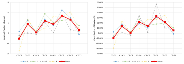
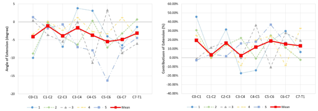
Figure 4(a) is based on measured vertebrae angles from neutral to flex- ion of each participant, and the red line with square markers is the average. According to the total flexion angle from C0 to T1, Figure 4b illustrates the percentage contribution of each pair of successive bones.
Figure 4: In a flexion head posture, there is a plot of (a) the measured vertebra angles from the neutral posture of each participant and group average; (b) the contribution percentage of each vertebra angle in each participant and their group average.Figure 5: In an extension head posture, there is a plot of (a) the measured verte- bra angles from the neutral posture of each participant and group average; (b) the contribution percentage of each vertebra angle in each participant and their group average.
Figure 5 shows the angles and percentage contributions of vertebrae angles in neutral to extension. It can be seen that the contribution of each verte- bra angle is quite different, and there are even vertebra angles with negative contributions. Both in flexion and extension, the angle of C5-C6 achieves maximum mean contribution. However, this trend may not be valid for one certain participant.
DISCUSSION
This study showed that MRI scanning is able to provide neck bone postures. Also, it turned out that different supports lead to different neck bone angles, and the shapes of support materials are important for spine relaxation. Also, the neck bone angles are quite different from person to person.
According to the review of Lindenmann (Lindenmann et al., 2022), there are some similar trends between our pilot study result and other works. Firstly, the angles of C0-C1 vary a lot compared to other angles. Then, the contribution of C7-T1 accounted for a relatively small proportion than most other angles.
In this study, we found a large difference between lying MRI scan and sit- ting CT scan conditions as the mean flexion and extension contributions are much more fluctuated than the other publications working on active neck postures in the review (Lindenmann et al., 2022). Firstly, the passive posture of neck bone motion with physical support may be different from postures activated by muscles. When muscles are actively engaged and con- tracted, their tension influences the positioning and movement of the bones. Because the muscles provide support, stability, and control over the bones, this active muscular engagement can result in more precise and controlled movements. Hence, the bone motion in passive postures is more likely to have larger fluctuations. Then, most other studies used active flexion and exten- sion maximum with a sitting posture rather than the passive lying posture. In lying positions, the pressure on the neck is generally reduced compared to sitting, as the weight of the head is distributed more evenly over the sup- porting surface, such as a pillow. Hence, the underlying mechanism could be quite different between sitting and lying for neck bones.
There are some limitations in this pilot study. Although all our participants had no symptoms of neck discomfort, four out of five participants showed some pattern of cervical kyphosis or military neck at their neutral posture. This may also influence the result. In addition, the bone edge of an MRI scan is not as clear as in CT. CT scans use X-rays, which are excellent at differentiating between tissues with large density differences, such as bone and soft tissue. On the other hand, MRI scans use powerful magnetic fields and radio waves to generate images, which are sensitive to variations in the water content and chemical composition of tissues. Hence, these may affect the accuracy of vertebra angle measurement from MRI scans.
CONCLUSION
In conclusion, we are able to measure the neck bone angle from the MRI scan, and the MRI scan can be a safer alternative choice to the harmful CT scanning in a lying posture. Also, we found out there is quite a large dif- ference between the passive lying posture and the active sitting posture of participants. It indicates that the research of the neck in a lying posture could use MRI scan facilities, like the neck posture with different pillows or other support materials.
A Pilot Study for Understanding the Lying Neck Posture With Flexion and Extension 111
ACKNOWLEDGMENT
Thanks to the University Research Facility in Behavioral and Systems Neuro- science at the Hong Kong Polytechnic University providing the guidance and facilities of the Human MRI scanning.
The work described in this paper was fully supported by a grant from the Research Grants Council of the Hong Kong Special Administrative Region, China (Project No. GRF/PolyU 15607922).
REFERENCES
AlAbdulwahab, S. S., Kachanathu, S. J., & AlMotairi, M. S. (2017). Smartphone use addiction can cause neck disability. Musculoskeletal Care, 15(1), 10–12. https: //doi.org/10.1002/msc.1170 Anderst, W. J. (2015). Cervical motion segment contributions to head motion during
flexion\extension, lateral bending, and axial rotation. The Spine Journal, 6. Anderst, W. J., Baillargeon, E., Donaldson, W. F., Lee, J. Y., & Kang, J. D.
(2011). Validation of a Noninvasive Technique to Precisely Measure In Vivo Three-Dimensional Cervical Spine Movement: Spine, 36(6), E393–E400. https: //doi.org/10.1097/BRS.0b013e31820b7e2f Botelho, R. V., & Ferreira, E. D. Z. (2013). Angular craniometry in craniocervical junction malformation. Neurosurgical Review, 36(4), 603–610. https://doi.org/ 10.1007/s10143-013-0471-0 Ishii, T., Mukai, Y., Hosono, N., Sakaura, H., Fujii, R., Nakajima, Y., Tamura,
S., Sugamoto, K., & Yoshikawa, H. (2004). Kinematics of the Subaxial Cervical Spine in Rotation In Vivo Three-Dimensional Analysis: Spine, 29(24), 2826–2831. https://doi.org/10.1097/01.brs.0000147806.31675.6b Ishii, T., Mukai, Y., Hosono, N., Sakaura, H., Nakajima, Y., Sato, Y., Sugamoto, K., & Yoshikawa, H. (2004). Kinematics of the Upper Cervical Spine in Rotation: In Vivo Three-Dimensional Analysis. Spine, 29(7), E139–E144. https://doi.org/10.1097/01. BRS.0000116998.55056.3C Lindenmann, S., Tsagkaris, C., Farshad, M., & Widmer, J. (2022). Kinematics of the
Cervical Spine Under Healthy and Degenerative Conditions: A Systematic Review. Annals of Biomedical Engineering, 50(12), 1705–1733. https://doi.org/10.1007/ s10439-022-03088-8 Parizel, P., Makkat, S., Van Miert, E., Van Goethem, J., Van Den Hauwe, L., & De Schepper, A. (2001). Intracranial hemorrhage: Principles of CT and MRI interpretation. European Radiology, 11(9), 1770–1783. https://doi.org/10.1007/ s003300000800 Wan, Z., Wang, W., Li, C., Li, J., Lin, J., Tian, F., Zhu, T., Wu, D., Guo, L., & Wang,
S. (2021). Validation and application of a novel in vivo cervical spine kinemat- ics analysis technique. Scientific Reports, 11(1), 24266. https://doi.org/10.1038/ s41598-021-01319-x
LISTED IN THE AHFE DIGITAL BOOKSTORE CATALOGUE
Explore the complete digital volume
Creativity, Innovation and Entrepreneurship
Read this individual research paper free through the original publisher, or explore the full digital proceedings book.
ISBN 978-1-964867-01-4
Explore this ISBN in the AHFE Bookstore ↗Listed in the July 2026 bookstore catalogue; search by ISBN to confirm current availability · Digital book purchase optional