Mei Ying Kwan, Kit Lun Yick, Joanne Yip, Chi Yung Tse (2022). Intervention of arch support: A quantitative study. In: Physical Ergonomics and Human Factors. AHFE Open Access, vol. 63, pp. 28–35. https://doi.org/10.54941/ahfe1002593
Hallux valgus is a common foot deformity characterized by hypermobility of the first metatarsal ray. The lower longitudinal arch is an intrinsic factor related to the lateral drift of the hallux. This paper conducted a quantitative study on the improvement of the first metatarsal and arch condition by arch support and used finite element analysis to simulate the bone displacement with the intervention of arch support. In this research, a foot arch support made of carbon fiber was developed and seventy-six female subjects were recruited for a two-month wear trial. Footprints of their dominant foot were measured to investigate the effect of the arch support on lifting the arch and correcting the hallux valgus pathology. Different foot parameters including foot length, foot breadth, heel breadth, arch angle, arch breadth, plantar arch index, foot type index, and hallux valgus angle were also compared. By using finite element analysis, the biomechanical effects of the arch support on the foot structure can be visualized. According to the results of the wear trial, the use of the arch support can significantly improve the arch curvature of the foot, while no significant correction of the hallux valgus angle was found. Among the arch parameters, the arch breadth and the foot type index are the key indicators to precisely characterize foot types and arch conditions. When a clear outline of the footprint is not available, arch breadth provides reliable association with the foot type index (R2 = 0.928). An arch breadth ≥ 4 cm is categorized as flatfoot. This article confirms the effectiveness of our arch support in lifting the arch over a two-month period and provides a scientific surrogate index to aid in diagnosis, which is important for therapeutic and diagnostic applications.
Hallux valgus (HV) is a foot deformity commonly seen in clinical practice. It is commonly associated with foot pain, which inhibits the mobility and phy- sical activity level of those who suffer from the deformity (Nix et al., 2012). HV angle is used as an indicator for objectively measuring the level of defor- mity. According to Hardy and Clapham (1951), it is the angle between the axis of the first metatarsophalangeal joints (MTP1) and that of the proximal phalanx of the big toe. HV deformity is identified when the HV angle exceeds 15 degrees (Richie Jr, 2020). Richie Jr (2020) proposed that flatfoot was an intrinsic risk factor related to the development of HV. In flatfoot, the foot is pronated and the arch collapses under the weight of the body. It may lead to
Intervention of Arch Support: A Quantitative Study 29
a change in the alignment of the first ray axis and is associated with lateral drift of the hallux or medial deviation of the MTP1. Custom-made orthotic insoles can be an effective means for arch support. Studies showed that wea- ring an arch-support insole provides the generation of propulsion force while walking and improve joint kinetics (Huang et al., 2020, Wang et al., 2020). In this study, an insole embedded with a rigid arch support made of carbon fiber was designed and developed for a two-month wear trial. The first aim of the present study was thus to evaluate the effect of our arch support inte- rvention by the footprint. We expect significant improvement in pathology in patients with flatfoot. The pathology of HV patients can also be alleviated accordingly.
It is important to diagnose HV and flatfoot scientifically to prevent further deterioration. Compared with ultrasonography, two-dimensional static foot- print analysis by Podograph is a cost-effective and reliable method (Queen et al., 2007). There is a significant correlation between their measurement (p <0.001) (Lo, 2014). In terms of arch measurements, researchers developed the foot type index as a reliable indicator with high sensitivity for diagnosing flatfoot (Pita-Fernández et al., 2015). The cut-off points for the diagnosis of flatfoot > 0.45 for the foot type index (Pita-Fernández et al., 2015). Howe- ver, traditionally collected footprints without clear outlines that data may be lost due to the inability to measure (Queen et al., 2007). This study compa- red several foot parameters, including foot length, foot breadth, heel breadth, arch angle, arch breadth, and plantar arch index, using the foot type index as a benchmark, to obtain the required measurements. This can help clinicians in choosing a better alternative to the foot type index and measuring the mis- sing data. Thus, the second part of this study is to investigate the relationship between the foot type index and selected arch measurements.
METHODOLOGY
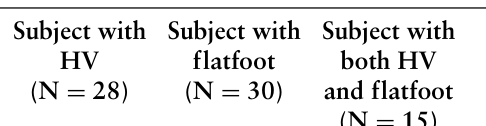
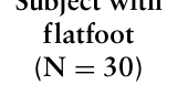
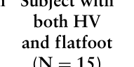
Participants Seventy-six elderly females volunteered for the study. Their average age was 81 years, with an average BMI of 24, an average height of 151 centimeters (cm) and an average weight of 54 kilograms. Participants were divided into four groups, namely (1) subject with HV (2) subject with flatfoot (3) subject with both HV and flatfoot and (4) control. Of the seventy-six participants, twenty-eight (36.84%) have HV, and thirty (39.47%) have flatfoot, of which fifteen (19.74%) have both HV and flatfoot. Thirty-three (43.42%) subjects without HV or flatfoot will serve as the control group.
They were asked to wear an arch support intervention for two months, with at least 20 hours per week. The arch support intervention is a san- dwich structure that an arch support made of carbon fiber was embedded between the Poron insole and midsole. Two-dimensional footprints were col- lected from each volunteer using Podograph before and after the wear trial. Written consent was obtained from all subjects before study commencement. All the study’s procedures were approved by the Human Subjects Ethics Sub- committee of Research Committee at University, and the study conformed to all policies regarding the use of human participants.
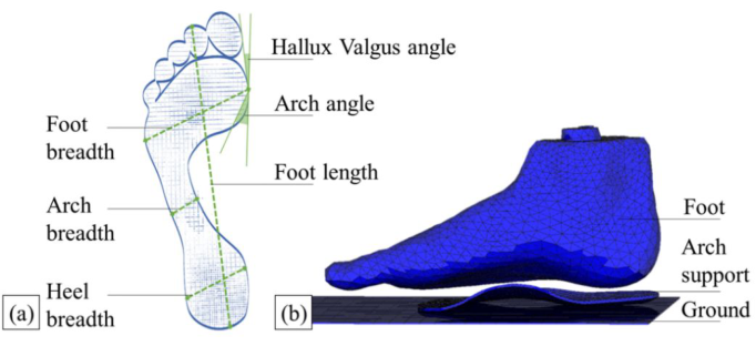
Measurements Two-dimensional footprints were collected from each volunteer in a barefoot weight-bearing standing position using a Podograph. During the test of foot- prints, the subjects should stand naturally with feet shoulder-width apart. The length, breadth and angle measurements of the dominant foot were suggested in Figure 1a. The foot type index was calculated as arch breadth divided by foot breadth. A lower index suggested that the arch is being supported (Pita- Fernández et al., 2015). The plantar arch index establishes a relationship between the central and posterior regions of the footprint. It was calculated as arch breadth divided by heel breadth. A lower index value means a higher arch.
Figure 1: The methodology in this study. (a) Details of foot anthropometric measure- ments; (b) The FE model.
Statistical Analysis Data on the footprint of the subjects will be assessed. The R project for stati- stical computing was used to analyze the data. The normality assumption will be checked by the normal QQ plot. Linear regression and Pearson’s correla- tions were adopted to analyze the association between foot type index and other foot measurements. Statistical differences were calculated with paired samples t-tests. The significance of the statistical analysis was set at a level of 0.05.
Finite Element Analysis The dominant foot MR images of a female subject with a normal weight BMI who has an 18-degree HV angle was taken in a neutral unloaded position to construct the finite element model. The geometry of the foot was taken from the model subject using a structured light handheld 3D scanner (Artec Eva, Luxembourg). The foot was put in a neutral and non-weight-bearing con- dition during scanning. The scanned data of the foot and the arch support were registered using Artec Studio 13 and then imported into a 3D model processing software (3ds Max, Autodesk). The bones, arch support, and foot
Intervention of Arch Support: A Quantitative Study 31
were constructed using finite element (FE) analysis software (MSC Marc/ Mentat) (see Figure 1b). The material properties were obtained with refere- nces and are listed in Table 1. The mesh size of the solid elements ranged from 3 mm to 10 mm, and their mesh type was Tetrahedral. Through the FE analysis, the displacement on the foot was systematically evaluated upon the arch support intervention.
(Wong et al., 2014) Arch support 80000 0.3 Solid Elastic-plastic isotropic Soft tissue 0.15 0.49 Solid Elastic-plastic isotropic (Lemmon et al., 1997) Bone 7300 0.3 Solid Elastic-plastic isotropic
(Nakamura et al., 1981) Ligament 260 0.4 Truss Elastic-plastic isotropic
(Siegler et al., 1988)
RESULT AND DISCUSSION
Paired samples t-tests have been carried out to evaluate the changes in foot parameters measured before and after the wear trial (see Table 2). The data were normally distributed. After the wear trial, there were significant chan- ges in all the arch measurements. It suggested that all the four groups of subjects showed statistically significant improvements in the arch angle, arch breadth, plantar arch index, and foot type index after the wear trial. Using the arch support design shows greater improvements on foot deformations in subjects with pathology compared to controls. It can be seen that the arch support plays a greater effect on the target patient. This conclusion matches our hypothesis. The phenomenon was particularly evident in subjects with both HV and flatfoot, who had a 7.933-degree improvement in the arch angle (p = 0.002), 0.149 reduction in the plantar arch index (p < 0.001), 0.657 cm reduction in arch breadth (p = 0.003), and 0.063 reduction in foot type index (p = 0.013).
The result proves that long-term wearing of arch support can help improve flatfoot. Previous study found that the arch support intervention increases the contact area of the midfoot to provide support for the medial arch. Arch support, which is composed of harder materials, can also provide better sup- port, resulting in shorter stance time in level walking (Perry et al., 2007). The shorter stance time could reflect the patient is gradually changed from a pathological gait to a normal gait and may increase gait speed while walking (Guo et al., 2017; Studenski et al., 2011). The arch support used in the current study, made of rigid carbon fiber, provides adequate support to the midfoot.
Table 2. Two-month effect of the wear trial.
Table available in the original PDF paper.
Parameters Subject with Subject with
Equation reproduced from the original PDF.Equation reproduced from the original PDF.Equation reproduced from the original PDF.
6.000 5.950 7.933 2.833
Arch angle (degree)
Mean of differences −0.108 −0.092 −0.149 −0.021 p-value <.001 <.001 0.002 0.002 Plantar arch index Mean of differences
−0.511 −0.448 −0.657 −0.105
p-value <.001 0.001 <.001 <.001 Arch breadth (cm)
Mean of differences −0.056 −0.046 −0.063 −0.010 p-value <.001 <.001 0.003 0.003 Foot type index Mean of differences
-1.536 1.033 −0.933 1.455
p-value <.001 0.004 0.013 0.013 HV angle (degree)
Mean of differences
0.205 0.152 0.257 0.265
p-value 0.086 0.190 0.334 0.334 Foot length (cm)
Mean of differences
0.048 0.037 0.150 0.018
p-value 0.014 0.023 0.009 0.009 Heel breadth (cm)
Mean of differences −0.043 −0.097 −0.210 −0.062 p-value 0.502 0.545 0.163 0.163 Foot breadth (cm) Mean of differences
p-value 0.712 0.302 0.193 0.193
In terms of HV angle, no significant changes were found in any group, but a slight improvement could be found in subjects with HV, with a 1.536- degree reduction in HV angle (p = 0.086). In addition to HV subjects, a 0.933-degree reduction in HV angle could be found in subjects with both HV and flatfoot (p = 0.334). The use of arch support to lift the arch of the foot tends to help HV correction. We believe that with adequate arch support, the anatomical alignment of the foot can be restored correctly (Farzadi et al., 2015, Kwan et al., 2021, Tehraninasr et al., 2008). However, the results of the two-month wear trial showed that the HV angle correction could not catch up with the improvement of the arch index. A longer treatment period may be required to have significant HV angle correction. The FE analysis results also objectively showed that when the simulated subjects stood with the arch support, there was no significant displacement of the foot bones. This may explain why arch support cannot improve HV quickly and effectively, and more aggressive intervention may be necessary.
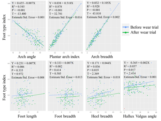
Linear Regression was computed to analyze the relationship between foot type index and other foot measurements and their interactions. Results were shown in Figure 2. The significantly negative relationship with foot type
Intervention of Arch Support: A Quantitative Study 33
Figure 2: Relationship between foot type index and other foot measurements (N = 76).
index and arch angle (p < 0.001, R2 = 0.545), and significantly positive relationship between foot type index and plantar arch index (p < 0.001, R2 = 0.878), and arch breadth (p < 0.001, R2 = 0.928) can be found. The coefficient of determination is the highest in arch breadth, followed by plan- tar arch index, and then arch angle. The improvements in the foot type index resulted in increased arch angle, with reduced arch breadth, plantar arch index, heel breadth, and HV angle. A significant positive relationship betw- een foot type index and heel breadth (p = 0.019, R2 = 0.036), and HV angle (p = 0.017, R2 = 0.037) were also found.
Correlations between foot type index and other foot arch related measu- rements were also analyzed. The arch angle, arch breadth, and plantar arch index were strongly correlated with foot type index. Among them, the corre- lation between foot type index and arch breadth was the strongest (p < 0.001, R = 0.960). The presented results suggest that all the arch indices studied are suitable for diagnosing flatfoot, while arch breadth is the most suitable mea- surement to substitute foot type index when necessary. If foot type index is not available, arch breadth, plantar arch index or arch angle provide useful foot information at the time of diagnosis. An arch breadth ≥4 cm, a plantar arch index ≥0.8 or an arch angle ≤29 degrees are considered as flatfoot, using a foot type index > 0.45 as an indicator.
The current study obtained only a two-month trial, and while it had signi- ficantly improved arch condition, it failed to treat HV pathology. Future studies could expand the duration of the wear trial. In addition to the duration of the trial, since the participants lived in elderly centers and were older retirees with more of their daily activities at the center, their activity levels were expected to be lower, which may also have affected the trial results.
CONCLUSION
It may be concluded from the results of this study that our arch support inte- rvention, with the use of hard arch support, can significantly improve the foot arch after a two-month wear trial. However, the correction on HV is not significant. Foot type index, as an indicator of two-dimensional static footprint analysis, has been used worldwide. Since the contour of the foot- print is not clear, it is important to find a closer method to replace or predict the foot type index. The presented results suggest that all the arch indices studied have strong correlations with foot type index, while among the mea- surements, arch breadth is the best predictor of the foot type index, it can be regarded as the most suitable measurement to substitute foot type index in research studies or when performing clinical diagnosis.
ACKNOWLEDGMENT
The authors would like to acknowledge the Departmental Grant of Insti- tute of Textiles and Clothing, The Hong Kong Polytechnic University (grant number PolyU RHRM) for funding this project.
REFERENCES
Chung, D. D. L. (1994) ‘CHAPTER 4 - Properties of Carbon Fibers’, in Chung, D.D.L. (ed.) Carbon Fiber Composites. Boston: Butterworth-Heinemann, pp. 65–78. Farzadi, M., Safaeepour, Z., Mousavi, M. E. and Saeedi, H. (2015) ‘Effect of medial arch support foot orthosis on plantar pressure distribution in females with mild- to-moderate hallux valgus after one month of follow-up’, Prosthet Orthot Int, 39(2), pp. 134–139. doi: 10.1177/0309364613518229 Guo, G., Guffey, K., Chen, W. and Pergami, P. (2017) ‘Classification of Nor-
mal and Pathological Gait in Young Children Based on Foot Pressure Data’, Neuroinformatics, 15(1), pp. 13–24. doi: 10.1007/s12021-016-9313-x Hardy, R. H. and Clapham, J. C. R. (1951) ‘Observations on Hallux Valgus’, Journal of bone and joint surgery. British volume, 33-B(3), pp. 376–391. doi: 10.1302/0301-620X.33B3.376 Huang, Y. P., Peng, H. T., Wang, X., Chen, Z. R. and Song, C. Y. (2020)
‘The arch support insoles show benefits to people with flatfoot on stance time, cadence, plantar pressure and contact area’, PLoS ONE, 15(8 August). doi: 10.1371/journal.pone.0237382 Kwan, M. Y., Yick, K. L., Yip, J. and Tse, C. Y. (2021) ‘Hallux valgus orthosis characteristics and effectiveness: A systematic review with meta-analysis’, BMJ Open, 11(8). doi: 10.1136/bmjopen-2020-047273 Lemmon, D., Shiang, T. Y., Hashmi, A., Ulbrecht, J. S. and Cavanagh, P. R. (1997)
‘The effect of insoles in therapeutic footwear—A finite element approach’, J Biomech, 30(6), pp. 615–620. doi: 10.1016/S0021-9290(97)00006-7 Lo, W. T. (2014) Development of plantar pressure relieving orthotic insoles for people with diabetes. Hong Kong Polytechnic University. Nakamura, S., Crowninshield, R. D. and Cooper, R. R. (1981) ‘An analysis of
soft tissue loading in the foot--a preliminary report’, Bull Prosthet Res, 10-35, pp. 27–34.
Intervention of Arch Support: A Quantitative Study 35
Nix, S. E., Vicenzino, B. T. and Smith, M. D. (2012) ‘Foot pain and functional limitation in healthy adults with hallux valgus: A cross-sectional study’, BMC Musculoskeletal Disorders, 13. doi: 10.1186/1471-2474-13-197 Perry, S. D., Radtke, A. and Goodwin, C. R. (2007) ‘Influence of footwear midsole
material hardness on dynamic balance control during unexpected gait termina- tion’, Gait and Posture, 25(1), pp. 94–98. doi: 10.1016/j.gaitpost.2006.01.005 Pita-Fernández, S., González-Martín, C., Seoane-Pillado, T., López-Calviño, B., Pértega-Díaz, S. and Gil-Guillén, V. (2015) ‘Validity of footprint analysis to deter- mine flatfoot using clinical diagnosis as the gold standard in a random sample aged 40 years and older’, Journal of Epidemiology, 25(2), pp. 148–154. doi: 10.2188/jea.JE20140082 Queen, R. M., Mall, N. A., Hardaker, W. M. and Nunley Ii, J. A. (2007) ‘Describing
the medial longitudinal arch using footprint indices and a clinical grading system’, Foot and Ankle International, 28(4), pp. 456–462. doi: 10.3113/FAI.2007.0456 Richie Jr, D. H. (2020) ‘Disorders of the First Ray: Part 1 Hallux Abductovalgus Deformity’. Cham: Cham: Springer International Publishing, pp. 105–137. Siegler, S., Block, J. and Schneck, C. D. (1988) ‘The Mechanical Characteristics of the
Collateral Ligaments of the Human Ankle Joint’, Foot Ankle, 8(5), pp. 234–242. doi: 10.1177/107110078800800502 Studenski, S., Perera, S., Patel, K., Rosano, C., Faulkner, K., Inzitari, M., Brach, J., Chandler, J., Cawthon, P., Connor, E. B., Nevitt, M., Visser, M., Kritchevsky, S., Badinelli, S., Harris, T., Newman, A. B., Cauley, J., Ferrucci, L. and Guralnik, J. (2011) ‘Gait Speed and Survival in Older Adults’, JAMA, 305(1), pp. 50–58. doi: 10.1001/jama.2010.1923 Tehraninasr, A., Saeedi, H., Forogh, B., Bahramizadeh, M. and Keyhani, M. R. (2008)
‘Effects of insole with toe-separator and night splint on patients with painful hal- lux valgus: A comparative study’, Prosthetics and Orthotics International, 32(1), pp. 79–83. doi: 10.1080/03093640701669074 Wang, Y., Lam, W. K., Cheung, C. H. and Leung, A. K. L. (2020) ‘Effect of red arch-support insoles on subjective comfort and movement biomechanics in vari- ous landing heights’, International Journal of Environmental Research and Public Health, 17(7). doi: 10.3390/ijerph17072476 Wong, D. W. C., Zhang, M., Yu, J. and Leung, A. K. L. (2014) ‘Biomechanics of first
ray hypermobility: An investigation on joint force during walking using finite element analysis’, Medical Engineering and Physics, 36(11), pp. 1388–1393. doi: 10.1016/j.medengphy.2014.03.004
LISTED IN THE AHFE DIGITAL BOOKSTORE CATALOGUE
Explore the complete digital volume
Physical Ergonomics and Human Factors
Read this individual research paper free through the original publisher, or explore the full digital proceedings book.
ISBN 978-1-958651-39-1
Explore this ISBN in the AHFE Bookstore ↗Listed in the July 2026 bookstore catalogue; search by ISBN to confirm current availability · Digital book purchase optional