Alejandra Gomez De Cadiz, Adrian Morales-casas, Claudia Marissa Aguirre Ramón, Ignacio Espíritu-garcía-molina, Cristina Herrera Ligero (2025). Connecting Image and Reality: The Role of 3D Printing in Surgical Planning. In: Human Interaction and Emerging Technologies (IHIET-AI 2025): Artificial Intelligence and Future Applications. AHFE Open Access, vol. 161, pp. 458–466. https://doi.org/10.54941/ahfe1005938
Training the healthcare service members of tomorrow or assisting in the surgical planning of today's interventions is challenging, as the current CT/MRI images require an experienced and trained eye to interpret the intricate anatomy of the human body correctly. Although the CT/MRI scans are highly detailed and widely implemented, they cannot address the level of understanding a trainee or expert could gain through the haptic perception or tactile learning experience offered by 3D printing. 3D printing enables hands-on understanding through highly detailed, patient-specific anatomical models from medical imaging, providing surgeons with an interactive way to visualise and practice complex procedures before entering the operating room. In surgical education, 3D-printed models, especially those simulating the texture of actual tissues or bones, provide essential tactile feedback that contributes to realistic training scenarios. This enhances surgical precision and reduces the likelihood of complications during surgery. Moreover, these models allow trainees to practice accurate replicas of human organs, improving their skills in a risk-free environment. Therefore, this paper presents some case studies focusing on 3D printing in surgical planning that can effectively highlight the technology's current advantages and limitations. The models, fabricated with flexible and radiotransparent materials, allow surgeons to simulate surgical scenarios, improving preoperative planning, instrument handling, and decision-making. Subjective validation by specialists demonstrated that these models accurately replicate the physical properties of the target anatomy, aiding in better visualisation and procedural practice. However, limitations were observed in current methodologies, such as challenges related to material elasticity, the durability of 3D-printed models, and difficulties in navigating tortuous anatomical paths during simulations. Further, there is room for improvement in the accuracy of specific anatomical features and the interaction with surgical instruments, where minor irregularities hinder smooth operation. According to the findings, future work should focus on refining the materials used in 3D printing to enhance the robustness and realism of the models, particularly in complex anatomical structures. Additionally, incorporating real-time imaging data with 3D printing could further improve the adaptability of these models for preoperative simulations. Expanding these technologies beyond their current use in vascular surgery could revolutionise other surgical fields, offering customised, patient-specific planning tools across various medical disciplines.
Connecting Image and Reality: The Role of 3D Printing in Surgical Planning 459
INTRODUCTION
Two-dimensional (2D) images, such as X-rays, magnetic resonance images (MRI), or computerized tomography (CT) scans, have been and still are valuable tools in understanding different pathologies and the planning of surgical interventions (Thiruchandran et al., 2025). In the early 2000s, the capability of turning 2D images generated by the listed medical image tools allowed the creation of complex, customized anatomical models first used to make dental implants and custom prosthetics (Lee Ventola, 2014). Since then, the application portfolio for 3D printing has evolved considerably. Applications like implant and tissue design, medical research, medical education and training, and surgical planning are exponentially growing (Lee Ventola, 2014; Tack et al., 2016).
Through 3D printing, the process of how modern surgeons address surgical planning is changing. In 2016, Martelli et al. (2016) concluded that although the 3D printing technology had become more affordable and user-friendly, the cost and time to produce desirable outcomes would limit its widespread use in hospitals. The limitations were mainly related to the need for more guidelines on practical experience with 3D printing in surgery, resistance towards losing control over the decisions that affect their patients, and accepting external technical support. Almost a decade later, the landscape changes in 2025, Thiruchandran et al. (2025) present a collection of case studies on surgical planning split into two main areas: surgical guides based on 3D models of patient’s anatomy and surgical instruments, 3D-printed instruments to assist with delicate procedures.
Therefore, modern surgical planning and training help to the limitations of CT and MRI, which require expert interpretation and lack tactile feedback crucial for skill development. 3D printing addresses these limitations by transforming imaging data into detailed, patient-specific anatomical models. These models enable interactive training and preoperative rehearsals, improving precision and reducing complications (Elkasabgy et al., 2020; Thiruchandran et al., 2025). This paper presents case studies highlighting 3D printing’s advantages, including enhanced surgical planning and tactile stimulation, while addressing material properties and cost limitations. Recommendations are provided for integrating real-time imaging and expanding 3D printing to other surgical fields.
Traditional imaging techniques like CT and MRI are invaluable for visualising complex anatomical structures but need to offer the hands-on interaction required for comprehensive training and surgical planning. Lee Ventola (2014) estimated that in the next 10 years, the 3D printing healthcare industry will grow from $11 million to $1.9 billion worth of industry. This prediction was not fulfilled as it was way too conservative. Its estimate is 52% below the actual market size; according to Global Market Insights, the current healthcare 3D printing market size is $2.9 billion (Healthcare 3D Printing Market Size and Share Report, 2030, 2024). In the next 10 years, 2032, the projected market value of 3D printing in healthcare applications is around $13.8 billion. Other reports have a much more optimistic perspective of revenue forecasted in 2030, set at $27.29 billion (Healthcare 3D Printing Market Size & Share Report, 2032, 2024).
Overall, it could be agreed that the forecast in revenue is, at least, as promising as the increasing number of case studies that highlight the utility of 3D-printed models in the healthcare sector. This paper explores 3D printing’s transformative role in surgical education and planning, emphasising its current applications, challenges, and future directions.
Current Applications of 3D Printing in the Surgical Environments Among the main medical applications for 3D-printed models of anatomical structures are educational applications, such as surgical training and simulation. Second, creating surgical instruments, therefore, preoperative planning applications. Last, patient-specific applications such as diagnosis and treatments (Winder & Bibb, 2005; Lee Ventola, 2014; Papotto et al., 2022; Thiruchandran et al., 2025). These applications underline the models’ effectiveness in risk-free training environments and their contribution to better surgical outcomes (Webb, 2000; Rengier et al., 2010).
In addition, 3D printed models help to enhance tactile learning by enabling the physical exploration of complex anatomies, which is critical for skill acquisition and confidence building. The 3D models fabricated from CT/MRI data replicate tissue textures, improving understanding of spatial and anatomical relationships (Mallon & Farnan, 2021; Thiruchandran et al., 2025). Therefore, the application in surgical training and simulation can refine techniques and decision-making processes (Papotto et al., 2022). Furthermore, such models support interprofessional education by facilitating collaborative learning in multidisciplinary teams, which is critical for optimising patient outcomes.
On the surgical preparation planning side, 3D printing has transformed the preoperative process by enabling the creation of custom surgical tools. These tools allow modern surgeons to perform specific procedures, or patient anatomies are now created rapidly and at a fraction of the cost compared to traditional manufacturing methods (Martelli et al., 2016; Tack et al., 2016; Elkasabgy, Mahmoud and Maged, 2020). The 3D printed tools enhance precision during surgery, minimising errors and reducing operation times. The orthopaedic and craniofacial surgeries, where exact measurements are critical, are a good case study for the use of 3D-printed guides to ensure proper alignment of instruments during the operative process (Wang et al., 2020). Lastly, 3D-printed models have also been shown to impact patient-specific applications for diagnosis and treatment significantly. In diagnostics, 3D- printed models enable clinicians to visualise and analyse complex anatomical anomalies in three dimensions, providing insights that may not be apparent in standard 2D imaging (Chen et al., 2022). This is especially valuable in cases involving congenital abnormalities, intricate vascular conditions, or rare tumour locations (Mallon & Farnan, 2021).
Moreover, 3D-printed implants and prosthetics are becoming increasingly common for treatment (Hieu et al., 2005; Bibb et al., 2009). These patient- specific devices are designed to fit perfectly within a patient’s anatomy, improving comfort and functionality. For example, custom cranial implants,
Connecting Image and Reality: The Role of 3D Printing in Surgical Planning 461
dental prosthetics, and orthopaedic devices have demonstrated superior outcomes to off-the-shelf alternatives (Lee Ventola, 2014). The technology also enables the creation of biocompatible materials and scaffolds for regenerative medicine, paving the way for future innovations in tissue engineering and organ printing (Mallon & Farnan, 2021).
Limitations and Challenges While 3D-printed models are promising to enhance surgical preparation and decision-making, addressing the material and technological limitations is critical to advancing their clinical utility. From a technological perspective, current 3D printing methodologies face challenges in achieving the necessary resolution and accuracy to precisely replicate the organic anatomical structures. Fused Deposition Modelling (FDM), for instance, often results in layer lines that may interfere with the smooth surfaces required for particular medical simulations (da Silva et al., 2021). Similarly, resin-based techniques such as Stereolithography (SLA) or Digital Light Processing (DLP) can produce high-resolution models. However, they may introduce brittleness due to the inherent properties of photopolymerised materials (Rashed et al., 2024). Other technologies, such as Selective Laser Melting (SLM) and Selective Laser Sintering (SLS), excel in creating flexible and lightweight models with high resolution. However, high production costs (particularly for SLM), material limitations, and post-processing requirements hinder widespread adoption.
The post-processing aspect of 3D printed models adds another layer of limitations, as it involves additional steps that may introduce errors or inconsistencies (Shahrubudin et al., 2020). Cleaning, curing, and finishing the printed parts can alter their dimensional accuracy and surface quality. These factors collectively hinder the scalability and practicality of 3D-printed models in routine clinical practice.
Regardless of such limitations, the potential of 3D printing in the healthcare sector becomes a valuable asset in the planning, simulation and training for complex interventions such as liver resections (Calle Gómez et al., 2025), orthopaedic surgery (Koshkin et al., 2024), fetal surgeries (Fils et al., 2024) or craniofacial reconstructions (Park, 2022) to name a few cases. Ultimately, 3D-printed models fabricated with flexible and radio-transparent materials allow surgeons to simulate surgical scenarios, improving preoperative planning, instrument handling, and decision-making. Despite the advancements and benefits of 3D-printed models in medical applications, significant limitations persist due to the intricate demands of their use. One notable challenge lies in the inherent mechanical properties of the materials employed in additive manufacturing. Flexible and radio- transparent materials, while advantageous for surgical simulation, often lack the required mechanical strength and durability to endure repetitive or high-stress scenarios, limiting their long-term usability (da Silva et al., 2021). Additionally, the elasticity of these materials may not adequately replicate the biomechanical properties of human tissues, which can impair the accuracy of surgical rehearsals (Rendas et al., 2022). Therefore, developing
and validating 3D-printed models represent a cornerstone for pre-surgical planning and training for complex interventions.
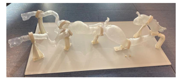
Case Studies of Surgical Planning and Training To overcome the limitations of 3D-printed models, developing and validating a model representing the aorta and its surrounding structures for pre-surgical planning and training in treating thoracic aortic aneurysms (TEVAR). The models were specifically designed to replicate anatomical conditions for TEVAR, which involve deploying prosthetic devices, such as stent grafts, into the aorta via femoral or transapical access. The prototype was designed to include the aortic valve, the aorta extending to the iliac bifurcations, and the left ventricle. Two variations of the left ventricle: one representing only the blood volume and another including the ventricular muscle, see Figure 1.
These models were fabricated using SLA (stereolithography) and RTV2 (room temperature vulcanising two components) silicon printing techniques. Flexible and semi-transparent resins were selected for the aortic structure. In contrast, elastic resins and silicone-based materials were used for the ventricular components to replicate realistic elasticity and transparency. The model also incorporated openings for femoral and transapical access, providing versatility for simulating different surgical approaches. To enhance functionality, design modifications were implemented after initial validation sessions. These included sealing internal iliac branches to facilitate water injection for lubrication and optimising the supports for robustness and ease of handling.
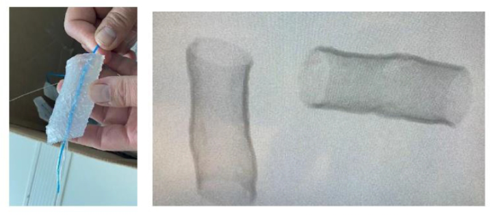
The model underwent a subjective validation process with surgical specialists at La Fe Hospital. Early assessments focused on material transparency, elasticity, X-ray transparency, and dimensional accuracy, see Figure 2.
Connecting Image and Reality: The Role of 3D Printing in Surgical Planning 463
Figure 2: Iterative validation process of the model with specialists and tests.
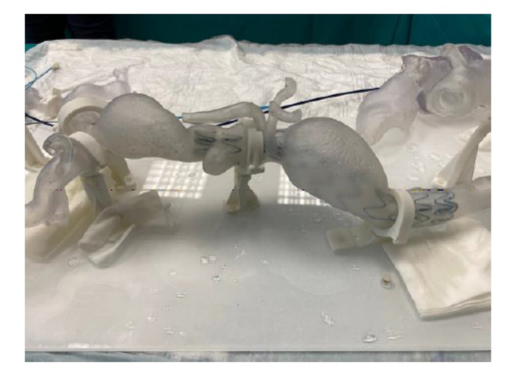
Iterative adjustments were made to accommodate surgical instruments and improve structural durability under manipulation. For instance, the iliac walls were thickened by 1.7 mm to allow smooth instrument insertion and redundant supports were removed to simplify handling surgical scenarios, demonstrating the model’s efficacy in reproducing anatomical tortuosity and surgical challenges. The final prototype successfully allowed the deployment of prosthetic devices at targeted locations, validating its utility for training and planning, see Figure 3.
Figure 3: Deployment of prosthetic devices at targeted locations.
Future Directions The development and validation of a 3D-printed anatomical model of the thoracic aorta and its surroundings have proven its potential in training vascular surgery and interventional radiology specialities. However, room for several improvements and research remains present.
Future model iterations could incorporate patient-specific anatomical data to enable personalised surgical simulations. By integrating imaging techniques such as computed tomography (CT) or magnetic resonance imaging (MRI), the models could provide more accurate anatomical representations tailored to individual cases. This advancement would enhance the realism and clinical applicability of the models in planning complex interventions such as the presented TEVR intervention.
Secondly, hybrid materials sections manufactured using RTV2 silicones with higher elasticity could be used to replicate human tissues’ biomechanical properties better. While the current models demonstrated satisfactory elasticity and transparency, incorporating materials that simulate tissue response under physiological conditions could improve the fidelity of surgical simulations.
Expanding the model’s scope to include additional anatomical regions, such as the ascending aorta and arch vessels, could enable simulations of more complex procedures. This expansion would address a broader range of surgical scenarios, including hybrid open-endovascular repairs.
By addressing these future directions, the resulting anatomical model could further improve surgical training, improve procedural outcomes, and ultimately contribute to better patient care in treating thoracic aortic aneurysms and related conditions.
CONCLUSION
In conclusion, the potential of combining 3D printing technology bridges the gap between traditional imaging and hands-on surgical training, offering detailed, patient-specific models for skill development and procedural training. To reinforce the statement, an innovative thoracic aorta and surrounding structures were presented as a training and preoperative planning tool for complex interventions such as thoracic endovascular aortic repair (TEVAR). This innovative model exemplifies how 3D printing and iterative design can advance surgical training and device testing. The insights gained from this project pave the way for developing more sophisticated and patient-specific models, potentially improving surgical outcomes and reducing risks in complex interventions. While challenges like material properties and cost remain, technological advancements and integration hold promise for broader medical applications.
ACKNOWLEDGMENT
The authors would like to thank the Cardiovascular Surgery Service at the Hospital “Universitario y Politécnico La Fe”, Valencia, Spain, for their invaluable support and collaboration throughout this project. Their
Connecting Image and Reality: The Role of 3D Printing in Surgical Planning 465
expertise, feedback, and dedication during the validation sessions were instrumental in refining and validating the 3D-printed anatomical models, ensuring their relevance and applicability to clinical practice. This work would not have been possible without their commitment to advancing surgical training and innovation in cardiovascular care.
REFERENCES
Bibb, R. et al. (2009) ‘Rapid manufacture of custom-fitting surgical guides’, Rapid Prototyping Journal, 15(5), pp. 346–354. Available at: https://doi.org/10.1108/ 13552540910993879. Calle Gómez, M. A. et al. (2025) ‘3D Printing-Assisted Surgical Planning Versus
Traditional Methods in Complex Liver Resections: A Systematic Review’, Salud, Ciencia y Tecnología, 5, pp. 1081–1081. Available at: https://doi.org/10.56294/ SALUDCYT20251081. Chen, W. L. et al. (2022) ‘Advantages of 3D-printing schemes for surgical planning pertaining to thoracic endovascular aortic repairs for type II aortic dissection’, Asian Journal of Surgery, 45(8), pp. 1629–1630. Available at: https://doi.org/10.1016/J. ASJSUR.2022.03.060. Elkasabgy, N. A., Mahmoud, A. A. and Maged, A. (2020) ‘3D printing: An
appealing route for customized drug delivery systems’, International Journal of Pharmaceutics, 588, p. 119732. Available at: https://doi.org/10.1016/j.ijpharm. 2020.119732. Fils, A. J. et al. (2024) ‘The Use of 3D Printing in Fetal Surgery for Surgical Planning: A Scoping Review’, Journal of Clinical Medicine 2024, Vol. 13, Page 4999, 13(17), p. 4999. Available at: https://doi.org/10.3390/JCM13174999. Healthcare 3D Printing Market Size And Share Report, 2030 (2024). Available
at: https://www.grandviewresearch.com/industry-analysis/healthcare-3d-printin g-market (Accessed: 16 December 2024). Healthcare 3D Printing Market Size & Share Report, 2032 (2024). Available at: https://www.gminsights.com/industry-analysis/healthcare-3D-printing-market? gad_source=1&gclid=Cj0KCQiAvP-6BhDyARIsAJ3uv7aQrl0Rgi7i2c7ROgswih Sfu7akRntY6DACNsy2GtsZI4DfgvH4ux4aAo9_EALw_wcB (Accessed: 16 December 2024). Hieu, L. C. et al. (2005) ‘Medical rapid prototyping applications and methods’,
Assembly Automation. Edited by J. Corney, 25(4), pp. 284–292. Available at: https://doi.org/10.1108/01445150510626415. Koshkin, A. B. et al. (2024) ‘Preoperative Planning Using 3D Printing as a Way to Improve the Outcomes of Surgical Treatment for Pilon Fractures’, Russian Sklifosovsky Journal ‘Emergency Medical Care’, 13(2), pp. 247–257. Available at: https://doi.org/10.23934/2223-9022-2024-13-2-247-257. Lee Ventola, C. (2014) ‘Medical Applications for 3D Printing: Current and Projected
Uses’, Pharmacy and Therapeutics, 39(10), p. 704. Available at: https://pmc.ncbi .nlm.nih.gov/articles/PMC4189697/ (Accessed: 9 December 2024). Mallon, A. and Farnan, T. (2021) ‘A case report detailing the use of 3D printing technology in surgical planning and decision making in ENT surgery-an axial 3D first in Northern Ireland’, International Journal of Surgery Case Reports, 87, p. 106407. Available at: https://doi.org/10.1016/J. IJSCR.2021.106407. Martelli, N. et al. (2016) ‘Advantages and disadvantages of 3-dimensional printing
in surgery: A systematic review’, Surgery, 159(6), pp. 1485–1500. Available at: https://doi.org/10.1016/j.surg.2015.12.017.
Papotto, G. et al. (2022) ‘Use of 3D printing and pre-contouring plate in the surgical planning of acetabular fractures: A systematic review’, Orthopaedics & Traumatology: Surgery & Research, 108(2), p. 103111. Available at: https://doi.org/10.1016/J. OTSR.2021.103111. Park, C. K. (2022) ‘3D-Printed Disease Models for Neurosurgical Planning,
Simulation, and Training’, Journal of Korean Neurosurgical Society, 65(4), p. 489. Available at: https://doi.org/10.3340/JKNS.2021.0235. Rashed, N. et al. (2024) ‘Engineering multifunctional UV-polymerizable imidazolidinyl urea-based methacrylate resin for 3D printing: Synthesis, characterization, and antimicrobial properties’, Chemical Engineering Journal, 488, p. 150737. Available at: https://doi.org/10.1016/J. CEJ.2024.150737. Rendas, P. et al. (2022) ‘Mechanical performance and bioactivation of 3Dprinted PEEK for high-performance implant manufacture: a review’, Progress in Biomaterials 2022 12:2, 12(2), pp. 89–111. Available at: https://doi.org/10.1007/ S40204-022-00214-6. Rengier, F. et al. (2010) ‘3D printing based on imaging data: review of medical
applications’, International journal of computer assisted radiology and surgery, 5(4), pp. 335–341. Available at: https://doi.org/10.1007/S11548-010-0476-X. Shahrubudin, N. et al. (2020) ‘Challenges of 3D printing technology for manufacturing biomedical products: A case study of Malaysian manufacturing firms’, Heliyon, 6(4), p. e03734. Available at: https://doi.org/10.1016/J. HELIYON.2020. E03734. da Silva, L. R. R. et al. (2021) ‘A comprehensive review on additive manufacturing of medical devices’, Progress in Additive Manufacturing 2021 6:3, 6(3), pp. 517–553. Available at: https://doi.org/10.1007/S40964-021-00188-0. Tack, P. et al. (2016) ‘3D-printing techniques in a medical setting: a systematic
literature review’, BioMedical Engineering OnLine, 15(1), p. 115. Available at: https://doi.org/10.1186/s12938-016-0236-4. Thiruchandran, G. et al. (2025) ‘Three-dimensional printing in orthopaedic surgery: A review of current and future applications’, Journal of Orthopaedics, 59, pp. 22–26. Available at: https://doi.org/10.1016/j.jor.2024.07.013. Wang, Y. et al. (2020) ‘A Review of the Application of Additive Manufacturing in
Prosthetic and Orthotic Clinics from a Biomechanical Perspective’, Engineering, 6(11), pp. 1258–1266. Available at: https://doi.org/10.1016/J. ENG.2020.07.019. Webb, P. A. (2000) ‘A review of rapid prototyping (RP) techniques in the medical and biomedical sector’, Journal of Medical Engineering & Technology, 24(4), pp. 149–153. Available at: https://doi.org/10.1080/03091900050163427. Winder, J. and Bibb, R. (2005) ‘Medical Rapid Prototyping Technologies: State of the
Art and Current Limitations for Application in Oral and Maxillofacial Surgery’, Journal of Oral and Maxillofacial Surgery, 63(7), pp. 1006–1015. Available at: https://doi.org/10.1016/j.joms.2005.03.016.
LISTED IN THE AHFE DIGITAL BOOKSTORE CATALOGUE
Explore the complete digital volume
Human Interaction and Emerging Technologies (IHIET-AI 2025): Artificial Intelligence and Future Applications
Read this individual research paper free through the original publisher, or explore the full digital proceedings book.
ISBN 978-1-964867-37-3
Explore this ISBN in the AHFE Bookstore ↗Listed in the July 2026 bookstore catalogue; search by ISBN to confirm current availability · Digital book purchase optional