[Objectives] This study focused on submaximal weightlifting (stopping at a targeted height) by elbow flexion and investigated the differences in muscle activity and joint dynamics under assisted and unassisted conditions. [Methods] Eight young adults lifted weights (equivalent to 15% of the biceps maximal voluntary contraction [MVC]) by isotonic elbow flexion with and without assistive force using our originally developed impedance-controlled assistive devices. The participants lifted weights to achieve a reference target on the screen (the angular position of the wrist) and terminated the movement at the specified target stopping position. During the task, we measured the dynamics of the elbow joint movement, as well as the co-contraction index of the biceps brachii (BB) and triceps brachii (TB) using electromyography (EMG). [Results and Discussion] The rectified EMG waveforms for the BB and TB exhibited a bimodal and unimodal waveform under the assisted and unassisted conditions, respectively. Under the assisted condition, the elbow joint angle tended to overshoot the target stopping position, suggesting that additional effort was required to accurately terminate the elbow flexion. [Conclusion] It was suggested that although humans could use an assistive force to reduce the muscle load, they have unique motor control to maintain joint movement accuracy.
Recently developed applications for human power augmentation have attempted to directly manipulate human joint movements using advanced sensing and motor control technologies (De Looze et al., 2016; Winter et al., 2021; Eden et al., 2022). The primary aim of such applications is to reduce the physical workload of manual tasks, such as caring for a bedridden person or repeatedly carrying a heavy load. To date, many previous studies have eval- uated the effectiveness of their applications by measuring the working muscle
activity using electromyography (EMG), or by evaluating its metabolism using the oxygen uptake and heart rate (Martini et al., 2019; Qu et al., 2021; Erezuma et al., 2022; Garcia et al., 2023).
However, to enhance the effectiveness of such applications, it is important to optimize co-cooperation between the users and the applications themselves (Bequette et al., 2020; Stirling et al., 2020; Yeoh et al., 2023). The assis- tive forces provided by such applications are a disturbance for dynamic joint movements. A user must first perceive the assistive force via various proprio- ceptor receptors and then instantly modify their motor control to effectively use that force as their own external force. To reduce muscle activity, the users must try to release the primary working muscle force. However, the release of force is known to be more difficult than the increase in force in terms of per- formance stability (Naik et al., 2011); moreover, Choi et al. (2019) revealed that the control strategies for the release of force can be either conservative or risky depending on the targeted step-down magnitude. Furthermore, as power assistance causes external perturbations of the joint, specific motor unit activity could be required to dampen the perturbations (Choi et al., 2020). Conversely, focusing on antagonist muscles during dynamic movements could provide clues to understanding human motor control during the appli- cation of assistive forces. Notably, the role of the joint movement is not only to exert force; it is also important to move the peripheral part of that joint smoothly and accurately to the proper position, a process which is regulated primarily by the antagonist muscles (Lewis et al., 2010; Hardesty et al., 2020). Considering that the motor control is modified during movement under assistive power, it is hypothesized that antagonist muscles contribute more, particularly in the latter parts of the joint movement.
Consequently, this study focused on submaximal weightlifting (stopping at a targeted height) by elbow flexion and investigated the differences in muscle activity and joint dynamics under assisted and unassisted conditions.
METHODS
Participants Eight healthy young adults (4 males and 4 females, age: 24.6 ±1.5 years, height: 167.8 ±7.1 cm, weight: 61.6 ±13.1 kg) participated in this study. All participants were right-handed. Before the experiments, written informed consent was obtained from all the participants. This study was also approved by the ethics committee of the Faculty of Design, Kyushu University (Approval number 474).
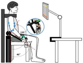
Experimental Setup A 1-DOF robotic arm was developed to provide an assistive force to partici- pants during elbow flexion. The robotic arm comprised a servo motor (whose rotational axis could be adjusted to align to the participant’s elbow joint), and an aluminum beam that could be attached to the participant’s wrist. Compli- ance control was used to regulate the assistive force based on a targeted angle shown to the participant and the current position of the robotic arm. Here,
Characteristics of Muscle Activity and Joint Dynamics During Weight Lifting 23
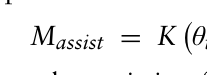
the spring behavior was used to calculate the assistive force to be provided by the robotic arm, expressed as follows:
(1)
θtarget −θarm
Equation reproduced from the original PDF.
where M_assist denotes the assistive force generated by the robotic arm, θ_arm denotes the rotational angle of the robotic arm, θ_target denotes the targeted angle shown to the participant, and K refers to the stiffness coefficient.
The stiffness coefficient determines the compliance of the arm. Moreover, the larger the difference between the targeted angle and the arm angle, the stronger the assistive force provided is. A stiffness coefficient of 0.2 N m deg-1 was used in this study.
Procedures (Tasks) Participants lifted weights (equivalent to 15% force of maximal voluntary contraction [MVC] during isometric elbow flexion at 90
) with and without assistive force using the aforementioned assistive device, in a sitting position. A monitor placed in front of the participant showed the initial, endpoint, actual, and reference positions of the wrist. The initial and endpoint angles of the elbow joint were set to 40
and 135
, respectively (the elbow in full extension being set to 0
). Participants were instructed to begin elbow flex- ion when a “start” cue sounded, continue elbow flexion with reference to movement of the reference position, stop flexion at the endpoint position, and maintain the position for 3 s. The reference position was programmed to start moving from its initial position on the “start” cue and stop at the end- point position after exactly 1.1 s. For the with assistive force condition, the targeted angle of the robotic arm was set to be 0.1 s ahead of the reference position shown to the participant, which creates a resultant assistive force. Each participant performed six trials consecutively under each condition. The order of the conditions was counterbalanced.
Figure 1: Schematic diagram of the assistive device.
Measurements During the task, the surface electromyography (EMG) activity of the biceps brachii (BB)—as the primary working muscle—and the triceps brachii (TB)— as the antagonist muscle—was measured using an analog-to-digital converter with a bio-amp (ADInstrument, PowerLab 16/30, ML880,Australia). The
mean values of the rectified EMG before and during the elbow flexion process from three of the six trials were calculated and normalized as a percentage of the EMG at the maximal voluntary contraction (%MVCBB and %MVCTB, respectively). Additionally, the level of the muscles’ co-contraction (CCratio) during the elbow flexion was calculated using the ratio of %MVCTB to %MVCBB.
Statistical Analysis For the mean values of %MVCBB and %MVCTB, a paired-t-test was used to compare the results under the assisted and unassisted conditions.
RESULTS
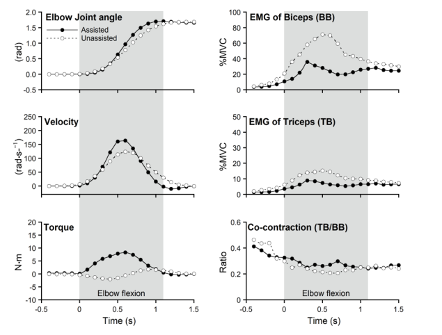
Figure 2 shows the temporal changes in the average values of all measured parameters under both conditions. Evidently, the elbow joint begins to flex slightly later than the sounding of the “start” cue and the peak velocity of flexion occurs approximately 0.5 s after the “start” cue for most partici- pants. However, the assisted condition exhibits a greater peak velocity than the unassisted condition. Under the assisted condition, the peak torque is also evident at the same time. As the elbow flexion stops, the unassisted condition exhibits a smooth stop, whereas the assisted condition shows the average elbow joint angle to be slightly higher than the endpoint target—that is, early arrival at and/or overshooting the endpoint position are evident in most participants.
Figure 2: Comparison of temporal changes in average values of each measured parameter under the assisted and unassisted conditions.
Characteristics of Muscle Activity and Joint Dynamics During Weight Lifting 25
The %MVC was lower under the assisted condition than under the unas- sisted condition for both the BB and TB from before the start to the end of the elbow flexion. Under the unassisted condition, the %MVC of both BB and TB peak at the same time as the peak in the elbow angular velocity (at approximately 0.5 s). However, under the assisted condition, they occur slightly earlier than under the unassisted condition (at approximately 0.3 s). After the peak, the %MVC of both the BB and TB slow down under both conditions; however, only under the assisted condition, the %MVCs increase slightly again before the elbow flexion stops. Furthermore, the decrease in %MVCTB is smaller than that in %MVCBB, resulting in a clear peak (at approximately 0.7 s) in the CCratio.
Table 1 presents the average %MVC of the BB and TB during elbow flex- ion (0 to 1.1 s) with and without the assistive force. Both the BB and TB exhibit considerably lower average %MVC values in the assisted condition than the unassisted condition.
Table 1. Average %MVC during the elbow flexion.
Table available in the original PDF paper.
%MVC of EMG (%) Reduction rate t-test (A) vs. (U) (%) (U-A)/U Assisted condition(A) Unassisted condition (U)
Biceps brachii 24.6± 9.1 51.8±19.2 51.2±13.6 p < 0.01 Triceps brachii 6.6±2.3 11.9±5.0 43.8±8.4 p < 0.01
DISCUSSION
The force provided by the robotic arm reduced the average %MVC during elbow flexion by 51.2% and 43.8% in both the biceps and triceps muscles, respectively (Table 1). Although it can be difficult to calculate what percent- age reduction in the %MVC could be expected from the mechanism of the assistive device used in this study, it is evident that the assistive force of this device was effective in reducing muscle activity. Conversely, the effect of the assistive force could also be regarded as the difference in the EMG of the BB and TB between the solid line with closed circles and the dashed line with opened circles shown in Figure 2; however, such differences depend on the time phase of the elbow flexion movement. Consequently, when discussing the effect of the assistive force on isotonic muscle contraction, it is necessary to divide it into specific time periods.
Immediately after the start of the movement, %MVCBB was lower than under the assisted condition in the non-assisted condition, probably because the participants learned how the assistive force was provided through prior rehearsals and suppressed muscle activity in advance. When the starting joint movement was initiated upon the “start” cue, the EMG started to activate prior to the start (Aruin et al., 1998; Bruttini et al., 2016). Because the %MVC was low (even during the pre-start phase of the assistive condition), it is evident that the joint movement was planned with an expectation that assistance would be provided.
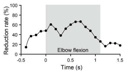
During the middle phase of the elbow flexion, the %MVC of both mus- cles and the torque increased before decreasing under both conditions. That is, the workload on the working muscles was at its maximum at around this time whilst lifting the weight and the torque from the device was well regu- lated based on the temporal variation of its workload. However, the assisted condition exhibited an earlier onset of the decrease (i.e., peak) than the unas- sisted condition. This implied that the assistive force was effective in reducing muscle activity after the peak. Interestingly, the rate of %MVC reduction— or the effectiveness of the assistive force—fell around that timing (Figure 3). This implied that there was some delay before it was successfully used for the assistive force, as it took some time to perceive it and modify the motor control strategy to adapt it.
During the deceleration phase, inertial forces were also acting, and the moment arm was shortening; consequently, the required muscle activity was also reduced (Gribble et al., 2003). Indeed, a decrease in the %MVCBB was evident under the unassisted condition. However, under the assisted con- dition, the antagonist muscle (the triceps in particular) exhibited a smaller decrease, resulting in a higher co-contraction index during the deceleration phase. In this study, the participants were instructed to stop the joint move- ment at a targeted position, suggesting that the triceps muscles were more active in generating some resistance to the assistive force and increased their co-contraction to stop at a precise position (Lewis et al., 2010; Komi et al., 2000). Nevertheless, the early arrival and overshooting of the endpoint tar- get, despite being complemented by such motor control, suggests that the participants struggled to stop smoothly.
Limitations This study examined only one combination between levels of a specific work- load and assistive force during a lifting task, which was performed only by elbow flexion with one degree of freedom. Additionally, there are many dif- ferent methods of controlling the assistive force for dynamic joint movements, and different parameters must be set for each. The findings of this study could be influenced by these factors.
Figure 3: Temporal changes in the reduction rate of %MVCBB under an assist force (calculated based on the data presented in Figure 2 (EMG of biceps)).
Characteristics of Muscle Activity and Joint Dynamics During Weight Lifting 27
CONCLUSION
In this study, it was evident that under the assisted condition, the reduction of muscle activity of the agonist and antagonist muscles and the precision of the joint movement were controlled based on the respective phase of elbow flexion.
ACKNOWLEDGMENT
This research was funded by JSPS KAKENHI (Grant Numbers 21H04898, 21K17791 and 24H00725).
REFERENCES
Aruin, A. S., Forrest, W. R., & Latash, M. L. (1998). Anticipatory postural adjustments in conditions of postural instability. Electroencephalography and Clinical Neurophysiology/Electromyography and Motor Control, 109(4), 350–359. Bequette, B., Norton, A., Jones, E., & Stirling, L. (2020). Physical and cognitive
load effects due to a powered lower-body exoskeleton. Human Factors, 62(3), 411–423. Bruttini, C., Esposti, R., Bolzoni, F., & Cavallari, P. (2016). Higher precision in pointing movements of the preferred vs. non-preferred hand is associated with an earlier occurrence of anticipatory postural adjustments. Frontiers in Human Neuroscience, 10, 365. Choi, J., Yeoh, W. L., Loh, P. Y., & Muraki, S. (2019). Force and electromyog-
raphy responses during isometric force release of different rates and step-down magnitudes. Human Movement Science, 67, 102516. Choi, J., Yeoh, W. L., Matsuura, S., Loh, P. Y., & Muraki, S. (2020). Effects of mechanical assistance on muscle activity and motor performance during isometric elbow flexion. Journal of Electromyography and Kinesiology, 50, 102380. De Looze, M. P., Bosch, T., Krause, F., Stadler, K. S., & O’sullivan, L. W. (2016).
Exoskeletons for industrial application and their potential effects on physical work load. Ergonomics, 59(5), 671–681. Eden, J., Bräcklein, M., Ibáñez, J., Barsakcioglu, D. Y., Di Pino, G., Farina, D.,...
& Mehring, C. (2022). Principles of human movement augmentation and the challenges in making it a reality. Nature Communications, 13(1), 1345. Erezuma, U. L., Espin, A., Torres-Unda, J., Esain, I., Irazusta, J., & Rodriguez-Larrad, A. (2022). Use of a passive lumbar back exoskeleton during a repetitive lifting task: effects on physiologic parameters and intersubject variability. International Journal of Occupational Safety and Ergonomics, 28(4), 2377–2384. Garcia, G., Arauz, P. G., Alvarez, I., Encalada, N., Vega, S., & Martin, B. J. (2023).
Impact of a passive upper-body exoskeleton on muscle activity, heart rate and discomfort during a carrying task. PLOS ONE, 18(6), e0287588. Gribble, P. L., Mullin, L. I., Cothros, N., & Mattar, A. (2003). Role of cocontraction in arm movement accuracy. Journal of Neurophysiology, 89(5), 2396–2405. Hardesty, R. L., Boots, M. T., Yakovenko, S., & Gritsenko, V. (2020). Computational
evidence for nonlinear feedforward modulation of fusimotor drive to antagonistic co-contracting muscles. Scientific Reports, 10(1), 10625. Komi, P. V., Linnamo, V., Silventoinen, P., & Sillanpää, M. (2000). Force and EMG power spectrum during eccentric and concentric actions. Medicine and Science in Sports and Exercise, 32(10), 1757–1762.
Lewis, G. N., MacKinnon, C. D., Trumbower, R., & Perreault, E. J. (2010).
Co-contraction modifies the stretch reflex elicited in muscles shortened by a joint perturbation. Experimental Brain Research, 207, 39–48. Martini, E., Crea, S., Parri, A., Bastiani, L., Faraguna, U., McKinney, Z.,... & Vitiello, N. (2019). Gait training using a robotic hip exoskeleton improves metabolic gait efficiency in the elderly. Scientific Reports, 9(1), 7157. Naik, S. K., Patten, C., Lodha, N., Coombes, S. A., & Cauraugh, J. H. (2011). Force
control deficits in chronic stroke: Grip formation and release phases. Experimental Brain Research, 211, 1–15. Qu, X., Qu, C., Ma, T., Yin, P., Zhao, N., Xia, Y., & Qu, S. (2021). Effects of an industrial passive assistive exoskeleton on muscle activity, oxygen consumption and subjective responses during lifting tasks. PLOS ONE, 16(1), e0245629. Stirling, L., Kelty-Stephen, D., Fineman, R., Jones, M. L., Daniel Park, B. K., Reed,
M. P.,... & Choi, H. J. (2020). Static, dynamic, and cognitive fit of exosystems for the human operator. Human Factors, 62(3), 424–440. Winter, A., Mohajer, N., & Nahavandi, D. (2021). Semi-active assistive exoskeleton system for elbow joint. 2021 IEEE International Conference on Systems, Man, and Cybernetics (SMC), 2347–2353. Yeoh, W., Choi, J., Loh, P. Y., Fukuda, O., & Muraki, S. (2023) Motor Character-
istics of human adaptations to external assistive forces. Journal of Robotics and Mechatronics, 35(3), 547–555.
LISTED IN THE AHFE DIGITAL BOOKSTORE CATALOGUE
Explore the complete digital volume
Physical Ergonomics and Human Factors
Read this individual research paper free through the original publisher, or explore the full digital proceedings book.
ISBN 978-1-964867-23-6
Explore this ISBN in the AHFE Bookstore ↗Listed in the July 2026 bookstore catalogue; search by ISBN to confirm current availability · Digital book purchase optional