Irina Kurnikova, Shirin Gulova, Natalia Danilina, Artyom Yurovsky, Vladimir Terekhov (2023). Computerized heart rate analysis in the selection of therapy for patients with arterial hypertension. In: Emerging Technologies in Healthcare and Medicine. AHFE Open Access, vol. 116, pp. 174–181. https://doi.org/10.54941/ahfe1004369
According to the World Health Organization (WHO), over 1 billion people are overweight and 600 million are obese, with metabolic syndrome (MS) affecting 35% of adults in the US and 20-25% in Europe. MS patients require appropriate therapy with comorbidity in mind, which requires further study and optimization. As part of the study, we conducted Holter ECG monitoring (HM) of patients with MS. MS was diagnosed on the basis of the MTP 3rd revision criteria. Additional criteria were AH, elevated triglyceride levels, decreased HDL cholesterol levels, impaired glucose tolerance (IGT), impaired fasting glycemia (EGS), and combined EGS/IGT disorders. MS was diagnosed based on 3 criteria: 1 main and 2 additional ones.Design. A total of 154 patients were examined in in-patient setting. They were subdivided into 2 main groups: Group I - patients with MS receiving β-blockers (n-97) to treat AH; Group II - patients with MS not receiving β-blockers (n-57).Each main group was divided according to the degree of obesity according to the WHO classification. Each patient underwent HM with programmed computer analysis of the wave spectrum of the obtained data and allocation of frequencies - 0.004-0.08 Hz (very low frequency - VLF); 0.09-0.16 Hz (low frequency - LF); 0.17-0.5 Hz (high frequency - HF) more than 0.5 Hz (ultra-low frequency waves - ULF); two coefficients are calculated - LF/HF (vagosympathetic balance coefficient) - ratio of low frequency waves power (LF) to high frequency waves power (HF), and centralization index (CI) - ratio of central regulation circuit activity to autonomic one (LF+VLF/HF).Results. Analysis found changes in HF, LF, and ULF domains of HRV spectrum, indicating transition to a more energy-intensive level of control and depletion of regulatory mechanisms. ULF(%) values above 6.9 require correction with β-blockers. The study found ULF% and VLF% values to be higher in the non-β-blocker group and administration of β-blockers resulted in normalization of indexes with the index of centralization and vagosympathetic balance. In patients receiving β-blockers, the values of these parameters corresponded to those of patients with normal body weight. In MS patients not receiving β-blockers, ULF% was 50% higher and VLF was 18% higher than in the normal weight group. The centralization index was elevated to 3.5. Administration of drugs to 17 patients in group II resulted in normalization of the indexes and achievement of the same values as in group I patients. At the dynamic follow-up for 2 years, Group I patients had no cardiovascular events. The 40 patients who refused to change therapy had no change in HM values and 27% of these patients had acute cardiovascular events at 2 years.Conclusion:Daily ECG monitoring with assessment of ULF%, VLF% and IC indices is a more subtle method of investigation, which allows to detect latent disorders of regulatory mechanisms (with seeming clinical well-being) in patients with disorders of these indices the risk of acute cardiovascular events development remains high. The control of ULF%, VLF% and IC index by HM-ECG method allows to change the therapy in time and to obtain a better result.
Arterial hypertension (AH) typically accompanies conditions like abdominal obesity, insulin resistance, and metabolic disturbances, rather than existing alone (Uspensky et al., 2017). Acknowledged as a 21st century pandemic, metabolic syndrome amplifies cardiovascular risk when combined with AH (Korotkova, 2014; Bloomgarden, 2006; Sprecher and Pearce, 2000; Isomaa et al., 2001). AH’s pathogenesis involves autonomic dysfunction, particularly hyperreactive sympathetic nervous response, making selective β-blockers an effective treatment (Agasarov, 2019; Kobalava and Tolkacheva, 2005). Metabolic disturbances exacerbate this dysfunction by inducing autonomic neuropathy.
Monitoring autonomic regulation and its response to therapy could be key in predicting therapeutic effect and drug selection. Heart rate vari- ability (HRV) analysis, a non-invasive method, assists in evaluating the body’s functional state and regulatory mechanisms (Baevsky et al., 2001). As per the European Society of Cardiology and the North American
Computerized Heart Rate Analysis in the Selection of Therapy for Patients 175
Society of Electrophysiologists, reduced HRV in patients with coronary artery disease (CAD) independently predicts higher risk of severe ventricu- lar arrhythmias and sudden death post-myocardial infarction (Shestakova, 2009; Bigger et al., 1993). Yet, the primary dysfunction in the autonomic nervous system in metabolic syndrome patients—whether sympathetic or parasympathetic— and the influence of β-blockers at various levels remain undetermined.
The aim of this study was to assess the influence of β-blockers on heart rate variability in patients with metabolic syndrome.
MATERIALS AND METHODS
The study was conducted at the Federal State Educational Institution of Higher Education “Patrice Lumumba Russian University of Friendship of Peoples” at the clinical bases of the Department of Hospital Therapy with courses in endocrinology, hematology, and clinical laboratory diagnostics during the period from 2020 to 2023. The work was carried out in accor- dance with the plan of scientific research work, observing the principles of medical bioethics (approval form of the ethics committee of the RUDN Medical Institute No. 5 of March 17, 2022). Criteria for including patients in the study:
age 40-74;
patients with obesity of varying degrees;
arterial hypertension in medical history;
patients with diabetes;
patients with dyslipidemia;
patient’s informed consent to participate in the study.
Criteria for excluding patients from the study:
age <40 and >74 years old;
absence of informed consent;
acute cardiovascular events at the time of the study;
persistent decompensation of diabetes;
chronic heart failure more than 2 degrees A total of 166 patients were examined, 150 patients in inpatient con- ditions, and 16 patients in outpatient conditions. The main reason for hospitalization of patients included in the study was high blood pressure, obesity of the 2nd and 3rd degrees, and type 2 diabetes.
All patients were divided into two major groups - patients with metabolic syndrome (n = 121), Observation Group 1, and patients with normal body weight (n = 45), Comparison Group 2. Metabolic syndrome was diagnosed based on the ATP III risk criteria (Dobrowolski et al., 2022).
Since patients with arterial hypertension (AH) often received antihyperten- sive therapy with beta-blockers, it seemed appropriate to divide patients into groups based on this characteristic. All patients with metabolic syndrome, depending on the therapy received, were divided into two subgroups: sub- group “A” - patients receiving beta-blockers, and subgroup “B” - patients not receiving beta-blockers.
Subsequently, in patients with metabolic syndrome (MS), the state of metabolic and autonomic regulation was also assessed at each stage of metabolic syndrome development, i.e., depending on the degree of abdominal obesity (A1, A2, A3, A4, and similarly in subgroup “B”).
The comparison group (subgroups “C” and “D”) included patients with normal body weight and arterial hypertension, but without abdominal obe- sity. This approach allowed us to assess the contribution of obesity to the dysfunction and regulation of the autonomic nervous system and the impact of beta-blockers on the state of these mechanisms, and therefore, on the devel- opment and progression of cardiovascular diseases in the compared groups of patients.
The complete examination included: medical history collection, physical examination, blood pressure measurement, standard biochemical tests, ECG recording, echocardiography, and 24-hour ECG monitoring.
Heart rate variability (HRV) was assessed with the help of the Valenta MN-02-8 hardware-software complex, calculating the generally accepted time and spectral HRV indicators according to the European Society of Car- diology and the North American Society of Electrophysiology. The studied HRV indicators by spectral characteristics are presented in Table 1. Patients underwent 24-hour ECG monitoring in hospital conditions.
Table 1. HRV indicators.
Table available in the original PDF paper.
Studied HRV (Heart Rate Variability) indicators for 24 hours of ECG recording Indicators Definition
ULF, ms2 Power in the ultra-low frequency range (less than 0.003 Hz) VLF, ms2 Power in the very low frequency range (0.003–0.04 Hz) LF, ms2 Power in the low frequency range (0.04–0.15 Hz) HF, ms2 Power in the high frequency range (0.15–0.40 Hz) LF/HF Ratio of low to high frequency power values in absolute terms
METHODS OF STATISTICAL PROCESSING OF RESEARCH RESULTS For statistical data processing, Statistica software (version 10) was used. Distribution testing was performed using the Shapiro-Wilk W-criterion. For quantitative variables with a normal distribution, the arithmetic mean (M) and standard deviation (SD) were calculated, and for quantitative vari- ables with an asymmetric distribution (Skewness>1) - the median (Me) and interquartile range (IQR). The Student’s t-test was used to verify the relia- bility and differences between two groups with a normal distribution, and the Mann-Whitney U-test was used for a non-normal distribution. For three independent groups, the Kruskal-Wallis criteria were used. Qualitative vari- ables are presented as absolute (n) and relative (%) values. The Pearson’s chi-square test (χ2) was used to compare groups in terms of the frequency of qualitative variables. Results were considered statistically significant at two-sided p values < 0.05. Comparison of two groups by a quantitative indi- cator, the distribution of which differed from normal, was performed using the Mann-Whitney U-test.
Computerized Heart Rate Analysis in the Selection of Therapy for Patients 177
RESULTS
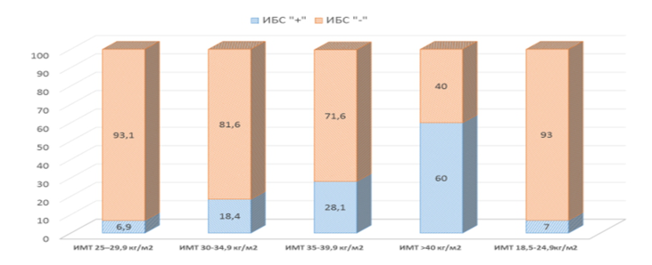
In the group of examined patients with metabolic syndrome (arterial hyper- tension and excess body weight or obesity), coronary heart disease (CHD) was noted in 25.9% of patients, which is almost twice as often as in the com- parison group (with arterial hypertension and normal body weight) — 15.6% of cases. We analyzed the frequency of CHD in Group A patients depending on the degree of obesity. And it turned out that results comparable to those of Group B were observed only in patients with excess body weight (BMI 25–29.9 kg/m2 - Group A1), where the frequency of CHD was 6.9%, but with each subsequent stage of obesity, this indicator increased progressively (Fig. 1).
Figure 1: Diagram of the prevalence of CHD depending on the degree of obesity.
In the group of patients with obesity of grade I, this indicator was already 18.4%, and in the group with morbid obesity (BMI ≥40kg/m2) — already 60%. All patients were under outpatient observation, so the main clinical and biochemical indicators, including the lipid spectrum of serum, were within acceptable values, and there were no significant differences in subgroups. In the echocardiographic study of patients with MS, 58 (75%) people had symmetric hypertrophy of the left ventricle, in the group of patients without MS slightly less - 27 (64%). The global and local contractile function of the left ventricle was normal in all patients. The average ejection fraction was 65.0±5.4%. As a result of the conducted 24-hour ECG monitoring, not only tempo- ral but also frequency indicators were evaluated in both compared groups (receiving and not receiving β-blockers) (Table 2, 3). More significant changes occurred when evaluating frequency characteristics.
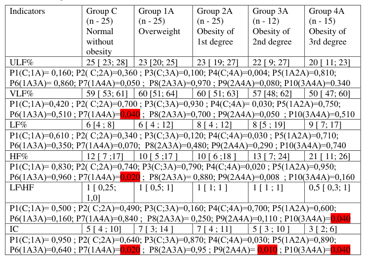
In the subgroups of patients with MS receiving β-blockers (group “A”), there were no significant differences in the indicators of the frequency and time spectrum of HRV depending on the degree of obesity. There were also no significant differences compared to the indicators of patients without obesity taking drugs.
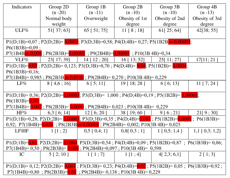
Table 2. Comparison of patient groups depending on the increasing body mass and taking beta-blockers.Table 3. Comparison of patient groups depending on body mass index not taking betablockers.
Note: Data are presented as median, 25th and 75th percentile, as (Me (IQR)) or arithmetic mean (M) and standard deviation of the mean (SD).
Computerized Heart Rate Analysis in the Selection of Therapy for Patients 179
In patients with metabolic syndrome who did not take β-blockers (group “B”), HRV changes different from the indicators of patients not taking them were detected already in the group of patients with excess body weight (B1). And in each group with a higher degree of obesity, the differences in the indicators became more and more significant (p < 0.001) compared to patients taking β-blockers (Table 3).
Starting from the second degree of obesity, the frequency spectrum in patients of group “B” shifted towards an increase in the ULF (%) and VLF (%) indicators. A high level of reliability of the correlation relationship with the body mass index we got for the indicators VLF% (r = 0.350, P < 0.003) and LF% (r = 0.56; P < 0.033), as well as with the coefficient of vagosympa- thetic balance LF/HF (r = 0.413; P < 0.0001). ULF (%) reflects the influence of higher suprasegmental centers of autonomic regulation. The average value of ULF indicators changed depending on the degree of obesity. The minimum values were found in patients with obesity of grade I (group B2), in which the indicator was 18.7±5.5%. The maximum ULF indicator was in patients with obesity of grade II (subgroup B3), the average value of which was 40.1±3.2%. Since ULF reflects the activity of higher regulation centers (their involvement in maintaining regulatory processes, the translation of regula- tory mechanisms from the level of “control” to the level of “centralization”), the state of exhaustion of reserves and breakdown of adaptation, which fully explains the increased risk of cardiovascular events in this group of patients. Therefore, it can be confidently said that in patients with metabolic syn- drome, the risk of developing CVD becomes high with the presence of grade II obesity and is largely due to a violation of autonomic regulation.
The average duration of patient observation was 2 years. During the obser- vation period, acute cardiovascular events were noted in the group of patients with metabolic syndrome receiving β-blockers - 4 cases (5.2%), in the group of patients not taking these drugs - 4 cases (9.1%), in the group of patients without obesity taking β-blockers (group C), this indicator was also better compared to the group taking other combinations of antihypertensive drugs (group D) - 4% versus 10%.
DISCUSSION
The main question to be answered in accordance with the purpose of the study is not only how obesity affects the functioning of such an important regulatory system as the autonomic nervous system, and, if “yes”, then from which degree of obesity this influence becomes significant, but also to assess the importance of autonomic dysregulation in increasing cardiovascular risk for this group of patients.
Some indicators characterizing the state of autonomic regulatory mecha- nisms can be used to predict the progression of metabolic syndrome (MS) and assess cardiovascular risk. In patients with abdominal obesity, starting from the 2nd degree, the values of ULF% and VLF% indicators reliably increased. LF reflects the activity of higher regulation centers (their involvement in maintaining the regulatory processes, the transfer of regulatory mechanisms from the level of “control” to the level of “centralization”), the state of exhaustion of reserves and the failure of adaptation, which fully explains
the increased risk of cardiovascular events in this group of patients. There- fore, it can be confidently said that in patients with metabolic syndrome, the risk of developing cardiovascular diseases becomes high in the presence of 2nd degree obesity and is largely due to the disruption of autonomic regula- tion. The data obtained from the study allow us to identify two stages in the development of arterial hypertension against the background of metabolic syndrome, increasing the risk of developing cardiovascular complications. The fact that the risk significantly increases in patients with 2nd degree obe- sity is known from other studies as well, but we would like to draw attention to the fact that similar risks appear in patients at the stage of transition from 1st degree obesity to 2nd degree. Disruptions of regulatory mechanisms begin earlier, and for a long time they proceed covertly, due to the increased ten- sion of adaptation mechanisms and reach the metabolic level only when the regulatory mechanisms are exhausted (centralization level).
β-blockers have a pronounced positive effect on the dynamics of the dis- ease and reduce the risk of developing cardiovascular diseases. The main indicators of heart rate variability in patients with MS taking beta-blockers did not have significant differences from the indicators of patients without obesity, which reduced the frequency of acute cardiovascular events to a comparable level.
CONCLUSION
1. Directly β-blockers have a pronounced positive effect on heart rate variability in patients with metabolic syndrome. 2. Patients with metabolic syndrome (especially those patients who have 1st degree obesity) are recommended to prescribe β-blockers (selective) for the prevention of cardiovascular diseases.
REFERENCES
L. G. Agasarov Mechanisms of correction of metabolic syndrome (literature review) / T. V. Aphanova, O. Yu. Kirgizova, V. K. Frolov // Bulletin of new medical technologies. 2019. No. 3. pp. 40–47. Baevsky R. M., Ivanov G. G., Chireikin L. V. et al. Analysis of heart rate
variability using various electrocardiographic systems (methodical recommenda- tions)//Vestnik arrhythmology 2001; 24:65–87. Bigger J. T. Jr, Fleiss J. L., Rolnitzky L. M. et al. The ability of several shortterm measures of RR variability to predict mortality after myocardial infaction // Circulation. - 1993. Vol. 88. pp. 927–934. Bloomgarden Z. T. Symposium: Debating the Metabolic Syndrome. Medscape Con-
ference Coverage, based on selected sessions // American Diabetes Association 66th Scientific Sessions; June 9–13, 2006, Washington, DC. Dobrowolski P. et al. Metabolic syndrome - a new definition and management guidelines: A joint position paper by the Polish Society of Hypertension, Polish Society for the Treatment of Obesity, Polish Lipid Association, Polish Association for Study of Liver, Polish Society of Family Medicine, Polish Society of Lifestyle Medicine, Division of Prevention and Epidemiology Polish Cardiac Society, “Club 30”Polish Cardiac Society, and Division of Metabolic and Bariatric Surgery Society of Polish Surgeons. Arch Med Sci. 2022 Aug 30;18(5): 1133–1156. Doi: 10.5114/aoms/152921. PMID: 36160355; PMCID: PMC9479724.
Computerized Heart Rate Analysis in the Selection of Therapy for Patients 181
Isomaa B., Almgren P., Tuomi T. et al. Cardiovascular morbidity and mortality associated with the metabolic syndrome // Diabetes Care. 2001. Vol. 24. No. 4. pp. 683–689. Kobalava Z. D., Tolkacheva V. V. Metabolic syndrome: principles of treatment //
Russian Medical Journal. 2005. Vol. 13, No. 7. pp. 451–458. Korotkova, S. B. Physical rehabilitation of patients with metabolic syndrome / S. B.
Korotkova, V. E. Aparin, I. V. Gridneva // Physical Culture and Health. 2014. No. 3(50). pp. 90–93. Metabolic Syndrome: Educational-methodical manual/ed. Y. P. Uspensky, Y. V.
Petrenko, Z. H. Gulunov, N. L. Shaporova, Y. A. Fominyh, R. M. Niyazov - St. Petersburg, 2017. 60 p. Shestakova M. V. Metabolic syndrome - a real threat to the health of the population of all countries. Medical Bulletin. 2009; (484):9–10. Sprecher D. L., Pearce G. L. How deadly is the “deadly quartet”? A post-CABG
evaluation // J. Am. Coll. Cardiol. 2000. Vol. 36. No. 4. pp. 1159–1165.
LISTED IN THE AHFE DIGITAL BOOKSTORE CATALOGUE
Explore the complete digital volume
Emerging Technologies in Healthcare and Medicine
Read this individual research paper free through the original publisher, or explore the full digital proceedings book.
ISBN 978-1-958651-92-6
Explore this ISBN in the AHFE Bookstore ↗Listed in the July 2026 bookstore catalogue; search by ISBN to confirm current availability · Digital book purchase optional