Irina Kurnikova, Guzal Akhmadullina, Shirin Gulova, Natalia Danilina, Ikram Mokhammed (2023). Methods of computer simulation in the development of technology for the functional assessment of the state of the liver in patients. In: Health Informatics and Biomedical Engineering Applications. AHFE Open Access, vol. 78, pp. 66–71. https://doi.org/10.54941/ahfe1003456
1 RUDN University, Moscow, Russia, RUDN University, Miklukho-Maklayast. 6, 117198 Moscow, Russia; Federal State Budgetary Educational Institution of Further Professional Education, Russian Medical Academy of Continuous Professional Education, Barricadnaya St., h.2/1, b.1 125993 Moscow, Russia ↗
2 Izhevsk State Medical Academy, Izhevsk, Russia, Izhevsk State Medical Academy, Kommunarov St. 281, 426034 Izhevsk, Russia ↗
Most of the diseases associated with carbohydrate and fat metabolism disorders (type 2 diabetes mellitus, obesity, metabolic syndrome) lead to changes in the structure and function of liver cells, and the formed liver dysfunction negatively affects the further progression of the disease. The process of liver damage develops with varying intensity and does not immediately lead to irreversible consequences; therefore, dysfunction should be detected as early as possible, and the data obtained should be used to assess the current state and predict their reversibility.Study purpose. To create a quantitative assessment method that allows to assess the current functional state of the liver and to determine the reversibility of existing functional disorders and the severity of structural changes, to assess the prognosis of the course of the disease and the effectiveness of restorative measures.Instruments and Data Collection Procedure. Examination of patients, in addition to conventional methods, included an assessment of the absorptive-excretory function of the liver and biliary tract patency using a scintillation gamma camera (Siemens Symbia T16) with subsequent processing on the SUPER-SEGAMS computer system (Hungary). Freshly prepared Bromesida, 99mТс was administered intravenously at the rate of 1.1 MBq per kg of the patient's body weight, with a normal content of bilirubin in blood. Series of scintigrams allow to assess the passage of the drug visually through the blood-liver-ducts-intestine system, to characterize the anatomical features and organic changes in the biliary system. Quantitative analysis of the "activity-time" curves obtained from the areas of interest (the right lobe of the liver - 2 zones, the left lobe of the liver, the common bile duct, the intestinal area, the heart area) makes it possible to study the absorptive-excretory function of the liver.Results. By the method of mathematical modeling, a formula was obtained - the index of the functional activity of hepatocytes:IFAH= (-1,1564 + 0,0653 × BMI - 0,0144 × Tmax) × 100,where:IFAH - index of functional activity of hepatocytes (liver cells);BMI - body mass index (kg/m2)Tmax - indicator of the absorption function of the liver (min) - the time to reach the maximum accumulation of the radiopharmaceutical in the liver. It is an indicator of the function of polygonal liver cells (normal = 8-12 min).Functional activity of hepatocytes: from 0 to 9.9 - normal functional activity of hepatocytes; from 10 to 19.9 - the risk of developing functional disorders; from 20 to 29.9 - reversible dysfunction of hepatocytes (steatosis); more than 30 - irreversible (organic) liver dysfunction (steatohepatitis). With negative IFAH values - the influence of extrahepatic factors, such as diseases that accelerate metabolism at the cellular level (thyrotoxicosis), taking drugs in violation of the prescriptions before these studies.The originality and novelty of the technique are confirmed by the patent – “A method for diagnosing fatty hepatosis” (patent RU 2 578 080 C2 dated February 19, 2016).The method was tested in clinical practice and the data obtained confirmed the high diagnostic accuracy (95%) of the proposed method for calculating the index of IFAH.Conclusion: the discussed method allows doctors to evaluate not only the current functional state of the liver, but also to determine the reversibility of existing functional disorders and the severity of structural changes; also could be used to evaluate the prognosis of the course of the disease and the effectiveness of restorative measures. It is characterized by relative ease of implementation. Only one parameter — Tmax is required, after which the study can be completed.
Currently, the most common liver disease is non-alcoholic fatty liver dise- ase (NAFLD), which is pathogenetically associated with metabolic syndrome, visceral obesity, and type 2 diabetes mellitus (Lazebnik, 2021). Early diagno- sis of NAFLD is still difficult because at the stage of steatosis, most often
Methods of Computer Simulation in the Development of Technology 67
there are no clinical manifestations and changes in the biochemical para- meters of the blood. All available methods of laboratory and instrumental diagnostics, including fibrotests, are not methods of early diagnosis, chara- cterize only the current functional state of the liver, do not allow identifying risk groups, assessing the reversibility of liver dysfunctions and drawing up an individual treatment plan. Diagnosis of NAFLD at the stage of steatosis is of great prognostic value because steatosis is an independent risk factor for the development of cardiovascular diseases and their complications (Tishkovsky, 2016; Statsenko, 2019; Lazebnik, 2021; Allahverdieva, 2018).
PURPOSE OF THE STUDY To create a quantitative evaluation method that allows assessing the current functional state of the liver, the reversibility of existing functional disorders and the severity of structural changes, assessing the prognosis of the course of the disease and the effectiveness of restorative measures.
MATERIALS AND METHODS
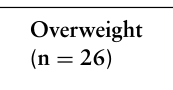
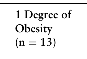
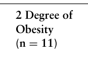
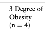
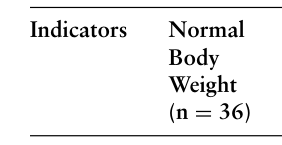
A single-stage single-center examination was carried out in 26 overweight patients and 28 people with various degrees of obesity (1 degree of obesity - 13 patients, 2 degree - 11 patients and 3 degree - 4 people). The comparison group consisted of persons with normal body weight (36 people).
The study was approved by the Biomedical Ethics Committee of the Izhevsk State Medical Academy of the Ministry of Health of the Russian Federation, Application No. 468 dated December 22, 2015. The examination was performed on the basis of the informed consent of the patient, in accor- dance with the order No. 163 (OST9/500.14.001-2002) paragraph P.4.6.1. Ministry of Health of the Russian Federation, provisions of the Bioethics Committee of the FSBEI HE “Izhevsk State Medical Academy of the Ministry of Health of Russia”.
Criteria for exclusion from the study: lack of patient consent to partici- pate in the study, renal failure (CKD C 4-5), the presence of viral hepatitis, markers of autoimmune liver diseases and storage diseases, the use of hepa- totoxic doses of alcohol. All patients underwent a biochemical blood test: fasting plasma glucose, total cholesterol, LDL cholesterol, HDL cholesterol, triglycerides, alanine aminotransferase, aspartate aminotransferase. Structu- ral changes in the liver were studied using ultrasound on the MyLabSeven machine (Italy). To assess the degree of hepatic steatosis, the classification of fatty degeneration of the liver by Batskov S.S. was used. (1996). The study of the absorption-excretory function of the liver was carried out using dynamic hepatobiliary scintigraphy using a scintillation gamma camera (Siemens Sym- bia T16) with subsequent data processing on the SUPER-SEGAMS system (Hungary). Patients were examined on an empty stomach in the supine posi- tion, in the anterior projection. A freshly prepared radiopharmaceutical (RP) Bromesida, 99mTc was administered intravenously at the rate of 1.1 MBq per kg of the patient’s body weight in a normal functional state of the liver. The detector of the gamma camera was installed as close as possible to the surface of the body, so that the liver and part of the intestine were included in the field of view of the detector. Registration of information was carried out at
a speed of 1 frame per minute for 60–90 minutes. 30 minutes after the start of the study, the patient was given a standard choleretic breakfast (two raw egg yolks).
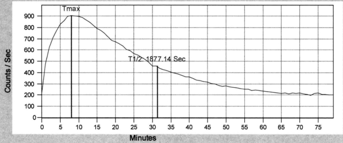
A series of obtained scintigrams made it possible to visually assess the passage of the drug through the blood-liver-ducts-intestine system, to cha- racterize the anatomical features and organic changes in the biliary system. Quantitative analysis of the “activity-time”curves obtained from the areas of interest (the right lobe of the liver - 2 zones, the left lobe of the liver, the com- mon bile duct, the intestinal area, the heart area) made it possible to assess the absorption-excretory function of the liver, the patency of the bile ducts, the functional state of the sphincter of Oddi and the presence of duodenogastric reflux (Fig. 1).
Known methods for diagnostic liver diseases (ultrasound, fibroscanning, ect.) describe the structure of the organ and changes after they have alre- ady occurred and are irreversible. The method of hepatobiliary scintigraphy allows to evaluate the function of the liver (absorption-excretory), therefore, it was chosen as the base for the development of a method for the early diagnosis of NAFLD.
Statistical processing of the obtained data was carried out using the STATISTICA 10.0 software package and MedCalc 12.5.0.0 software. The normality of the distribution of actual data was verified with the Shapiro- Wilk test. Values that do not follow the normal type of distribution were presented as median, lower and upper quartiles; relative - in the form of the frequency of occurrence of the trait and 95% confidence interval calculated by the Wilson method. The significance of differences between independent groups of quantitative variables that do not obey the normal type of distribu- tion was calculated by a nonparametric method using the Mann-Whitney U test, qualitative variables - by Pearson’s Chi-Square (χ2) method with Yate’s
Figure 1: Hepatobiliary scintigram characterizing the absorption-excretory function of the liver depending on time. Note: Tmax - the time to reach the maximum accumu- lation of radiopharmaceuticals in the liver; T1/2 - an indicator that reflects the rate of removal of the radiopharmaceutical from the liver parenchyma by 50% relative to the maximum.
Methods of Computer Simulation in the Development of Technology 69
correction. To determine the magnitude of the relationship between pheno- mena, mathematical modeling was used by constructing 3D graphs. ROC analysis was used to determine the predictive significance of the indicators. Quantitative interpretation of the results was carried out according to ROC curves with an assessment of the AUC. The level of statistical significance was taken as p<0.05.
RESULTS
Ultrasound examination revealed signs of liver steatosis in all patients with 2 and 3 degrees of obesity. In patients with 1 degree of obesity, ultrasound signs of fatty liver infiltration were detected in 69.2% of cases out of 13 patients, with overweight - in 30.8% of cases out of 26 patients, with normal body weight - in 13.9% of cases out of 36 patients.
According to dynamic hepatobiliary scintigraphy in overweight patients, a statistically significant slowdown in the time to reach the maximum accu- mulation of radiopharmaceuticals in the liver (Tmax 16.0 (14.04-18.02) min, p = 0.003) and the rate of radiopharmaceutical excretion from the liver pare- nchyma (T1/2 31.24 (21.7-66.0) min, p = 0.013) compared with subjects with normal body weight (Tmax 12.53 min, and T1/2 23.8 min respectively). In patients with the first degree of obesity, similar data were obtained (Tmax 16.02 min and T1/2 29.9 min respectively). In patients with the second and third degree of obesity, a tendency to slow down the absorption-excretory function of the liver was revealed in Table 1.
Analysis of the data of a radioisotope study of liver function in patients with a normal ultrasound picture of the liver revealed a statistically signifi- cant slowdown in the absorptive function of the liver in overweight patients compared with patients with normal body weight (Tmax 16.0 (13.25-17.87) min and Tmax 12.5 (11.1-15.2) min, p = 0.024). The data obtained confirm the possibility of early diagnosis of liver damage in overweight and obese patients using dynamic hepatobiliary scintigraphy.
Table 1. Indicators of dynamic hepatobiliary scintigraphy in persons depending on body weight with a normal ultrasound picture of the liver, Me [Q1;Q3].
Table available in the original PDF paper.
Equation reproduced from the original PDF.Equation reproduced from the original PDF.Equation reproduced from the original PDF.Equation reproduced from the original PDF.Equation reproduced from the original PDF.
P (p=0,013) (p=0,011) Note: P – statistical significance of differences in values compared with data from patients with normal body weight.
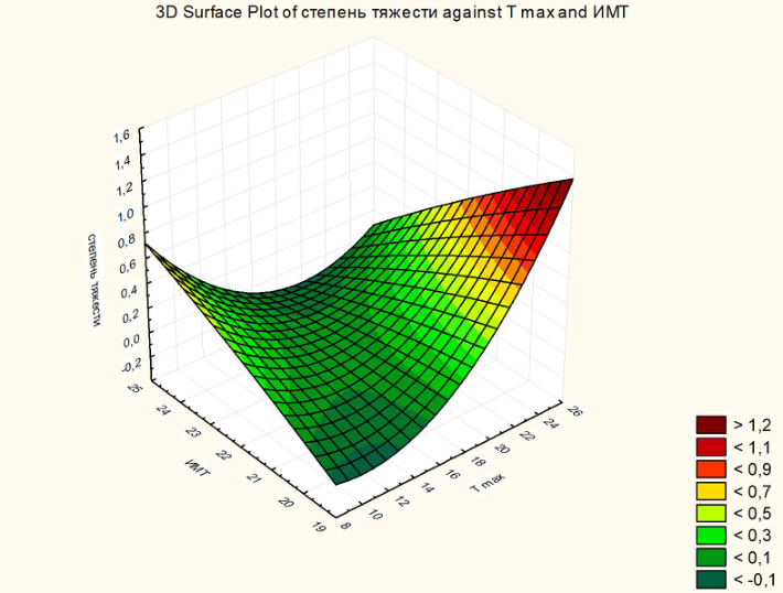
Mathematical modeling with the construction of a 3D graph established a relationship between the degree of fatty infiltration of the liver (according to Batskov S.S., 1996) with BMI and the absorptive function of the liver. With an increase in the patient’s body weight and a slowdown in the absorption function, the degree of fatty infiltration steadily increases (Fig. 2).
The obtained results of the study made it possible to calculate the formula for the diagnosis of functional disorders of the liver: IFAH = (−1,1564 + 0,0653 × BMI −0,0144 × Tmax) × 100,
where: IFAH – index of functional activity of hepatocytes; BMI – body mass index (kg/m2); Tmax - indicator of the absorption function of the liver (min).
According to the index value obtained, the conclusion about the functional activity of hepatocytes was substantiated: from 0 to 9.9 - normal functional activity of hepatocytes; from 10 to 19.9 - the risk of developing functional disorders; from 20 to 29.9 - reversible dysfunction of hepatocytes (steato- sis); more than 30 - irreversible (organic) liver dysfunction (Kurnikova et al., 2016). Subsequently, IFAH was calculated for all examined patients with a normal ultrasound picture of the liver, which revealed a risk of developing functio- nal disorders in 32.3% of patients with normal body weight, and reversible functional disorders in 19.3% of patients. In overweight patients, reversible functional disorders were detected in 20% (95% CI: 7,05–45,19) of cases,
Figure 2: Correlations between the severity of liver damage, BMI and Tmax.
Methods of Computer Simulation in the Development of Technology 71
and irreversible in 80%(95% CI: 54,81– 92,95). All obese patients had irre- versible functional impairments according to IFAH calculation. In patients with 1 degree of obesity, irreversible functional disorders of the liver were detected in 100% of cases in the absence of morphological and laboratory changes. All patients with III degree of obesity showed changes in the ultra- sound picture of the liver and biochemical parameters of the blood. These results confirm that ultrasound diagnostics cannot be a method of early diagnosis of liver damage.
CONCLUSION
The method for diagnosing fatty hepatosis developed using mathematical modeling allows diagnosing liver damage before the appearance of ultra- sound changes characteristic of fatty liver infiltration and predicting the course of the process. Using the calculation of IFAH, it is possible to identify a group of patients at risk of developing functional disorders of the liver, which will allow timely start of preventive measures. To calculate IFAH, it is requi- red to calculate only one indicator during dynamic hepatobiliary scintigraphy (Tmax), which will reduce radiation exposure and examination time.
REFERENCES
Allahverdieva, Ya. S. Modern possibilities of magnetic resonance technologies in the diagnosis of liver obesity / Ya. S. Allahverdieva, S. V. Vorobyov, N. I. Mineev // Medical Bulletin of the North Caucasus. - 2018. - No. 13(4). – P. 695-701, doi – https://doi.org/10.14300/mnnc.2018.13140. Lazebnik, L.B.Non-alcoholic fatty liver disease in adults: clinic, diagnosis, treatment.
Recommendations for therapists, third version / L. B. Lazebnik, E. V. Golovanova, S. V. Turkina et al. // Experimental and clinical gastroenterology. - 2021. - No. 185(1). – P. 4-52, doi: 10.31146/1682-8658-ecg-185-1-4-52. Patent 2578080 dated March 20, 2016 “Method for diagnosing fatty hepatosis”, authors Kurnikova I. A., Akhmadullina G. I., Babin S. N., Evstyagina K. A. Statsenko, M. E. Non-invasive diagnosis of non-alcoholic fatty liver disease: sim-
ple “tools” are already in the hands of a practical doctor / M. E. Statsenko, S. V. Turkina, M. A. Kosivtsova, N. N. Shilin // Vestnik VolgSMU 2019; 2(70): 134–139. Tishkovsky, S. V. Non-alcoholic fatty liver disease and type 2 diabetes mellitus are links in the same process / S. V. Tishkovsky, E. V. Davydchik, L. V. Nikonova // Journal of GrSMU. - 2016. - No. 1. - S. 92–98.
LISTED IN THE AHFE DIGITAL BOOKSTORE CATALOGUE
Explore the complete digital volume
Health Informatics and Biomedical Engineering Applications
Read this individual research paper free through the original publisher, or explore the full digital proceedings book.
ISBN 978-1-958651-54-4
Explore this ISBN in the AHFE Bookstore ↗Listed in the July 2026 bookstore catalogue; search by ISBN to confirm current availability · Digital book purchase optional