Fernando Moreira da Silva (2023). Color processing and human perception. In: Intelligent Human Systems Integration (IHSI 2023): Integrating People and Intelligent Systems. AHFE Open Access, vol. 69, pp. 223–230. https://doi.org/10.54941/ahfe1002840
Color processing is a complex phenomenon, which involves distinct variables, not being a relatively simple human capacity, as was long thought. Neurosciences have been helping to achieve new discoveries, recontextualizing existing knowledge and raising new questions.The brain is the organ responsible for decoding electrical signals into experiences that make sense for humans to perceive the world. Color vision is closely associated with visual processing and human perception. Cones, visual receptors in the human eye specialized for color vision, transform electromagnetic waves into electrical stimuli, which in turn are conducted to the human cortex through the geniculostriate pathway. The visual cortex is divided into at least five areas, present in the occipital lobe and designated according to their structure and function (V1, V2, V3, V4, and V5), each of these areas playing a specific role when it comes to visual processing. In previous studies, we have shown that areas V1 and V2 are mainly responsible for initial visual processing. However, more recent investigations have led to the conclusion that color processing is largely associated with the V4 area, since this area becomes significantly more active when performing tasks in which color processing is necessary, in addition to lesions in this region causing achromatopsia, dysfunction linked to chromatic identification and perception. We have been developing a quasi-experience with humans, in order to help the understanding of brain reactions to different color dimensions, especially color processing and human perception and cognition, comparing the results obtained with those of other projects previously developed. The study has also focused on color constancy, that is, the human tendency to perceive a given object as having the same color regardless of changes in lighting, angle or distance. This paper presents the work developed so far with the participation of various groups of individuals, with different ages and genders, as well as the results obtained by the application of the chosen methodology. Although at first glance it seems like a relatively simple human ability, color processing is a very complex phenomenon, involving distinct variables, many of which are still a mystery to researchers. It is hoped that this investigation may add knowledge, especially at the level of color processing and human perception, with a view to its future use and application in projects focused on the use of color.
Color vision is closely associated with visual processing and human perce- ption, being the brain the organ responsible for decoding electrical signals into experiences that make sense for humans to perceive the world.
The brain visual cortex is divided into at least five areas, present in the occipital lobe and designated according to their structure and function (V1, V2, V3, V4, and V5), each of these areas playing a specific role when it comes to visual processing.
In previous studies, we have shown that V1 and V2 areas are mainly responsible for initial visual processing. However, more recent investigations have led to the conclusion that color processing is largely associated with the V4 area, since this area becomes significantly more active when performing tasks in which color processing is necessary.
In this paper, we present the achieved results of a research that we have been developing through a quasi-experience with humans, in order to help the understanding of brain reactions to different color dimensions, especially color processing and human perception and cognition, comparing the results obtained with those of other projects previously developed.
VISUAL PROCESSING AND VISUAL CORTEX Color vision is closely associated with visual processing. In the human eye, it is possible to identify two different types of visual receptors, located in the retina: cones and rods. Rods are responsible for processing and captu- ring stimuli in poorly lit environments, while cones are specialized for color vision; they pick up and reflect electromagnetic waves based on their length. The cones transform electromagnetic waves into electrical stimuli, which in turn are carried to the human cortex through the geniculostriatal conduction pathway before reaching the visual cortex itself. The geniculostriatal condu- ction pathway takes its name because it passes through the lateral geniculate nucleus, located in the thalamus. The information from the retina is carried to the brain by a million optic nerve fibers, and nearly half of the visual cor- tex is used to process these signals. The cornea, pupil and lens provide the perfect refraction for projecting the image outside onto the retina. The retina is a thin sheet of neurons, made up of five cell types that are arranged in three cell layers separated by two synaptic layers. Photoreceptor cells in the outermost layer absorb light and convert it into a neural signal, an essential process known as phototransduction. These signals are transmitted synopti- cally to bipolar cells, which connect to ganglion cells in the innermost layer of the retina. These are the retinal output neurons, and their axons form the optic nerve. In addition to this vertical pathway, from sensory neurons to neurons that carry signals out of the retina, the retinal circuit includes many lateral connections provided by horizontal cells in the outer synaptic layer and by amacrine cells in the inner synaptic layer. (Moreira da Silva, 2013).
The human retina therefore has cone receptors, specialized in color perce- ption; this is because the cones have light-sensitive photopigments, which allows them to recognize and respond to light. There are three types of cone receptors: those that reflect red light, those that reflect green light, and those that reflect blue light. All other colors are perceived through combinations of these three primary colors – yellow, for example, is a combination of red and green waves. In cases of color blindness, which is a type of color vision defect resulting from a condition called dichromacy, these cones are not able to
Color Processing and Human Perception 225
perform their expected function, and therefore certain colors of the spectrum are not visible to these people. The most common type of color blindness is protanopia, in which red light-reflecting cones are not present on the indi- vidual’s retina, thus making it impossible to distinguish between shades of red and green. In rarer cases, the individual is unable to perceive any type of color. Dichromacy can be explained by the absence of a type of cone receptor in the individual’s retina. Color blindness is not, therefore, a dysfunction ori- ginating in the human brain or cortex, but resulting from alterations found in the individual’s own eye.
Visual perception involves an interaction between the retina, thalamic nuclei and various areas of the cerebral cortex. The retina defines the limits of vision. The mechanisms of visual processing can be observed in the rece- ptive field properties of individual neurons and in the functional organization of the cortex. The cerebral cortex is the outer gray matter layer of the cere- bral hemispheres, about 2 to 4 mm thick, and contains numerous neuron bodies. This layer has numerous and complex folds, whose elevations are called gyri and depressions are called sulci. The cerebral cortex has several important gyri in each lobe, which perform different functions. The occipi- tal lobe, which is found at the back of the head and is at the sensory level related to the primary visual cortex, forms the caudal aspect of the cere- bral hemisphere. The grooves on its lateral surface exhibit great anatomical variability, resulting in two or three occipital gyri (superior and inferior or superior, middle and inferior). On the medial surface of the occipital lobe, the calcarine sulcus separates the cuneus, which lies above the sulcus, from the lingual gyrus below. The regions of both gyri that are adjacent to the calca- rine sulcus form the primary visual cortex (Brodmann’s area 17), responsible for the integration and perception of visual information: the visual cortex.
The visual cortex is divided into at least five areas, present in the occipital lobe and designated according to their structure and function. These areas are more commonly known as V1, V2, V3, V4, and V5. Each of these areas plays a specific role when it comes to visual processing. For example, areas V1 and V2 are primarily responsible for early visual processing, and V3 primarily responds to moving shapes (Nolte, 2009).
Several studies point out that color processing is largely associated with the V4 area. We are aware that lesions in this region cause achromatopsia, a con- dition that affects the ability to identify or even perceive colors. Furthermore, through functional magnetic resonance studies, which we also used in our study, it was possible to prove that the V4 area becomes significantly more active when performing tasks in which color processing is necessary. The V4 area is not only responsible for color vision, being also an area relevant to spatial vision, for example. However, it certainly plays an important role in the brain’s processing of colors. Recent studies in the field seem to prove that in addition to V4, other areas of the brain are also involved in color vision. As an example, we can mention the dorsolateral prefrontal cortex, which may be related to color categorization, or the insula, which is possibly involved in hue discrimination between two similar colors (Nogueira, 2021).
226 Moreira da Silva
VISUAL PROCESSING AND COLOR CONSTANCE The functional properties of neurons in the visual cortex are highly dyna- mic and can be changed by visual experience or perceptual learning. Visual processing in the cortex is subject to the influence of cognitive functions, especially attention, expectation and discrimination of image elements.
The brain processes the visual scene in parallel, both through the visual pri- mitive aspect (contrast, brightness, color, movement, line orientation, depth) and through the more detailed features. Integration of binocular afferent starts in the primary visual cortex. Perceptions of shapes, contours and depth are calculated from the relative positions of objects on the retinas. Images that are in front of or behind the fixation plane fall into slightly different positions in the two eyes. The visual system attempts to measure the surface characteristics of the object by comparing the light arriving from different parts of the visual field. Thus, the perception of brightness and color are highly context-dependent (Lemos Pereira, 2018).
Color constancy refers to the human tendency to perceive a given object as having the same color regardless of changes in lighting, angle or distance, i.e. the ability of the human eye to recognize objects with a stable color under various lighting conditions. For example, any human is able to determine that a tomato is red, even in situations where the lighting is not favorable. Thus, it seems safe to say that the human brain takes into account much more than just raw visual stimuli in color processing, also considering contextual aspects to better create a mental representation of an object. This happens in a matter of milliseconds, and completely automatically. Color consistency is not reduced to visual chromatic adaptation: the visual system does not only take into account general lighting, but the local contrast between objects to assign a color to them. Ewald Hering was the first to recognize this property of color vision, which shows that “there is no direct correspondence between wavelength and perceived color”.
If we did not have color consistency, the appearance of objects would be subject to drastic changes depending on environmental variables, which, in turn, would make it difficult to distinguish and categorize these objects. The greater the level of familiarity an individual has with a particular object, the better will be his color constancy. This is only possible due to chromatic adaptation, which can be defined by the ability to adjust to ambient lighting and see objects as they really are (Nogueira, 2021).
Color constancy also plays a key role in human perception of color. This implies that we perceive that colors “always” are the same (under natural conditions), that is, for example, red for us will always be red. However, this constancy is partial, as the perception of color changes a little when the lighting changes.
COLOR AND HUMAN PERCEPTION Color Psychology studies our brain’s ability to identify a color and transform it into a sensation or feeling. Depending on the color, the Human Being has the ability to perceive the dimension of an object or environment differently, since colors have the power to stimulate our brain in various ways.
Color Processing and Human Perception 227
When we talk about persuasion, we talk about emotion. And colors have the ability to define our mood, our emotions and even our desires. Color Psychology recognizes eight primary emotions, and each emotion is represen- ted by a color that plays a role in influencing human behavior: Anger; Fear; Sadness; Disgusted; Surprise; Curiosity; Acceptance; Happiness. Colors have the ability to arouse emotions in human beings, even managing to influence our state of mind and mood.
There is a strict link between synesthesia and color, which contributes to the cognitive factor of color. Contributions appear from various points of the cognitive sciences that solidify the opinion that, in the construction of the concepts that words imply, human cognition starts from the transformation of embodied experiences into mental images. Damásio (2010) describes the results of brain functioning in mappings of the world that build experienced images, mappings that do not involve only singular and isolated perceptions, but complex sets: the brain maps the world around it and maps its own ach- ievements. Each of these mappings encompasses multiple aspects up to the stage of constituting “the images in our minds”, which are the momentary maps of the brain of everything and anything, within our body and around it, real or previously recorded in memory.
This linguistic-cognitive functioning implies that words (because they are intrinsically linked to mental images and constructed conceptualizations) originate cognitive mappings from perceptions and their respective men- tal correlations. Thus, associating, in a mental image, a color with the value of “acute” is seen as arising from synesthetic perceptions at the pre- linguistic level of the conceptual organization. It seems evident, therefore, that synesthesia is seen as the result of non-primary and not very “logical” relationships.
In brain architecture, the area of vision is the one with the greatest perce- ptual and cognitive power, so the linguistic-cognitive cluster of colors is very important in the semantic processing of languages. The colors triggered by a sentence result from cognitive and semantic associations that speakers build. The brain and the mind cannot be seen as constituted by autonomous and discrete “domains”, individualizing the metaphors of metonymy and synesthesia. In conceptual organization processes, everything is connected in a network and, although these three mechanisms can be methodologi- cally differentiated, the reality is more complex and interdependent (Teixeira, 2022). As far as human perception of color is concerned, there are four important factors: o Wavelength and illumination, ie how objects reflect light; the effect of the surrounding area, also called simultaneous contrast; the level of ada- ptation of the observer, that is, the presence of light or darkness; and color memory, given that prior knowledge of the characteristic color of certain objects influences our perception. Visual perception is determined by neural processing at all stages of the visual system. At the physiological level, there is a selective discoloration of visual pigments in the chromatic adaptation that involves specific neurons in the V4 area of the brain, which, as already mentioned, is located in the extrastriate cortex (secondary visual cortex). Stri- ated neurons respond to visual stimulation, this response being related to the
228 Moreira da Silva
wavelength (which determines the type of color we see), and the response of V4 neurons is related to human color perception.
EXPERIMENT IN COLOR PROCESSING Following previous investigations, an experiment was developed to under- stand not only color processing, but also the areas of the brain that were activated during the quasi-experience.
Participants were asked to view sequences of colored squares that varied in terms of color (green, blue, red, yellow) and perceptual distance (1 or 2 hue steps). They tried to identify the colors that were displayed on the screens, always obeying the same sequence.
The main aim of the present research focused on the effects of the cate- gorization decision, main effects of hue distance and interaction between categorization and hue distance. This type of experiment has been the tar- get of the work of other researchers, such as Liu, Lu & Seger (2019). We predicted that the areas involved in visual processing would be sensitive to hue distance, with greater adaptation to identical than non-identical stimuli, as well as that the frontal lateral regions would be sensitive to color category.
Fifty-two individuals participated in the quasi-experience (26 males, 26 females, age range 26-42), with normal or corrected-to-normal vision and reported no impairment in color perception. All participants gave informed consent before starting the experiment.
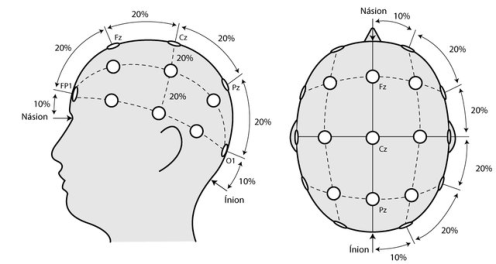
The colors were presented on a screen, in the form of monochromatic squares, isolated or in color pairs. Electroencephalography (EEG) was also used in order to acquire electrical signals in the scalp. For this purpose, caps made of an elastic spandex fabric with pure tin electrodes embedded in the fabric were used. The electrodes on the caps were positioned according to the international 10–20 electrode placement system. The EEG allows the graphic recording of the brain’s spontaneous electrical activity over a period of time originated in the brain, through multiple electrodes applied to the scalp. The generic EEG acquisition system consists of a set of electrodes, amplifiers and a data recording medium. Since all skulls are different, a standard method of electrode placement had to be found. The 10/20 International System was chosen (figure 1).
In 1958, Dr. Herbert Jasper proposed a system based on the placement of 21 electrodes, taking into account the size of the patient’s skull and also the distances between the capture points. The system was named 10-20, pre- cisely because it uses distance values that correspond to 10 or 20% of the skull section, measured from reference points. The electrodes were named according to the region of the brain they were covering: Fronto-polar (Fp), Frontal (F), C (Central), P (Parietal), T (Temporal) and O (Occipital). In addi- tion, they were also numbered to make it possible to distinguish the right and left sides, in which even numbers are used on the right, and odd num- bers on the left. On the other hand, those positioned exactly on the midline received the ending Z (Zero): Fz, Cz, Pz. Those located in the ears (Auricu- lars) were given the name A1 and A2, following the same numbering rule (Bezerra, 2022).
Color Processing and Human Perception 229
Figure 1: Representation of the 10/20 international system (Rodrigues, 2018).
RESULTS
The process started with a training task; the average number of attempts needed to reach the desired objectives was 7. During the color visualization task, the participants judged whether the colored pair came from one- or two-color categories. Participants had a high overall accuracy rate: 94%.
The occipital, caudate and anterior regions of the insula were active when more than one hue was presented, indicating a role in perceptual proces- sing and attentional monitoring. The dorsolateral prefrontal cortex showed greater activity when two colors were present than a single color, indicating a role in color category coding. The cognitive control regions of the intra- parietal sulcus and the pre-supplementary motor area were sensitive to the interaction of decision and distance in perceptual space, indicating a role in the combination of these functions during decision making. These results support theories that colors are categorically represented at high levels of the cognitive hierarchy and that the visual cortex is sensitive to hue rather than color category. We realized that the dorsolateral frontal regions were sensi- tive to color categories and not to perceptual distance, with greater activity when two color categories were present.
We also asked participants to judge whether a given sequence of chroma- tic stimuli had been extracted from one- or two-color categories. This task allowed us to identify neural regions underlying the perceptual and decision processes in color categorization.
The systematicity of the results seems to prove that, in our cognitive processes of conceptual construction and categorization, humans are more synesthetes than traditionally thought. In fact, it seems that the mind seeks, as much as possible, equivalences between perceptions from the most diverse areas, trying to associate colors not only with objects, but also with feelings, ideas and emotions.
CONCLUSION
Although at first glance it seems like a relatively simple human capacity, color processing is a complex phenomenon, involving different variables – many of which are still a mystery to researchers. In addition, new discoveries are
230 Moreira da Silva
constantly being reached, recontextualizing previous knowledge and giving rise to new questions that need answers. It is these questions that guide us to important new milestones in the neurosciences.
As a consequence, this is an area that still has enormous potential, and whose study possibilities are far from being exhausted. It is intended to con- tinue this work, trying to discover more about color processing and cognition, as well as new areas and activities of the human brain that are linked to color recognition.
ACKNOWLEDGMENT
The author would like to acknowledge the support given by CIAUD – Resea- rch Centre in Architecture, Urbanism and Design, Lisbon School of Archite- cture, Universidade de Lisboa, and by FCT – Foundation for the Science and Technology, Portugal. This work is financed by national funds through FCT - Fundação para a Ciência e a Tecnologia, I.P., under the Strategic Project with the references UIDB/04008/2020 and UIDP/04008/2020.
REFERENCES
Bezerra, J. P. (2022). Website: https://www.brainlatam.com/blog/afinal-como-epossivel-ler-o-eeg-em-diferentes-lugares-no-mundo-1115. Damasio, A. (2010). Self comes to mind: Constructing the conscious brain. Panthe-
on/Random House. Lemos Pereira, C. (2018). Processamento Visual (Parte 2) – Nível Intermediário.
Website: https://cienciasecognicao.org/neuroemdebate/arquivos/4620 Liu, Z., Lu, W., & Seger, C. A. (2019). Perceptual and categorical processing and representation in color categorization. Brain and Cognition, 136. Moreira da Silva, F. (2013). Colour and inclusivity: a visual communication design
project with older people. Lisboa: Caleidoscópio, ISBN 978-989-658-228-9. Nogueira, A.C., (2021). Neurociência das cores. Website: https://www.ufjf.br/lanc/2021/03/04/neurociencia-das-cores/. Nolte, J. (2009). The human brain: an introduction to its functional anatomy.
Philadelphia, PA: Mosby/Elsevier. Rodrigues, P. G. (2018). Extração de Características em Interfaces Cérebromáquina utilizando Métricas de Redes Complexas, Thesis for Master in Biomedical Engineering, Universidade ABC, São Bernardo do Campo, DOI: 10.13140/RG.2.2.34910.13122. Teixeira, J. (2022). Cores, Significado Linguístico e Cognição: das Imagens Mentais
e Sinestesia à “Sintonímia”, in Linguagem – Cognição – Cultura: Teorias, aplica- ções e diálogos com foco na Língua Portuguesa (Português Europeu e Português do Brasil), Hanna Batoréo (coord.), Universidade Aberta, Ciência e Cultura, 17, pp. 191–212, ISBN 978-972-674-922-6.
LISTED IN THE AHFE DIGITAL BOOKSTORE CATALOGUE
Explore the complete digital volume
Intelligent Human Systems Integration (IHSI 2023): Integrating People and Intelligent Systems
Read this individual research paper free through the original publisher, or explore the full digital proceedings book.
ISBN 978-1-958651-45-2
Explore this ISBN in the AHFE Bookstore ↗Listed in the July 2026 bookstore catalogue; search by ISBN to confirm current availability · Digital book purchase optional