Robert Dizor, S M Mizanoor Rahman, Anil Raj (2022). Gait Analysis for Rehabilitation using Rigid and Flexible Exoskeletons. In: Intelligent Human Systems Integration (IHSI 2022): Integrating People and Intelligent Systems. AHFE Open Access, vol. 22. https://doi.org/10.54941/ahfe1001013
Diseases of the musculoskeletal and nervous systems have afflicted humans since recorded history. Similarly, injuries and related trauma of one form or another have impaired human ambulation or even made it impossible to stand, walk, run or even to sit or squat. Stretchers, crutches, wheelchairs, and exoskeletons have been developed to help improve the mobility of these disabled individuals, but often require assistance from others to some degree, limiting patient autonomy. To ascertain which assistive devices might be better suited to a particular patient with an ambulation disability or weakness, the healthcare providers must perform an assessment of the individual’s gait to first under-stand the underlying symptomatic deficits, diseases, or injuries. This paper reviews how exoskeletons can with respect to the gait cycle assist the weak and elderly as well as patients with specific diseases or injuries that impact ambulation.
Intelligent Human Systems Integration (IHSI 2022): Integrating People and Intelligent Systems https://openaccess.cms-conferences.org/#/publications/book/978-1-7923-8988-7
INTRODUCTION
Early powered exoskeleton prototypes developed by the U.S. Defense Advanced Re- search Projects Agency (DARPA) and other military organizations sought to assist soldiers in the field who had to travel long distances as quickly as possible and carry heavy loads that would include their nutrition, weaponry, ammunition, and necessary survival equipment (Lockheed-Martin, 2022). Soon afterward commercial versions like EKSO were being utilized in the industrial sector to assist the employee who had to lift or carry heavy boxes (Ekso, 2022). These devices, which use traditional rigid exoskeleton structures, have also proven useful to patients during post-stroke recov- ery and rehabilitation to address various degrees of paralysis and difficulty standing and walking (Sczesny-Kaiser, 2019). Rigid exoskeletons, however, often have poor human-machine interaction (HMI), and they can be cumbersome to the user (Asbeck, 2014). Flexible exoskeletons evolved from the more rigid exoskeletons, and these have gained the interest of the medical community. At first, these were rigid exoskeletons that were made softer with protective materials for safety and comfort, but later the exoskeletons were improved with structural fabrics and varying stiffness structural components that minimize rigidity (Asbeck, 2014). These flexible devices often uti- lize actuating mechanisms, such as cables and pneumatic devices, instead of relying upon only servomotors at the joints for the movement control (Veneman, 2006). The development of flexible assistive exoskeleton devices has begun accelerating, finding use cases for medical rehabilitation, elderly care, as well as sports training, space extravehicular activity, industrial safety, and military soldiers’ protective gear in the battlefield [Lockheed-Martin, 2022, Ekso, 2022, Sczesny-Kaiser, 2019). This paper first reviews gait analysis methods needed to assess the elderly, patients with neuro- logical or musculoskeletal disease and athletes seeking improved performance, and then examines rigid and flexible exoskeletons approaches for medical rehabilitation. Finally, the advantages and current shortcomings are discussed with respect to how the elderly and patients could one day use an exoskeleton to achieve a functional gait.
GAIT ANALYSIS The human lower extremity anatomy and associated motion is complex, which has often led to oversimplification in analysis and engineering designs for exoskeletons. For example, many exoskeletons and orthotic designs simplify the motion of the knee as a single degree of freedom (DOF) joint, ignoring the tibial and femoral rotation that occurs during flexion and extension (Christof, 2010). Anatomists, physiologists, and other medical researchers have described the human lower extremity anatomy and function in detail for decades (Mayich, 2014). By improving their understanding of this existing information, engineers can make significant improvements in func- tion, mobility, and comfort when designing lower extremity exoskeletons both for patients who require assistance and/or rehabilitation, as well as for augmenting
Intelligent Human Systems Integration (IHSI 2022): Integrating People and Intelligent Systems https://openaccess.cms-conferences.org/#/publications/book/978-1-7923-8988-7 healthy individuals or athletes seeking to extend their capabilities (Archer, 2006, Van der Krogt, 2012). More recently, gait analysis that breaks the gait stride into both temporal and spatial parameters has been applied to the modeling and control of bi- pedal movement in humanoid robots or exoskeletons using what we have learned from human ambulation (Uzair, 2019, In-Sik, 2014).
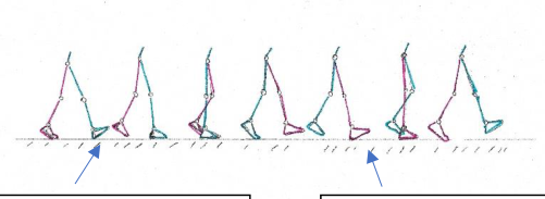
Temporal Analysis Human gait can be described as a series of alternating strides or movements of the lower extremities in a coordinated and rhythmic forward motion (Mayich, 2014). This movement occurs with minimum expenditure of effort and energy when able bodied individuals walk or run. Abnormalities of the gait can be detected and assessed in patients with injury or disease by identifying changes in kinetic force and kinematic (spatial/temporal) data (Mayich, 2014, Archer, 2006). A single gait cycle consists of a foot heel contact to the ground with the ipsilateral or observed leg, a contralateral or opposite foot heel contact in a similar fashion which then completes with a second heel contact of the ipsilateral foot as shown in Figure 1. Gait analysis requires at least one cycle (but preferably more) to assess the gait while ambulating (Baker, 2013).
Figure 1. One complete gait cycle. This begins with an initial ipsilateral (green) heel strike or contact and ends with a second ipsilateral heel strike.
Analysis of the kinematics of ambulation reveals two temporal phases to the gait cycle as shown in Figure 2. At the beginning of the gait cycle, the stance phase starts when the ipsilateral heel contacts the ground (sometimes called the heel strike) and ends when the same ipsilateral foot finally leaves contact with the ground (sometimes called the toe-off or push-off), making up approximately 60% of the gait cycle in a normal healthy adult (Baker, 2013). The swing phase begins when the stance phase ends, thus the swing phase starts with the ipsilateral toe-off and ends with the ipsilat- eral leg heel contact or foot strike. The swing phase for the ipsilateral leg makes up the remaining 40% of the gait cycle and consists of an acceleration, mid-swing, and deceleration components (Baker, 2013). The swing phase influences both balance and agility because the contralateral leg is in a single support configuration.
The stance and swing phases alternate between the ipsilateral and contralateral legs as the individual walks or runs. When the right foot and leg are in the stance phase,
Intelligent Human Systems Integration (IHSI 2022): Integrating People and Intelligent Systems https://openaccess.cms-conferences.org/#/publications/book/978-1-7923-8988-7 the left foot and leg will be in a complementary swing phase for a portion of the right leg stance phase. Consequently, in a complete gait cycle, the individual alternates between having either one foot or two feet on the ground at any instant (Baker, 2013). To describe this, we break the ipsilateral stance phase into initial and terminal double support phases (two feet on the ground) separated by a single support phase (only one foot in contact with the ground) while the contralateral leg and foot are in its swing phase. The initial double support phase begins the gait cycle and stance phase for the ipsilateral foot. This ends with the toe-off and swing phase of the contralateral foot. The ipsilateral single support phase begins with the contralateral swing phase and continues through the end of the contralateral swing phase and heel contact. This be- gins the terminal or second double support phase of the ipsilateral foot stance phase and ends with the toe-off. The ipsilateral leg and foot are in swing phase (contralateral single support phase) till the ipsilateral heel contact ends the complete cycle as is shown in Figure 1.
Often, gait problems or pathology will shorten the ipsilateral stance phase of the gait cycle because the patient cannot fully bear his or her weight on that lower ex- tremity (Phillips, 2017). This results in limping that may or may not be obvious to the casual observer. This will ultimately shorten the contralateral swing phase as well, forcing a longer stance time on the stronger contralateral lower extremity and a shorter stance time on the affected ipsilateral foot. When analyzing foot pathology, physical therapists break the stance phase as is shown in Figure 2 into smaller sub- phases: 1) Contact (heel strike) phase, 2) Flat-Foot phase, 3) Mid-stance phase, and 4) Propulsive phase, which is further divided into active and passive phases (Phillips, 2017).
Ipsilateral (green) heel strike Toe-Off Midstance
Figure 2. The stance phase begins with the ipsilateral (green) heel strike and ends with ipsilateral toe-off.
Figure available in the original PDF paper.
The initial contact subphase begins the stance phase with the heel strike. Any pa- thology of the heel will alter or even sometimes eliminate this heel contact subphase (Phillips, 2017). If the lead lower extremity or foot cannot tolerate the impact, the patient may put the foot down in a toes-first manner attempting to reduce the force on the heel at ground contact (Phillips, 2017). During the ipsilateral heel contact sub- phase, the contralateral foot is still in contact with the ground contributing to double support (Baker, 2013). The heel contact subphase ends after the contralateral foot toe- off. Next, the stance phase then continues to transition into the flat-foot or loading
Intelligent Human Systems Integration (IHSI 2022): Integrating People and Intelligent Systems https://openaccess.cms-conferences.org/#/publications/book/978-1-7923-8988-7 subphase. The lead or ipsilateral foot now carries the total weight of the individual in the single support phase while still maintaining forward momentum and balance. This loading of the ipsilateral foot begins with the heel contact and ends with the first metatarsal head contacting the ground in the lead foot. After the flat-foot subphase, we have the mid-stance subphase. The contralateral foot is now in its mid swing phase. The mid-stance subphase includes increased loading of the foot that is now in complete contact with the ground, but also ends when the lead foot heel finally comes off the ground. The flat-foot and mid-stance subphases transition the lead ipsilateral foot from shock absorption due to the heel strike and initial contact to a stability and balance function. Finally, the propulsive subphase concludes the stance phase by be- ginning and ending with active and then passive components (Baker, 2013). During the active part of propulsion, the mid foot typically supinates (inverts slightly at the toes and metatarsals) and becomes stiffer as the toe-off begins and the heel lifts off the ground. The active part is when the lead foot is still in single support. The passive part begins with the contralateral heel strike beginning double support and ends just as the lead foot toe-off is complete ending the terminal double support and beginning the swing phase of the ipsilateral foot.
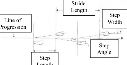
Spatial Analysis Because normal gait is relatively symmetric, practitioners can use analysis of gait spatial and temporal parameters to reveal abnormalities or injuries that disturb the symmetry. This focus on symmetry helps to inform diagnoses and treatment plans for rehabilitation or selection of compensatory orthotics. The gait spatial parameters are usually evaluated by step length, stride length, step width, and foot angle as is shown in Figure 3 (Baker, 2013).
First, a line of progression is drawn from the midpoint of the individual at the start of the gait cycle to the same endpoint at the end of the gait cycle. This line is the vector for the individual’s path while walking or running. The step length equals the distance measured along the line of progression from the heel strike (posterior heel contact) of the previous footfall to the heel strike of the opposite foot. The stride length is measured between heel strikes of the same foot and represents the distance covered by one complete gait cycle. The midline of the footprint bisects the foot with a line drawn from the heel (at heel strike) to roughly the second and third metatarsal heads at the base of the second and third toes, which will vary by the shape and size of the foot. From this footprint midline, the step width is usually measured between and orthogonal to the line of progression. In a healthy individual, the left and right step widths measure the same distance from the vector line of progression. Many conditions can cause asymmetry of step width, such as diabetic osteomyelitis (bone infection), prior injury or missing toes, or asymmetric polydactyly (extra toes) (Baker, 2013). The step angle is measured between the footprint midline and the line of pro- gression with 0 degrees defined as foot midline parallel to the line of progression. The angle is positive when the toes are angled or pointing laterally and negative when pointing inward or medially (pigeon-toed).
Intelligent Human Systems Integration (IHSI 2022): Integrating People and Intelligent Systems https://openaccess.cms-conferences.org/#/publications/book/978-1-7923-8988-7
Stride Length Step Width
Line of Progression
Step Angle Step Length
Figure 3. Spatial analysis terms visually depicted within the gait cycle.
Gait Analysis Technology Gait analysis has traditionally employed dedicated motion capture lanes outfitted with multiple video cameras that track retroreflective markers positioned on key anatomi- cal locations on the individual under evaluation (In-Sik, 2014). These cameras have a high-speed frame rate and the markers and their positions on the hips and lower extremities are standardized including the size of the marker and its anatomical loca- tion (Helen Hayes Markers) (Collins, 2009). Many sensors are positioned within a platform installed in the floor of an ambulation test area or within a treadmill. Data collection can then be made from several strides taken by the patient. The platform approach does not work as well for athletes. Similarly, in patients with a disability or weakness, it is better to evaluate running or walking to fatigue on a treadmill. In these cases, walking or running shoes with embedded force sensors can provide data ob- tained with a more natural gait, and can even capture temporal data outside of the lab, however, the in-lab platform approach is better for spatial data analysis (Tahir, 2020). While synchronization and alignment of multiple 3D tracking cameras can be com- plicated, this motion capture method provides additional insight into balance and agil- ity to complement force and pressure mapping (Collins, 2009, Tahir, 2020).
RIGID EXOSKELETONS The first powered exoskeletons consisted of rigid, metal structural frames with a lim- ited range of motion for the lower extremity joints (Kazerooni, 2006). One of first exoskeletons attempted, the “Hardiman” prototype (General Electric, Co., Boston, MA) not only would have dwarfed the individual pilot or user in size and mass, but also proved to be difficult to control and thus dangerous for use (Kazerooni, 2006) . More recently, the Berkeley Lower Extremity Exoskeleton (BLEEX, University of California, Berkeley, CA) allowed the user to carry 34 kg (Kazerooni, 2006). Alt- hough the original rigid exoskeletons were cumbersome and complex, iterative im- provements to mechanical design and advances in computer technologies and elec- tronics have enabled real-world use of solutions such as the Hybrid Assistive Limb
Intelligent Human Systems Integration (IHSI 2022): Integrating People and Intelligent Systems https://openaccess.cms-conferences.org/#/publications/book/978-1-7923-8988-7
(HAL, Cyberdyne, Inc., Tsukuba, Japan) for limited rehabilitation of patients (Sczesny-Kaiser, 2019). These are being used to rehabilitate patients, but they require therapists in a clinic setting to help the patient. Honda Corporation developed their “Bodyweight Support Assist Device” initially in 2005, and they have continued to improve this lower extremity exoskeleton (Honda, 2022). In S Korea, EXPOS was similarly developed near 2005, and has likewise continued to improve their exoskel- eton for rehabilitation or walking assistance (Kyoungchul, 2006).
These rigid exoskeletons employed a stiff metal or composite frame that could support the body weight of the individual, including paraplegic patients as well as those patients requiring walking assistance and rehabilitation due to stroke from cer- ebral vascular injury. However, if they do not account for the complexity of human lower extremity joint motion while walking, running, or even moving from a seated into a standing position, the rigid exoskeleton could induce injury and extract a high metabolic cost during its use (Sczesny-Kaiser, 2019, Neuhaus, 2017). Current and planned rigid exoskeleton designs, like the Mina rigid exoskeleton (Florida Institute for Human and Machine Cognition, Pensacola, FL), however, have begun to provide increased degrees of freedom (DOF) (Neuhaus, 2017).
In practice, it takes a significant amount of time and metabolic energy for patients to prepare, adjust and employ a rigid exoskeleton prior to use (Neuhaus, 2017). The range of mechanical adjustments limit use of a rigid exoskeleton to individuals who can wear and use the exoskeleton within a given time (for clinic-based exoskeletons). The heavy exoskeleton frame and motors require significant power, and current bat- tery technologies still have undesirable bulk and mass with a limited available run time. For safety and servicing, the controls, thermal management, and power are often positioned within a backpack that can change the patient’s center of gravity and mass (Neuhaus, 2017, Longhan, 2019). This can create balance issues possibly leading to an increased chance of falling. The rigid exoskeleton joints do not easily accommo- date the rotational movement of the tibia and femur, and the subsequent poor fit and joint tracking can cause muscular, joint, skin, or tendinous injury (Longhan, 2019). Osteoporosis is not uncommon in the elderly, and thus exoskeleton induced fractures are also a significant concern and must be prevented in this population (Seriolo, 2013). Because of the disturbance to balance and kinematics, users need crutches to help stabilize themselves and to reduce the chance of falling; crutches are required for users with paraplegia due to the lack of sensory feedback that accompanies their lack of muscle control (Neuhaus, 2017, Longhan, 2019).
FLEXIBLE EXOSKELETONS The first flexible exoskeletons focused on reducing the fatigue associated with using exoskeletons with rigid frames. Harvard University (Cambridge, MA) developed more than one flexible exoskeleton design that reduced the metabolic cost needed to walk and run (Sangjun, 2018). A Canadian company, Bionic Power Inc., even pro- duced a military soft exoskeleton that partially regenerated some energy for charging equipment (BionicPower, 2022). By using energy harvesting and lighter weight flex- ible materials, power demand on the batteries decreases, which can further reduce
Intelligent Human Systems Integration (IHSI 2022): Integrating People and Intelligent Systems https://openaccess.cms-conferences.org/#/publications/book/978-1-7923-8988-7 exoskeleton mass or increase operational time for a given battery chemistry. The de- creased metabolic demand on the patient also facilitates longer therapy sessions or training time for athletes. As this concept continues to mature, more patients could benefit from exoskeleton-assisted rehabilitation to accelerate recovery and a shorten time to resume their normal daily activities (Longhan, 2019, Sangjun, 2018). The compliance of soft, adjustable, flexible exoskeletons can accommodate small misa- lignments or extreme joint angles (e.g., when jumping or climbing) without transmit- ted adverse forces to the limb, and thus reducing risk of harm or injury to the patient. This better accommodates the tibial and femoral rotation associated with walking and standing, and thus improves the human-machine interface (Christof, 2010).
There are some inherent challenges to the flexible exoskeleton. With traditional rigid exoskeleton frames, motor actuators at the joints can control the movement and simplify the control kinematics. While flexible exoskeletons also employ motors they can be smaller and weaker and transmit power via Bowden cables and pneumatics from remote mounting points where stiffer structural elements can be used. By mov- ing the servomotors from the joints, cables help make the exoskeleton lighter and more flexible. This also helps distribute the system mass while managing user bal- ance. Pneumatic bladders that mimic muscle is another method to provide flexion or extension at joints (Veneman, 2006).
Another key point with flexible exoskeletons is that while their lack of a rigid frame reduces weight, this also means that forces to propel the exoskeleton and pa- tient forward must be transmitted to and from the ground through the bony skeleton and muscle tissue of the user (Yandell, 2017). This means that the impact from each step will not be absorbed by a rigid exoskeleton, but instead will be transmitted to the patient’s foot and leg. This could be a problem for the weak and elderly with osteo- porosis and the associated increased risk of fracture, however, it could provide the strain necessary for bone remodeling to increase functional strength and health of the lower extremities. Likewise, the forces involved with each step requiring flexion and extension of the lower extremity joints will be transmitted through the individual to the ground. This could increase metabolic demand of the user and accelerate fatigue if the design does not provide high efficiency in a lightweight flexible exoskeleton.
CONCLUSIONS AND FUTURE WORK Exoskeletons for patient rehabilitation are still in the early stages of development for clinical use, but there has already been significant improvement. The walk-through of gait analysis presented will hopefully provide insight and clarity to future devel- opment of more human-centered exoskeleton designs. The lighter flexible exoskele- ton with its decreased metabolic fatigue needs to be combined with the structural strength benefits of the traditional exoskeletons. Carbon fiber and other composite structural elements can provide this lightweight support and bespoke stiffness tailored to the desired kinematics (Honda, 2022). Carbon nanotube technology may prove useful for lightweight, unobtrusive tension cables (Honda, 2022). Ankle and leg forces must be efficiently translated into guided and controlled movement in future work, not only so the user can walk, but also so that the patient can stand from a sitting position, then walk and run without suffering any injury. Ideally, the flexible
Intelligent Human Systems Integration (IHSI 2022): Integrating People and Intelligent Systems https://openaccess.cms-conferences.org/#/publications/book/978-1-7923-8988-7 exoskeleton will be as easy to put on as sports garments and equipment. Ideally, don- ning a soft muscular exoskeleton would complement the user’s endoskeleton, becom- ing incorporated with the patient’s legs, just as if they had full and complete control of their lower extremities (Pazzaglia, 2016).
The actuator system must mimic the human neuromuscular biological system for true patient acceptance. This means that EEG (electroencephalogram) or EMG (elec- tromyogram) sensing could be utilized to naturally follow through on the patients’ intention to sit, stand or walk. A brain-computer interface (BCI) has been shown to effectively allow humans with a spinal cord injury as well as monkeys to control electronic devices (King, 2013). EMG sensor arrays embedded within the exoskele- ton would be positioned over the muscle group of interest, though they could be im- planted for permanent use as well if small enough. A future study could focus on the use of a reflex-like machine learning algorithm that autonomously augments or re- places any reflex as needed to complement the patient’s intention. This must operate in the background without thought by the user or BCI just as human reflexes do in the healthy individuals. Having reflexes within the exoskeleton or limb replacement would improve response time and subsequently improve safety and comfort. Provid- ing the patient with immediate and direct control as well as proprioceptive feedback from the exoskeleton will be key to realizing the embodiment of the exoskeletons sought by developers and healthcare providers for their patients (Pazzaglia, 2016).
REFERENCES
Archer, K.R., et al, (2006). “Gait symmetry and Walking Speed Analysis following lower extremity trauma,” Physical Therapy, Vol. 86, pp. 1630-1640. Asbeck, A.T., et al, (2015). “A biologically inspired soft exosuit for walking assis-
tance,” The International Journal of Robotics Research, Vol. 34, No. 6, pp. 744-762. Baker, R, (2013). Measuring Walking: A Handbook of Clinical Gait Analysis. London: Mac Keith Press. BionicPower, (2022). “Power Walk,” https://www.bionic-power.com/ Christof, R., et al, (2010). “Torsional profile versus gait analysis: Consistency be-
tween the anatomic torsion and the resulting gait pattern in patients with rota- tional malalignment of the lower extremity,” Gait and Posture, Vol. 32, pp. 405- 410. Collins, T.D., et al, (2009). “A six DOF marker set for gait analysis and Comparison with a modified Helen Hayes Marker set,” Gait and Posture, Vol. 30, pp. 173- 180. Ekso exoskeletons, (2022). https://www.eksobionics.com Honda Corporation, (2022). “Bodyweight Support Assist”,
https://www.asimo.honda.com/innovations/ In-Sik, L., et al, (2014). “Gait optimization of biped robots based on human motion analysis.” Robotics and Autonomous Systems, Vol. 62, pp. 229-240. Kazerooni, H., et al, (2006). “Hybrid control of the Berkeley Lower extremity exo-
skeleton (BLEEX),” The international journal of Robotics research, Vol. 25, No. 5-6, pp. 561-573.
Intelligent Human Systems Integration (IHSI 2022): Integrating People and Intelligent Systems https://openaccess.cms-conferences.org/#/publications/book/978-1-7923-8988-7
King, C. et al, (2013). “Operation of a brain-computer interface walking simulator for individuals with spinal injury,” Journal of NeuroEngineering and Rehabili- tation 2013, Vol. 10:77, pp.1-14. Kyoungchul, K., Doyoung J., (2006). “Design and control of an exoskeleton for the
elderly and patients,” IEEE/ASME Transactions on Mechatronics, Vol. 11 (4), pp. 428-432. Lockheed-Martin, (2022). https://www.lockheedmartin.com/en-us/products/exoskeleton-technologies.html Longhan, X., et al, (2019). “An Unpowered Flexible lower limb exoskeleton: Walk-
ing Assisting and Energy harvesting,” IEEE/ASME Transactions on Mecha- tronics, Vol. 24 (5), pp. 2236-2247. Mayich, D.J., et al, (2014). “Gait analysis in Orthopedic foot and ankle surgery - Topical Review, Part I: Principles and Uses of Gait Analysis,” Foot and Ankle International, Vol. 35(1), pp. 80-90. Neuhaus, P., (2017). “IHMC's experience competing in the Cybathlon compared to
the DARPA robotics challenge,” Journal of Neuro-engineering and rehabilita- tion, Vol. 14:112, pp. 1-6. Pazzaglia, M., Molinari, M., (2016). “The Embodiment of Assistive Devices – from wheelchair to exoskeleton,” Physics of Life Reviews, Vol. 16, pp. 163-175. Phillips, A., McClinton, S., (2017). “Gait deviation associated with plantar heel
pain,” Clinical Biomechanics, Vol. 42, pp. 55-64. Sangjun, L., et al, (2018). “Autonomous multijoint soft exosuit with augmentationpower-based control parameter tuning reduces energy cost of loaded walking,” Journal of Neuro-engineering and Rehabilitation, Vol. 15:66, pp. 1-9. Sczesny-Kaiser, M., et al. (2019). “A Randomized and Controlled Crossover Study
Investigating the Improvement of Walking and Posture Functions in Chronic Stroke Patients Using HAL Exoskeleton - The HALESTRO Study (HAL- Exoskeleton STROke Study).” Frontiers in Neuroscience, Vol. 13, pp.1-13. Seriolo, B., et al, (2013). “Osteoporosis in the elderly,” Aging Clin Exp Res, Vol.
25, pp. S27-29. Tahir, A.M., et al, (2020). “A systematic approach to the design and characterization of a smart insole for detecting vertical Ground reaction forces (VGRF) in Gait Analysis,” MDPI, Sensors, Vol. 20 (957), pp. 1-25. Uzair, K., Zhiyong, C., (2019). “Natural gait analysis for a biped robot: jogging ver-
sus walking.” Science China, Information Sciences, Vol. 62, pp. 1-3. Van der Krogt, M.M., et al, (2012). “How robust is human gait to muscle weakness,” Gait and Posture Vol. 36, pp. 113-119. Veneman, J.F., et al, (2006). “A series elastic- and Bowden-cable-based actuation
system for use as torque actuator in exoskeleton-type robots,” The Interna- tional Journal of Robotics Research, Vol. 25, No. 3, pp. 261-281. Yandell, M.B., et al, (2017). “Physical interface dynamics alter how Robotic Exosuits augment human movement: implications for optimizing wearable assis- tive devices,” Journal of NeuroEngineering and Rehabilitation, Vol. 14:40, pp. 1-11.
Intelligent Human Systems Integration (IHSI 2022): Integrating People and Intelligent Systems https://openaccess.cms-conferences.org/#/publications/book/978-1-7923-8988-7
LISTED IN THE AHFE DIGITAL BOOKSTORE CATALOGUE
Explore the complete digital volume
Intelligent Human Systems Integration (IHSI 2022): Integrating People and Intelligent Systems
Read this individual research paper free through the original publisher, or explore the full digital proceedings book.
ISBN 978-1-7923-8988-7
Explore this ISBN in the AHFE Bookstore ↗Listed in the July 2026 bookstore catalogue; search by ISBN to confirm current availability · Digital book purchase optional