Robert Dizor, S M Mizanoor Rahman, Anil Raj (2022). Exoskeleton Design Considerations based on the Lower Extremity Musculoskeletal Anatomy. In: Intelligent Human Systems Integration (IHSI 2022): Integrating People and Intelligent Systems. AHFE Open Access, vol. 22. https://doi.org/10.54941/ahfe1001014
This paper focuses on understanding the musculoskeletal anatomy of the lower extremities and how the joint movement is controlled by the muscles. The lower extremity has been subdivided into three major sections: the hip joint, the knee joint, and the ankle joint. For each of these joints, attention is given to which muscles control the motion or degrees of freedom with respect to these joints. The muscles are grouped by their major motion like flexion or extension and additional detail is provide with respect to secondary and more complex motion. Drawings were made showing the location of the major flexors and extensors for the three major joints. Furthermore, the muscles were grouped within tables by their motion and major innervations. This information is based on the medical anatomical and physiology texts included within the references. These drawings and tables can be helpful for quick reference and understanding the motion for each of the lower extremity joints, thus improving surface EMG sensor placement and positioning when designing lower limb prosthetics or exoskeletons.
Intelligent Human Systems Integration (IHSI 2022): Integrating People and Intelligent Systems https://openaccess.cms-conferences.org/#/publications/book/978-1-7923-8988-7
INTRODUCTION
Early lower extremity limb replacement prosthetics like artificial legs were goal oriented but limited in their function and utility and could not fully mimic or truly replace the limb (Ferris, 2017). Likewise, early exoskeletons first developed for the military and commercial sector to assist healthy individuals carry heavy loads but were limited in their ability to provide comprehensive function (Ekso, 2022). Later, lower extremity exoskeletons and prosthetics incorporated hinge joints at the ankle and knees and more degrees of freedom (DOF) at the hip joint to in- crease user range of motion (Ferris, 2017, Pinto-Fernandez, 2020). These exo- skeletons and prosthetics, however, still did not fully emulate the human anatomy and kinematics; consequently, they produced discomfort and placed a significant metabolic demand on the user, both of which limited the time that the user could wear these devices (Ferris, 2017, Pinto-Fernandez, 2020).
Flexible exoskeletons evolved from rigid exoskeleton concepts and have more recently gained the interest of the medical community. Initially, rigid exoskele- tons were made softer by covering them with protective material for safety and comfort, but later incarnations were improved with structural fabrics and flexible members replacing the rigid structural components (Asbeck, 2015). Instead of relying only upon servomotors at each joint for movement, other actuating mech- anisms such as cables and pneumatic drives have allowed shifting mass and bulk from the lower extremities for both rigid and flexible exoskeletons (Veneman, 2006). Increasing the DOF at each joint has improved exoskeleton function by allowing more accurate tracking of the anatomy during movement. Exoskeleton designs have also begun to incorporate control methods developed in the pros- thetic limb replacement domain, namely surface electromyography (EMG). EMG sensors have been used to detect proximal (intact) muscle activity, and more recently machine learning (ML) has been used to identify specific voluntary movement commands (Asma, 2020). This paper focuses on understanding the musculoskeletal anatomy of the lower extremities and how the joint movement is controlled by the muscles. Also, attention is given to understanding their in- nervation, and thus improving surface EMG placement and positioning when de- signing lower limb prosthetics or exoskeletons.
THE LOWER EXTREMITY JOINTS AND THEIR MOTION Although orthopedic surgeons, anatomists, and physical therapists for decades have detailed the lower extremity human anatomy and function which generates known complex motion, exoskeleton and prosthetics designers have often over- simplified their mechanisms while engineering feasible devices with available technologies (Ferris, 2017, Moore, 1992). For example, the function and motion of the knee and ankle have often been oversimplified by ignoring the tibial and femoral rotation at the knee throughout the leg swing or the importance of ever- sion and inversion within the ankle for stable foot plant (Christof, 2010). Im- proved understanding of the lower extremity movement can make a significant difference in the function, mobility, and comfort when designing exoskeletons or
Intelligent Human Systems Integration (IHSI 2022): Integrating People and Intelligent Systems https://openaccess.cms-conferences.org/#/publications/book/978-1-7923-8988-7 prosthetics (Ferris, 2017, Moore, 1992, Pazzaglia, 2016).
The Hip Joint The hip joint (see Table 1) is a three DOF ball and socket synovial joint between the acetabulum (socket) of the hip joint and the head of the femur (ball) (Moore, 1992). The hip joint permits flexion and extension, abduction and adduction, and rotation (internal/medial and external/lateral rotation) as well as circumduction (circular rotation of the leg which includes using flexion, extension, abduction, and adduction) (Moore, 1992, Zaffagnini, 2016, Soucie, 2011). While the narrow neck of the femur between the ball and shaft serves to allow an increase in range of motion (ROM) of the hip joint that would not be present if the femoral neck was as thick as the head of the femur, the periarticular fibrocartilage tissue and labrum limit the full ROM and contribute to increased strength and stability (Moore, 1992. Chung, 2000). As a result, the average healthy adult hip joint can flex ventrally up to 125 degrees and extend dorsally to approximately 115 de- grees, whereas elderly patients have less joint ROM than younger adults and males have less ROM than females (Zatsiorsky, 2002, Kashitaro, 2017, Hem- merich, 2006). Exoskeleton and prosthetic designers can measure an individual’s ROM manually with a goniometer, a simple plastic or metal measuring device with two arms forming a pivot with an angle scale. They may also often use an electronic goniometer or inertial measurement unit (IMU). Other motion capture methods may be utilized allowing recording of multiple joint angles with limb position while in motion (Moore, 1992, Zaffagnini, 2016, Soucie, 2011).
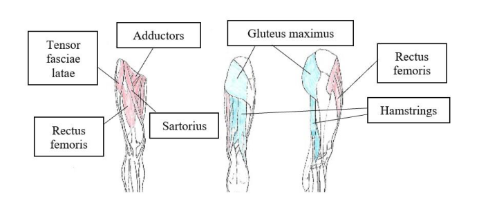
Figure 1. Anterior, posterior, and lateral views (right to left).
Hip joint flexors (red) and extensors (blue).
Three different muscle groups contribute to flexion of the hip joint (Figure 1): the flexors (quadriceps), adductors in the thigh, and the abdominal flexors, (Zaf- fagnini, 2016). The most important muscle is the centrally positioned rectus fem- oris muscle of the quadriceps femoris group (Moore, 1992). It is the largest of the hip flexors and is located within the anterior fascial compartment of the thigh, which simplifies the positioning of surface EMG sensors when assessing neuro- muscular activity and voluntary hip joint flexion (Soucie, 2011).
Intelligent Human Systems Integration (IHSI 2022): Integrating People and Intelligent Systems https://openaccess.cms-conferences.org/#/publications/book/978-1-7923-8988-7
Table 1: Summary of Muscle Action for the Lower Extremity (Moore, 1992, Zaffagnini, 2016).
Table available in the original PDF paper.
The generally accepted range of motion measurements are in parentheses. Older individuals have slightly less ROM than young people. Males have less ROM than females (Center for Disease Control, 2022, Soucie, 2011) The large femoral nerve innervates the rectus femoris and travels with the fem- oral artery and vein as a neurovascular bundle (see Table 2) passing into the an- terior compartment of the thigh via Hunter’s Canal just deep to the sartorius mus- cle (Moore, 1992). Two important secondary flexors, the sartorius and iliacus muscles, both originate in the pelvis, but the iliacus inserts medially on the lesser trochanter of the proximal femur (below the femoral neck) while the sartorius crosses the knee joint to insert on the medial surface of the proximal tibia (Moore, 1992, Zaffagnini, 2016). These two muscles also contribute to control of the DOF at the hip (Soucie, 2011). The iliacus travels with the psoas major and psoas mi- nor muscles, which both originate from the abdominal vertebral column, and all
Intelligent Human Systems Integration (IHSI 2022): Integrating People and Intelligent Systems https://openaccess.cms-conferences.org/#/publications/book/978-1-7923-8988-7 three flexors help flex the trunk as well as the thigh. Within Hunter’s Canal, the femoral nerve innervates both the iliacus and sartorius muscles, just medial to the rectus femoris and quadriceps, providing an accessible location for surface EMG sensor placement (Chung, 2000). The psoas muscles, however, are directly in- nervated by lumbar and sacral spinal nerve branches within the abdomen and pelvis before they form the femoral and sciatic nerves (Moore, 1992).
Many of the muscles that provide smaller contributions to flexion also provide adduction and abduction. Those found in the medial compartment move the leg inward and those found in the lateral compartment move the leg outward, respec- tively. Some of these muscles also contribute to medial rotation (adductors) or lateral rotation of the femur (abductors) in addition to their limited contribution to flexion (Zaffagnini, 2016). Normal ROM for both adduction and abduction, as well as both medial and lateral rotation can reach 45 degrees from neutral (Zatsiorsky, 2002, Kashitaro, 2017). The obturator nerve innervates the adductors within the medial compartment, but branches of the sciatic and femoral nerves contribute to muscle group activation as well (Moore, 1992). Surface EMGs po- sitioned over these medial compartment nerves and muscles would help charac- terize and assess adductor muscular activity associated with medially flexing of the thigh (Soucie, 2011). The gluteus medius and minimus muscles in the poste- rior pelvis together with the tensor fascia latae muscle abduct the thigh at the hip. Surface EMG sensors placed over the superior gluteal nerves that innervate these lateral compartment abductors can provide a measure of abduction activity (Moore, 1992). Lateral (external) rotation is associated with the abductors, and medial (internal) rotation correlates with the adductors respectively, but these complex motions include some degree of flexion or extension as well. It is a com- bination of the many muscles described within the tables that contribute to the DOFs of the hips in addition to flexion and extension (Moore, 1992).
The posterior compartment hamstring muscles (see Table 2) primarily extend the thigh at the hip joint with assistance from the gluteus maximus (Zaffagnini, 2016). The biceps femoris (short and long heads, hence biceps) is the largest and most lateral of the three hamstring muscles. This muscle originates within the pelvis and extends inferiorly to cross the knee and insert on the proximal lateral tibia, enabling it to also contribute to lateral rotation of the knee. In a similar fashion the medial hamstrings (semitendinosus and semimembranosus) both originate in the pelvis and insert below the knee at the proximal medial tibia to provide both thigh extension and medial knee rotation functionality (Moore, 1992). Surface EMGs positioned over the posterior compartment can assess the hamstring movement for extension (Zatsiorsky, 2002). The large sciatic nerve within the posterior compartment innervates the hamstrings (Chung, 2000).
The Knee Joint The knee joint is the largest and most complicated joint of the lower extremity (see Table 1). Although structurally it resembles a hinge joint, it is a condylar synovial joint consisting of the two condyles of the femur and tibia that also in- cludes a saddle joint between the patella and the femur (Moore, 1992). The pa- tella, a sesamoid bone within the distal continuation of the quadriceps tendon proximal to its insertion on the proximal tibial tuberosity, resides anterior to the
Intelligent Human Systems Integration (IHSI 2022): Integrating People and Intelligent Systems https://openaccess.cms-conferences.org/#/publications/book/978-1-7923-8988-7 condyles of the femur, located distal to the patellar ligament (Eckhoff, 2005). The patella acts as a fulcrum that increases the leverage of the quadriceps muscles (rectus femoris, vastus lateralis, vastus medialis, and vastus intermedius) when extending the lower leg (Moore, 1992). The knee joint is held together by numer- ous ligaments and a fibrous capsule in addition to the musculature which act to stabilize and constrain the DOF of the knee (Eckhoff, 2005). The knee joint mo- tion consists primarily of flexion and extension, however gliding and rotation occur between the tibial and femoral components throughout the leg swing (Kashitaro, 2017). At full extension, a slight medial rotation (internal rotation) of the femur occurs relative to the tibia pulling the supportive ligaments taut, thus helping to stabilize the knee while standing (Moore, 1992). The knee joint can flex up to 130 degrees to allow climbing, squatting, or kneeling, and can extend past zero (hyperextension) by a few degrees when standing (Soucie, 2011).
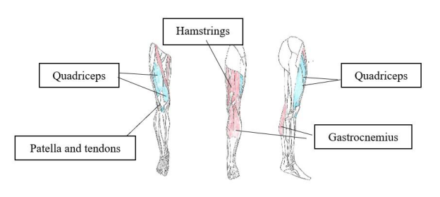
Figure 2. Anterior, posterior, and lateral views (right to left).
Knee joint flexors (red) and extensors (blue).
Within the posterior compartment (calf) of the shank of the leg (see Figure 2), both superficial (gastrocnemius, soleus, and plantaris) and deep (popliteus) mus- cles contribute to flexing the knee (Moore, 1992). When compared to the deeper muscles, surface EMG sensors positioned over the upper calf can more easily detect superficial muscle activation during knee flexion with a better signal to noise ratio (Posa, 2020). The inner and outer heads of the gastrocnemius muscle originate at the medial and lateral femoral condyles, respectively, and share a common insertion on the calcaneus (heel) via the calcaneal (Achilles) tendon. Crossing both the knee and ankle joints, this muscle also contributes to plantar flexion of the foot by pulling back and up on the calcaneus (Moore, 1992). The plantaris muscle also crosses the knee and ankle, originating along the supracon- dylar line of the lateral distal femur and inserting on the calcaneus. The lateral femoral attachment allows for medial (internal) rotation of the shank (lower leg) by pulling the posterior calcaneus laterally (Kashitaro, 2017). The popliteus, the only deep group muscle of the calf with knee flexion function, originates from the lateral condyle of the femur and the popliteal ligament posteriorly. It inserts on the upper posterior side of the tibia and medially rotates the shank as well as flexing the knee. The popliteus ligament, an extension of the semimembranosus hamstring muscle, acts as a protective membranous covering for popliteal fossa blood vessels and nerves that course behind the knee joint. All posterior calf mus- cles are innervated by the tibial nerve (Moore, 1992).
Intelligent Human Systems Integration (IHSI 2022): Integrating People and Intelligent Systems https://openaccess.cms-conferences.org/#/publications/book/978-1-7923-8988-7
The hamstring musculature of the posterior thigh not only extends the thigh, but because it inserts on the tibia, it also helps to flex the knee, and rotate the shank medially and laterally (Chung, 2000). The gracilis adductor muscle in the medial thigh originates in the pelvis like the sartorius and inserts medially on the upper tibia, assisting knee flexion and medial rotation of the shank, as well as both flexion and adduction at the hip (Moore, 1992). Surface EMG sensors placed on the lower posterior thigh can detect knee flexion activation. Likewise, EMG sensors placed on the lower anterior thigh would capture the primary contribution for knee extension from the quadriceps femoris (Chung, 2000). As we can see, muscles within the lower extremity often have multiple functions adding to the complexity of motion control and indicates a need for multiple EMG sensors to improve accuracy.
The Ankle Joint The ankle (talocrural) joint is a hinge-type synovial joint in the transverse plane between the tibia and fibula superiorly and the trochlea (pulley) of the talus infe- riorly which supports plantar flexion and dorsiflexion (Moore, 1992). Inferior to the talocrural hinge joint, the intertarsal joints work together to allow for inver- sion and eversion at the subtalar joint. The intertarsal joints form a plane between the talus and calcaneus (heel). The transverse tarsal joint is the collective term for these intertarsal joints formed by the talus, calcaneus, navicular, and cuboid tarsal bones of the proximal foot (Chung, 2000). The talocalcaneonavicular joint is a ball and socket joint that functions together with the transverse tarsal (midtarsal) joint to manage inversion and eversion of the joint. The plantar flexion and dor- siflexion transverse axis is centered just superior to the sagittal axis of the inver- sion and eversion movements (Zatsiorsky, 2002).
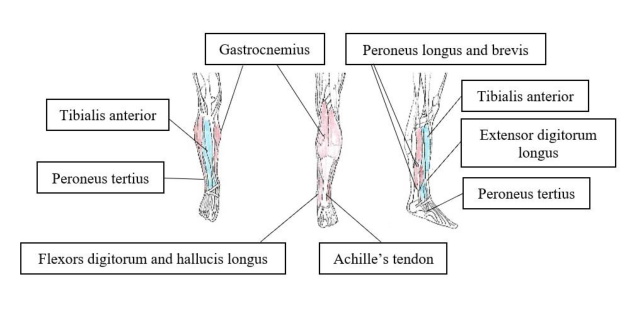
Most of the muscles within the calf (see Figure 3) contribute to plantar flexion at the ankle talocrural hinge joint (see Table 1). These include two of the super- ficial muscle group (gastrocnemius and soleus) and three of the deep muscle group (flexor hallucis longus (FHL), flexor digitorum longus (FDL), and tibialis posterior (TP)) (Moore, 1992). Because the FHL inserts on the distal phalanx of the great toe and the FDL inserts on the distal phalanges of the lesser toes, these muscles also provide plantar flexion of the toes. The gastrocnemius and soleus join and form the Achilles tendon at the insertion on the calcaneus (Rivest, 2008). Of the posterior calf muscles (see Table 1), only the TP contributes to inversion of the ankle due to its many insertions on the tarsal and metatarsal bones. These posterior calf muscles are all innervated by the tibial nerve. Surface EMG sensors only need to be positioned on the calf over the distal gastrocnemius and soleus muscles just proximal to the Achilles tendon to assess plantar flexion (Takashi, 2019).
Intelligent Human Systems Integration (IHSI 2022): Integrating People and Intelligent Systems https://openaccess.cms-conferences.org/#/publications/book/978-1-7923-8988-7
Figure 3. Anterior, posterior, and lateral views (right to left).
Ankle joint plantar flexors (red) and dorsiflexors (blue).
The peroneus longus (PL) and peroneus brevis (PB) muscles are within the lateral compartment of the lower leg and contribute to plantar flexion. The PL originates proximally at both the lateral tibial condyle and head of the fibula and descends laterally, then passes under the lateral malleolus of the ankle, and finally crosses under the foot to insert on the base of the first metatarsal bone. This al- lows it to generate ankle and foot eversion in addition to plantar flexion. The PB is shorter and smaller than the PL originating along the lower lateral fibula fol- lowing under the longer PL. The PB however inserts medially at the base of the fifth metatarsal bone contributing to plantar flexion and some degree of eversion. These muscles are innervated by the superficial peroneal nerve and surface EMG sensors positioned over these muscles within the distal lateral leg above the ankle could help assess eversion (Chung, 2000).
All the anterior compartment leg muscles (see Table 2) are innervated by the deep peroneal nerve, and all contribute to dorsiflexion. Surface EMG sensors po- sitioned just lateral to the ventral tibia (shin) can assess dorsiflexion (Zatsiorsky, 2002). The tibialis anterior muscle descends medially passing under the medial malleolus of the ankle to insert on the first metatarsal, thus enabling both inver- sion and dorsiflexion action. The extensor hallucis longus inserts on the dorsal side of the distal phalanx of the great toe and can both invert the foot and ankle and extend the great toe (Chung, 2000). The extensor digitorum longus inserts at the dorsal bases of the middle and distal phalanges of the lesser four toes contrib- uting to foot and ankle eversion in addition to extension of the lesser toes (Moore, 1992). The smaller peroneus tertius follows the PB to the base of the fifth meta- tarsal and minimally contributes to eversion. The two medial anterior leg muscles invert and the lateral two muscles evert the ankle suggesting that medial and lat- eral surface EMG sensors placed along the anterior lower leg could differentiate between inversion and eversion, respectively, in addition to dorsiflexion (Zatsiorsky, 2002).
Intelligent Human Systems Integration (IHSI 2022): Integrating People and Intelligent Systems https://openaccess.cms-conferences.org/#/publications/book/978-1-7923-8988-7
Table 2. Summary of Muscle Innervation for the Lower Extremity (Moore,
Table available in the original PDF paper.
1992)
Intelligent Human Systems Integration (IHSI 2022): Integrating People and Intelligent Systems https://openaccess.cms-conferences.org/#/publications/book/978-1-7923-8988-7
EMG Positioning It is not sufficient to place a single EMG sensor over the largest muscle contrib- uting to a particular DOF, if the goal of exoskeleton design is to be comfortable and completely in sync with the user’s intentions. Each motion, whether rotating the hip joint to change direction while walking, bending the knee while climbing stairs, or plantar flexing the ankle while jumping, requires a variable complex combination of muscle movements that is best defined when sensing the many muscles that contribute to the finer movements of the lower extremities (Zatsiorsky, 2002). Larger muscles will have more muscle fiber recruitment when contracting compared to smaller muscles; thus, there will be an associated stronger bioelectric signal near the belly of the muscle than near the thinner ten- don and ends of the muscle (Takashi, 2019). Since individuals may vary in size and shape, elderly or weak individuals will differ in body habitus from stronger more athletic or younger individuals. Likewise, different diseases like Myasthe- nia Gravis can have predictable but differing patterns as well (Posa, 2020). Con- sequently, positioning the sensors where the bioelectric signals are detectable with sufficient confidence is important to understand the associated movements of the lower extremities (Takashi, 2019).
CONCLUSIONS AND FUTURE WORK Exoskeleton or prosthetic limb replacement designs that do not take into consid- eration the actual musculoskeletal anatomy and kinematics can quickly fatigue the individual or patient secondary to high metabolic demand and discomfort (Ferris, 2017). Understanding the complex innervation, movement, and motion of the lower extremity joints and how they function can aid in the design of better and more efficient exoskeletons and prosthetic joints that meet the users’ expec- tations and reduce metabolic fatigue. The discussion of skeletal details, the mus- cular innervation, the range of motion and the muscles contributing to each DOF should help facilitate and improve placement and positioning of surface EMG sensors within the exoskeleton and prosthetics designs. The exoskeleton or pros- thetic designer should consider the numerous and complex combinations of mus- cle activity creating their EMG sensor-driven wearable robotic solutions.
A future study could examine different cohorts or groups that require an exo- skeleton or limb replacement. A sensor array embedded within an ankle sock of the lower extremity could obtain EMG sensor data while walking or performing different activities of daily living, thus generating training data for a machine learning (ML) algorithm (Takashi, 2019). The ML algorithm could then be used to discern which bioelectric signal patterns are associated for specific motions of the lower extremities for these different groups of individuals. The presence or lack of reflexes could be studied as well. Finally, the ML could then auto tune to the user for more precise and personalized identification of an individual’s voli- tional motion and intention. Using the EMG detected bioelectric signal patterns, the ML software could manage control of actuators that aid absent or weakened muscles when completing simple or complex movement. Prosthetic limbs or ex- oskeletons that use surface EMG arrays to sense key intentional movement would improve the comfort and usability for the user with the goal of having full and complete control of their extremities (Pazzaglia, 2016).
Intelligent Human Systems Integration (IHSI 2022): Integrating People and Intelligent Systems https://openaccess.cms-conferences.org/#/publications/book/978-1-7923-8988-7
Electroencephalogram (EEG) or brain computer interface (BCI) treadmill gait rehabilitation (BMI) controlled exoskeletons and prosthetics have shown feasi- bility (Garcia-Cossio, 2015). However, duplicating the speed and function of hu- man spinal reflexes is required to make these wearable robotic designs more use- ful outside the rehabilitation facility. Future work could incorporate surface EMG arrays with ML algorithms that learned to mimic movement and reflexes of the lower extremity within exoskeletons and prosthetic limb replacements. Surface EMG sensors are now sufficiently small enough to fit within clothing (Babusiak, 2018). Miniaturized versions of these sensors could even be implanted within significantly weakened muscles to improve the signal to noise ratio, detect more subtle intentional muscle activation, and improve exoskeleton or prosthesis con- trol performance. Providing the patient with direct control and proprioceptive feedback while maintaining comfort will be key to realizing the embodiment of the exoskeleton sought by developers and healthcare providers for their patients (Ferris, 2017).
REFERENCES
Asbeck, A.T., et al, (2015). “A biologically inspired soft exosuit for walking assistance,” The International Journal of Robotics Research, Vol. 34, No. 6, pp. 744-762. Asma, N., et al, (2020). “Low-cost Active Dry-Contact Surface EMG Sensor
for Bionic Arms,” IEEE International Conference on Systems, Man, and Cybernetics (SMC), October 11-14, Toronto, Canada, pp. 3327-3332. Babusiak, B., Borik, S., Balagova, L., (2018). “Textile Electrodes in Capacitive Signal Sensing Applications,” Journal of Measurement, Vol. 114, pp. 69- 77. Bruening, D.A., et al, (2020). “Whole body kinematic sex differences persist
across non-dimensional gait speeds,” PloS One, Vol. 15(8), pp. 1-16. Center for Disease Control, (2022). “Normal Joint Range of Motion Study,” https://www.cdc.gov/ncbddd/jointrom Christof, R., et al, (2010). “Torsional profile versus gait analysis: Consistency
between the anatomic torsion and the resulting gait pattern in patients with rotational malalignment of the lower extremity,” Gait and Posture, Vol. 32, pp. 405-410. Chung, K.W., (2000). Gross anatomy: Board review series. Philadelphia: Lippincott Williams & Wilkins. Eckhoff, D.G., et al, (2005). “Three-Dimensional Mechanics, Kinematics, and
Morphology of the Knee viewed in virtual reality,” Journal of Bone and Joint Surgery, Vol. 87, pp. 71-80. Ekso exoskeletons, (2022). https://www.eksobionics.com Ferris, D., Schlink, B., (2017). “Robotic Devices to Enhance Human Movement Performance,” Kinesiology Review, Vol. 6, pp. 70 -77. Garcia-Cossio, E. et al, (2015). “Decoding Sensorimotor Rhythms during Ro-
botic-Assisted Treadmill Walking for Brain Computer Interface (BCI) Ap- plications,” PloS One, Vol. 10(12), pp. 1-21. Hemmerich, A., et al, (2006). “Hip, Knee, and Ankle Kinematics of High Range of Motion Activities of Daily Living,” Journal of Orthopedic Re- search, Vol. 24, pp. 770–781.
Intelligent Human Systems Integration (IHSI 2022): Integrating People and Intelligent Systems https://openaccess.cms-conferences.org/#/publications/book/978-1-7923-8988-7
Kashitaro, H., et al, (2017). “Hip, knee, and ankle kinematics during activities of daily living: a cross-sectional study,” Brazilian Journal of Physical Therapy, Vol. 21(3), pp. 159-166. Moore, K.L., (1992). Clinically Oriented Anatomy. Williams & Wilkins. Pazzaglia, P., Molinari, M., (2016). “The Embodiment of Assistive Devices –
from wheelchair to exoskeleton,” Physics of Life Reviews, Vol. 16, pp. 163-175. Pinto-Fernandez, David et al, (2020). “Performance Evaluation of Lower Limb Exoskeletons: A Systematic Review,” IEEE Transactions on Neural Sys- tems and Rehabilitation Engineering, Vol. 28, No. 7, pp. 1573-1583. Posa A, Temneanu A, Emmer A, Langer T, Kornhuber M., (2020). “Jitter pat-
terns in conventional concentric needle electromyography recordings of regenerating motor units,” Muscle & Nerve, Vol. 62, pp. 593–596. Rivest, L.P., et al, (2008). “A Directional Model for the estimation of the rotation Axes of the Ankle,” Journal of the American Statistical Association, Vol. 103, No. 483, pp. 1060-1069. Soucie, J.M., et al, (2011). “Range of Motion measurements: Reference Values
and a database for comparison studies,” Haemophilia, Vol. 17(3), pp. 500- 507. Takashi, I. et al, (2019). “Sock-Type Wearable Sensor for Estimating Lower Leg Muscle Activity Using Distal EMG Signals,” Sensors, Vol. 19, pp. 1- 18. Veneman, J.F., et al, (2006). “A series elastic- and Bowden-cable-based actua-
tion system for use as torque actuator in exoskeleton-type robots,” The In- ternational Journal of Robotics Research, Vol. 25, No. 3, pp. 261-281. Zaffagnini, S., et al, (2016). “Soft tissues contribution to hip joint kinematics and biomechanics,” Hip Int, Vol. 26 (Suppl 1), pp. S23-S27. Zatsiorsky, V.M., (2002). Kinematics of human motion. Human Kinematics.
Champaign, IL.
Intelligent Human Systems Integration (IHSI 2022): Integrating People and Intelligent Systems https://openaccess.cms-conferences.org/#/publications/book/978-1-7923-8988-7
LISTED IN THE AHFE DIGITAL BOOKSTORE CATALOGUE
Explore the complete digital volume
Intelligent Human Systems Integration (IHSI 2022): Integrating People and Intelligent Systems
Read this individual research paper free through the original publisher, or explore the full digital proceedings book.
ISBN 978-1-7923-8988-7
Explore this ISBN in the AHFE Bookstore ↗Listed in the July 2026 bookstore catalogue; search by ISBN to confirm current availability · Digital book purchase optional