Sean Gallagher, Richard F. Sesek, Jerry Davis (2018). Effects of Force and Repetition on Inflammation due to Eccentric Muscle Contractions. In: Advances in Physical Ergonomics and Human Factors: Part II. AHFE Open Access, vol. 2. https://doi.org/10.54941/ahfe100070
Recent evidence strongly suggests that force and repetition interact in a consistent manner to affect musculoskeletal disorder (MSD) risk, likely due to an underlying fatigue failure process in affected tissues. This pilot study evaluated whether a force-repetition interaction was present with respect to inflammatory responses of elbow flexor muscles after eccentric exercise. 24 subjects performed eccentric contractions with 6 assigned to each of the following groups: low-force, low-repetition (LFLR), low-force, high-repetition (LFHR), high-force, low-repetition (HFLR) or high-force, high-repetition (HFHR). High- and low-force eccentric contractions were set 120% and 60% of maximum isometric capacity, respectively. Low and high repetition rates were defined as two and eight repetitions/set. A baseline magnetic resonance image (MRI) was taken of their non-dominant arm prior to the eccentric exercise, as were relaxed elbow angle and maximum isometric strength. Dependent measures included MRI data on edema in the muscle (day 2 versus day 0), and relaxed elbow flexion angle and isometric strength (obtained days 0, 1, 2, 4, and 8 post-exercise). Significant force-repetition interactions were found for relaxed elbow angle at days 2 and 4 post-exercise (p < 0.05), and for days 2 and 4 for isometric strength (p < 0.05). MRI data demonstrated a non-significant force-repetition interaction (p > 0.05), but a tendency towards the expected interaction pattern.
Musculoskeletal disorders (MSDs) represent one of the leading causes of long-term pain and physical disability world-wide (Bjorkland, 1998; Woolf and Pfleger, 2003). In 2011, MSDs accounted for 33% of lost work time workplace injuries and illnesses in the United States (U.S.). with a median of 11 days absence from work (Bureau of Labor Statistics, 2012). Work-related MSDs in the U.S. are associated with 130 million health care encounters and are estimated to cost over $50 billion annually (NIOSH, 2013). In 2011, the number of occupational injuries involving days away from work due to hand and wrist injuries were 140,460 and 47,550, respectively, with incidence rates of 13.9 and 4.7 per 10,000 workers, respectively (Bureau of Labor Statistics, 2012).
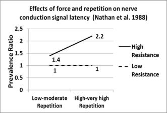
It has long been recognized that forceful exertions, repetitive motion, and non-neutral body postures are important MSD risk factors (Punnett and Wegman, 2004). However, a recent systematic review of occupational-related MSD epidemiology demonstrated a consistent pattern of interaction between force and repetition. This was observed across a number of disorders, including carpal tunnel syndrome, tendinitis, epicondylitis, hand pain and low back disorders (Gallagher and Heberger, 2013). As illustrated in figure 1, low-force tasks generally demonstrate a small or modest increase in MSD risk with increased repetition, while high-force tasks consistently exhibit a large escalation in MSD risk with increased repetition. The authors of this review article provided a theoretical basis for the interaction between force and repetition, suggesting that this interaction pattern would be anticipated if musculoskeletal tissues incur damage as the result of fatigue failure during the performance of occupationally- related tasks. This systematic review was recently awarded the 2013 international ergonomics association/liberty mutual medal for occupational safety and ergonomics.
Additional support for the fatigue failure hypothesis of MSD causation can be found in data from a rat model where Sprague-Dawley rats were exposed to one of the following conditions: low-force, low-repetition (LFLR), low-force, high-repetition (LFHR), high-force, low-repetition (HFLR) or high-force, high-repetition (HFHR) exertions (Barbe et al., 2013). After exposure to these regimens for a twelve-week period, tissue pathology and serum cytokine data were obtained from the rats. Tissue pathology results for tendon damage, cartilage damage and bone volume all demonstrated significant force-repetition interactions of the pattern consistent with an underlying fatigue failure process. Furthermore, many serum cytokines exhibited the same pattern of force-repetition interaction, including TNF-alpha, Interleukin Ia, Interleukin Ib, and Macrophage Inflammatory Protein 2. Inflammation and histopathology are often congruent and the magnitude of an inflammatory response to tissue insult or overuse appears indicative of the extent of injury in the tissues (Carp et al. 2007), thus these cytokine results also support the presence of an underlying fatigue failure process in the development of tissue damage and MSDs.
It was of interest to these authors to examine whether a fatigue failure process might be evaluated in human tissues in vivo. One available method is to examine the inflammation and muscle damage associated with eccentric muscle contractions. Eccentric exercise is known to lead to sarcomere “popping” (i.e., muscle fiber damage), which is repaired over the course of 1-2 weeks. Subjects who experience such eccentric bouts of exercise will experience a short-term inflammatory response (peaking 2-4 days post-exercise) that is healed over the period of 7-14 days, and will gain a training benefit that lasts up to 6 months. If a similar bout of exercise is experienced after healing has
Figure 1. Epidemiology studies demonstrate a force-repetition interaction for MSD risk (Gallagher and Heberger, 2013)
occurred, the inflammatory response will be greatly reduced. Thus, the purpose of the current study was to examine whether inflammatory responses to various levels of force and repetition during eccentric exercise exhibit a force- repetition interaction response in terms of inflammatory responses.
METHOD
Subjects Twenty-four subjects (18 males and 6 females) were recruited to participate in this study. The average age of the male subjects was 25.5 (± 3.2) years and their height and weight were 174.0 (± 6.1) cm and 83.0 (±14.5) kg, respectively. Female subject average age was 23.7 (± 2.8), height was 162.2 (± 11.1) cm, and weight was 60.0 (± 11.8) kg. Subjects were randomly assigned to one of four force-repetition categories. 6 subjects were assigned to each of the following groups: low force, low repetition (LFLR). low force, high repetition (LFHR), high force, low repetition (HFLR) or high force, high repetition (HFHR). Subjects were remunerated for their participation, and all procedures were reviewed and approved by the Auburn University Institutional Review Board (IRB).
Experimental Design Subjects were assigned to one of the four force-repetition conditions (6 subjects per condition) and had a baseline MRI taken prior to the performance of the required exercise on day 0. Other baseline measures taken included relaxed elbow angle of the subject non-dominant arm, isometric strength with the non-dominant arm at a 90-degree angle, and measurement of pressure sensitivity at four positions on the biceps muscle. MRI data acquisition procedures are detailed below. A follow-up MRI was obtained on day 2 post-exercise. In addition, measures of relaxed elbow angle, isometric strength, and pressure sensitivity were obtained during follow-up visits on days 1, 2, 4, and 8 post-exercise. It was hypothesized that these measures would demonstrate a specific pattern of interaction between force and repetition.
Eccentric Exercise The subjects performed three tests of isometric maximum isometric elbow flexion strength at 90 degrees of elbow flexion, each trial separated by 2 minutes. The peak isometric strength from these trials was used to set force levels for the eccentric trials. The subjects then performed the eccentric contraction regimen, which consisted of eight sets of contractions at different force and repetition levels using their non-dominant arm. High force eccentric contractions were set at 120% of the maximum isometric capacity, while low force contractions were set at 60% of
Figure 2. Subject performing eccentric contractions on Biodex dynamometer.
Figure available in the original PDF paper.
the maximum isometric capacity. Low repetition tasks were defined as two repetitions per set while high repetition tasks were eight repetitions per set. Each subject performed 8 sets of the prescribed exercise (each set lasted approximately 1 minute). Subjects were provided feedback on the amount of force they were producing on a video screen and were instructed to closely match the required torque for the condition. Sets were separated by a rest period of 30 seconds. The duration of each eccentric contraction was 2 seconds and the dynamometer arm (see Figure 2) moved at a speed of 60 degrees per second.
MRI Data Acquisition All magnetic resonance imaging was performed on a 3 Tesla clinical scanner (Siemens AG, Erlangen, Germany) with a flexible RF coil wrapped loosely around the subject’s arm and centered on the elbow joint. A phantom filled with 0.05 molar copper sulfate solution was placed under the coil and next to the subject’s arm to become a reference contrast region in each acquired image. For each MRI session, multi-slice, two-dimensional structural images were acquired using a T2-weighted, turbo spin echo sequence with parameters: repetition time (TR)=4700 ms, echo time (TE)=65 ms, echo train length=10, readout bandwidth (BW)=205 Hz/pixel, number of averages=5, in-plane matrix=192×192, 32 contiguous 3.0 mm thick slices, for voxel size=0.65×0.65×3.0 mm3, and overall volume of view=125x125x96 mm3.
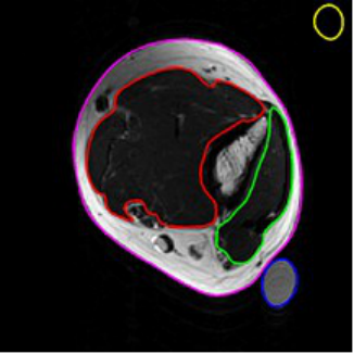
On Day 0, subjects had a baseline MRI on their non-dominant arm and a follow-up MRI on day 2. The T2-weighted images (useful for examining for evidence of edema) were evaluated for changes in signal due to edema related to the exercise. As can be seen in Figure 2, various regions were segmented for a contrast-noise ratio (CNR) analysis. These included the elbow flexor group in red (muscles affected by the eccentric exercise), the elbow extensor group in green (unaffected by exercise), the entire arm circumference (in pink), as well as the copper sulfate solution (blue) and a background region (yellow). The CNR ratio was calculated by comparing the CNR for the elbow flexors to the elbow extensors at Day 2 (post-exercise) versus the same comparison on Day 0 (pre-exercise).
Figure 2. MRI slices from HFHR subject. Left panel is baseline scan and the right panel is two days post-exercise. Note the increased signal in the elbow flexors in the right panel (indicative of edema, CNR=9.9).
Isometric Strength In addition to the baseline data, isometric strength assessments were performed on days 1, 2, 4, and 8 post-exercise. Isometric strength is considered one of the best measures of the degree of damage and subsequent recovery of the exposed muscle groups in the period after eccentric exercise (Prasartwuth et al., 2005). Three tests of isometric strength of the affected (non-dominant) arm were obtained on each of the follow-up days, with each trial being separated by 2 minutes as with the baseline isometric data collection. The percentage decrease in isometric strength for each follow-up day was calculated with respect to the baseline value.
Data Analysis MRI data were analyzed using a custom MATLAB program, which permitted Contrast to Noise Ratio (CNR) analysis of muscles affected by the eccentric exercise (elbow flexors) versus those not affected by the exercise (elbow extensors). Edema in the inflamed muscle presented as increased signal in the MRI image (lighter color), and differences in the CNR was calculated at baseline and at day 2 in the same subject, with the former being subtracted from the latter to evaluate the effect of the exercise on the inflammatory response. Subjects were requested to avoid taking non-steroidal anti-inflammatory drugs (NSAIDs) for at least the first two days to avoid the inflammation reducing characteristics of these drugs, which could moderate the edema response. Subjects reported universal compliance with this request.
RESULTS
MRI CNR results
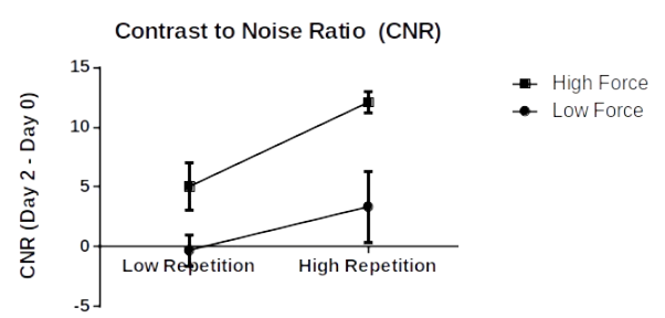
Figure 2 illustrates the results of the CNR comparisons from day 2 post-exercise versus baseline for the four force/repetition conditions. Results demonstrate the anticipated pattern of interaction between force and repetition; however, the interaction did not achieve statistical significance (p > 0.05). Force was found to be a statistically significant main effect (p < 0.05).
Figure 2. MRI Contrast to Noise Ratio results.
Relaxed Elbow Angle
Figure 3 provides the data pertaining to resting elbow angle responses for the various force-repetition groups. Significant force-repetition interactions were observed on Days 1 and 2 (p < 0.05).
Day 1 Day 2
Day 4
Figure 3. Resting elbow angle results for Days 1, 2, and 4 post-exercise.
Figure available in the original PDF paper.
Isometric Strength
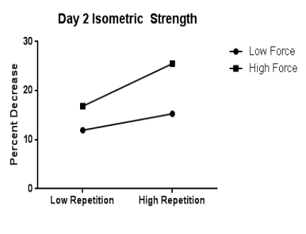
Figure 4 presents the results of isometric strength testing during the follow up days post-exercise. A significant force-repetition interaction was observed on day 4 post-exercise (p < 0.05).
Figure 4. Percentage isometric strength decrease on Days 2 and 4 post-exercise.
DISCUSSION
As described in the introduction, data from epidemiological studies and animal studies have demonstrated a consistent pattern of interaction between force and repetition and MSD risk, tissue pathology, and cytokine responses. As detailed previously (Gallagher and Heberger, 2013; Barbe et al. 2013), these responses would be anticipated if an underlying fatigue failure process was an etiological cause of MSDs. The current pilot study was performed to investigate whether force-repetition interactions were evident with respect to various measures related to muscle damage and inflammation after exposing subjects to various force and repetition levels in eccentric exercise. This was considered a method of investigating whether a fatigue failure process could be demonstrated in vivo.
All of the dependent measures described in this paper demonstrated a pattern of response suggestive of an interaction between force and repetition. Some of these achieved statistical significance while others did not. The fact that some measures did not achieve statistical significance may have been due in part to a lack of statistical power. The “between subjects” nature of the experimental design is not ideal in terms of statistical power due to the considerable between-subjects variability. However, in this case a within-subjects design is not feasible since exposure to eccentric exercise provides a training effect that makes within-subjects comparisons unreliable. This represents a limitation to the current pilot study that can be addressed with larger sample sizes in future studies.
Significant force-repetition interactions were observed for relaxed elbow angle (days 2 and 4 post exercise) and isometric strength decrease (day 2). The interaction between force and repetition and relaxed elbow angle is thought to be due to an influx of calcium from the damaged sarcoplasmic reticulum, and appears to be indicative of the amount of damage sustained by the affected muscle due to eccentric contractions. Isometric strength is considered one of the best measures of the state of muscle damage and recovery, and also showed a force-repetition interaction on day 2 post-exercise. Both of these findings illustrated the pattern of force-repetition interaction expected should an underlying fatigue failure pattern be present.
The CNR analysis from the MRI images showed the general pattern anticipated, but significant between subjects variability was present and did not result in a significant force-repetition interaction from being detected. Several issues may have interfered with the MRI analyses. For one thing, some subjects had difficulty remaining still throughout the time required for MRI data acquisition, resulting in movement artifacts. This could be addressed in the future by reducing the time of the MRI data collection by reducing the number of slices obtained.
CONCLUSIONS
Based on the findings of this study, the following conclusions are drawn:
1. Changes in relaxed elbow angle and isometric strength after eccentric exercise exhibited force-repetition interactions congruent with what would be expected if muscle damage was due to fatigue failure of the affected tissues.
2. Results of MRI analysis on edema in the muscles after eccentric exercise showed the general pattern of force-repetition interaction anticipated from fatigue failure theory, but did not achieve statistical significance.
3. The results of this study provide support for the theory that MSDs are the result of a fatigue failure process. The pattern of response for all dependent measures was similar to those observed for MSD risk, tissue pathology results, and inflammatory cytokine responses; however, not all measures demonstrated statistical significance.
4. Use of eccentric exercise as a method of examining fatigue failure of muscle tissues in vivo appears fruitful as a research technique and further research may incorporate measures such as creatine phosphokinase and inflammatory cytokines.
5. Future studies using this technique may need to incorporate a larger number of subjects due to the significant between-subjects variability observed with certain dependent measures.
ACKNOWLEDGEMENTS
This research was made possible by a grant from the National Institute for Occupational Safety and Health (NIOSH) Deep South Occupational Safety and Health Education and Research Center [Grant UAB-000500836-001]. The authors would like to acknowledge Michael Porter, Patrick Almas, Yousif Abulhassan, and Mradul Sangal for their assistance in the conduct of this research.
REFERENCES
Barbe, M. F., Gallagher, S., Massicotte, V. S., Tytell, M., Popoff, S. N., Barr-Gillespie, A. E. (2013), The interaction of force and repetition on musculoskeletal and neural tissue responses and sensorimotor behavior in a rat model of work-related musculoskeletal disorders. BMC Musculoskeletal Disorders, 14, 26 pages. Bjorklund, L. (1998), The Bone and Joint Decade 2000-2010. Inaugural meeting 17 and 18 April 1998, Lund, Sweden. Acta
Orthopaedica Scandinavica Supplementum, 281, 67-80. Bureau of Labor Statistics (2012), Nonfatal occupational injuries and illnesses requiring days away from work, 2011.
http://www.bls.gov/news.release/osh2.nr0.htm News Release USDL-12-2204, November 8, 2012. Carp, S.J., Barbe, M.F., Winter, K.A., Amin, M., Barr, A.E. (2007), Inflammatory biomarkers increase with severity of upperextremity overuse disorders. Clinical Science, 112, 305-314. Gallagher, S., Heberger, J.R. (2013), Examining the interaction of force and repetition on musculoskeletal disorder risk: a
systematic literature review. Human Factors, 55, 108-124. NIOSH (2013), Use of workers’ compensation data for occupational safety and health: proceedings from June 2012 workshop. .
Utterback DF, Schnorr TM, eds Cincinnati, OH: US Department of Health and Human Services, Public Health Service, Centers for Disease Control and Prevention, National Institute for Occupational Safety and Health, DHHS (NIOSH) Publication No 2013-147, pp. 1-232.
Punnett, L., Wegman, D.H. (2004), Work-related musculoskeletal disorders: the epidemiologic evidence and the debate. Journal of Electromyography and Kinesiology, 14, 13-23. Woolf, A.D., Pfleger, B. (2003), Burden of major musculoskeletal conditions. Bulletin of the World Health Organization, 81,
646-656.
LISTED IN THE AHFE DIGITAL BOOKSTORE CATALOGUE
Explore the complete digital volume
Advances in Physical Ergonomics and Human Factors: Part II
Read this individual research paper free through the original publisher, or explore the full digital proceedings book.
ISBN 978-1-4951-2105-0
Explore this ISBN in the AHFE Bookstore ↗Listed in the July 2026 bookstore catalogue; search by ISBN to confirm current availability · Digital book purchase optional