Lyn Phillips (2021). Pressure Ulcer Prevention: Keep it Safe, Keep it Simple!. In: Advances in Human Aspects of Healthcare. AHFE Open Access, vol. 13. https://doi.org/10.54941/ahfe100475
Pressure Ulcer (PU) Prevention Guidelines typically recommend that vulnerable patients be physically repositioned every 2-4 hours, although the risk of caregiver injury is rarely discussed. Some guidelines, concerned with the fabric and construction of slings, continue to mandate the removal of lift equipment from beneath the patient after use and, despite a weak evidence base, this might lessen repositioning frequency and discourage safe practice. A pragmatic solution may be a flexible, breathable, quick-drying, low-friction lift sheet, designed to work in synergy with a pressure-redistributing mattress and which replaces the standard sheet. A series of standardised laboratory tests compared key performance characteristics of two sheet textiles: a 100% cotton hospital bed sheet and the Maxi Transfer™ sheet, a novel synthetic lift sheet. Results showed that when compared to the cotton sheet, the synthetic sheet was more breathable, had lower heat retention properties, superior wicking and better synergy with the therapeutic mattress. Regular repositioning, the cornerstone of PU prevention, is most likely to occur when clinicians have immediate access to lifting equipment. Replacing the standard bed sheet with an advanced textile, lifting device, may positively impact concordance with repositioning protocols, improve tissue microclimate and so improve patient outcomes and, importantly, caregiver safety.
Pressure ulcers and caregiver back injury – two very challenging, costly and common injuries, which are totally interrelated and yet rarely discussed in the same context. Pressure ulcers are defined as tissue lesions that occur over a bony prominence as a result of exposure to prolonged pressure with or without shear [NPUAP and EPUAP, 2009], an inevitable consequence of unmanaged immobility. The only certain way to prevent pressure ulcers is to move the patient (or the pressure beneath the patient) and this requires some form of physical intervention on behalf of the caregiver. Routine preventative care requires regular patient repositioning, the handling of a significant physical ‘load’ that is typically repeated at two to four-hourly intervals day and night. As contemporary healthcare favours a very short stay in acute care facilities, followed by early discharge into the community, the dependency level of in- patients tends to be high, with most patients requiring at least some level of assisted mobility. Recent trends in patient demographics, resulting in older, sicker and heavier patients, means that nurses and therapists, more so than ever before, have to physically manage greater loads, with greater frequency and probably with fewer staff. This paper will focus only on the interventions required for pressure ulcer prevention, for which physical repositioning is a fundamental requirement.
PRESSURE ULCER PATHOLOGY AND PREVALENCE Pathophysiology
In brief, the pathophysiology of pressure injury can be described as the interaction between, firstly, intrinsic and extrinsic factors, which affect the ability of muscle, fat and skin to withstand pressure (tissue tolerance) and, secondly, the direct impact of mechanical loading on the tissue. This mechanical loading is defined by the duration, magnitude and direction of ‘load’ (pressure) applied to the skin [NPUAP and EPUAP, 2009]. In reality, pressure is almost never truly perpendicular and, due to the nature of soft tissue, shear is generally present, even if at a cellular level. As shear causes distortion and narrowing of blood vessels plus, in extreme cases, damage to the cytoskeleton, it is generally considered a priority for management alongside pressure. The importance of microclimate management is also increasingly understood, in that heated skin has a raised metabolic demand at a time when compression is reducing blood flow (perfusion) [Brienza and Geyer, 2005], it also has a tendency to naturally sweat. Damp skin, whether from sweat or incontinence, is less able to withstand friction and ultimately pressure and shear. One additional aspect of prevention, which is critical but not always clearly defined in local and national policy, is time. Cellular studies indicate that tissue injury may occur in as little as 2-hours [Bansal, 2005] this may mean that the patient may require preventative care in the peri-admission phase of their hospitalisation. Protecting a patient, in those critical first few hours around admission, is a matter of determining their ability to reposition themselves sufficiently and frequently enough to avoid injury. If they can’t move adequately, then a physical repositioning regimen must be initiated without delay.
Prevalence Despite a much greater understanding of the pathophysiology of pressure ulcers, and the increasing availability of effective preventative interventions, pressure ulcer prevalence has proved largely resistant to change. Sadly, as many as one in every five hospital patients across Europe [Woodburg and Houghton, 2004; Vanderwee, 2007; Gallagher, 2008; Van Gilder, 2008; Buttery and Phillips, 2009; Moore and Cowman, 2012] will experience some form of injury, with most pressure ulcers occurring under clinical supervision [Posnett, 2009; Buttery and Phillips, 2009]. These nosocomial, or healthcare-acquired, pressure ulcers most commonly occur over the sacrococcygeal area and heel and are classified by severity into one of five main categories. Category 1 wounds manifest as sub-dermal microvascular damage beneath intact skin and Category 2, shallow and painful, yet superficial, ulcers where the skin is broken or blistered but the damage is limited to the epidermis. In more severe cases, damage may be extensive, with full thickness dermal loss (Category 3) or involve damage or destruction to the underlying structures such as muscle, tendon and bone (Category 4). At times the full extent of the injury may be obscured by slough, infective or necrotic tissue, in which case they are classified as unstageable. If damage is suspected to have occurred within the deeper tissue, but without obvious signs at the skin surface, these are classified as ‘deep tissue injury’. Superficial ulcers (Category 1 & 2) are thankfully the most common, as the long-term sequelae are less serious. However, pressure ulcers that develop into full-thickness tissue loss can result in long-term disability or, in the worst cases, lead to sepsis, organ failure and death.
Cost of pressure ulcers In addition to causing significant suffering to patients, pressure ulcers represent a substantial economic burden to healthcare providers with the estimated cost of treating the most severe wounds ranging from more than £14,000 in the UK [Dealey, 2012] to almost $130,000 in the USA [Brem, 2010]. In the past decade it has been estimated that the United Kingdom spends up to £2.1 billion, or 4% of the nation’s healthcare budget, on pressure ulcers; a significant amount is attributed to nursing time [Bennett, 2004]. This situation is not restricted to just one healthcare system, with other countries reporting a similar economic burden; the Netherlands [Severens, 2002] has a conservative estimate of 1%, Hungary up to 0.8% [Gulásci, 2001], while expenditure in the USA approaches $11.6 billion per annum [Zulkowski, 2005]. None of these figures include the lost ‘opportunity’ cost, which arises when acute care beds are occupied by patients necessarily retained in hospital for treatment [Graves, 2005], nor the cost of litigation; a risk which naturally increases as patients and care-givers come to expect that the majority of these wounds might have been preventable with simple, but timely, nursing interventions. To this point, a group of actuaries in the USA, analysed negligence claims for over 560,000 ‘medical errors’ [Shreve, 2010]. By comparing the treatment costs with a similar non-injured cohort, the authors established that the USA spends more than US$3.9 billion per year treating pressure ulcers: a condition that topped the league table for medical errors! These figures do not include the cost of litigation, liability insurance and compensation payouts but only the costs attributed to treatment. By highlighting that over 90% of the ulcers investigated may have been avoided with reasonable care, this study reaffirmed the long-held notion that most pressure ulcers can and should be prevented.
Pressure ulcer prevention The premise that pressure ulcers can and should be avoided in all but exception circumstances has been rarely challenged in the past. Indeed, the first international collaborative Pressure Ulcer Guideline [NPUAP and EPUAP, 2009] reinforces many existing local and national protocols by clearly setting out the basic requirements for the identification and management of vulnerable patients. Such guidelines are based upon the most rigorous review of international contemporary clinical research and expert opinion and are focussed on the fundamental principles of prevention. Essentially any patient who is confined to bed or chair should be considered ‘at risk’, as should any patient who may have physical or sensory impairment; the latter diminishing the natural protective stimulus to move spontaneously. Also at risk are patients who, because of illness or medication, have an altered mental status and may be unaware, unable or unmotivated to move. The most important test of ‘risk’ is whether or not the patient can, and will, make a purposeful and frequent change of position.
The critical role of repositioning Fortunately, immobility and altered sensation (sedated, unconscious, paralysed) are two of the easier risk factors to indentify and can be mitigated with relative speed: simply – move the patient (reposition) and/or manage the pressure beneath the patient (support surface). Ideally, both interventions should be adopted, with the frequency of repositioning governed by the patient’s overall condition, skin response and type of support surface used [NPUAP and EPUAP, 2009]. In the simplest terms, if pressure ulcers are caused by exposure to prolonged pressure +/- shear as a result of immobility, then a primary intervention must be to move the patient and to move them in a way that, as far as possible, completely off-loads the tissue and without causing shear to the tissues in the process. Techniques must also avoid causing friction to the epidermis, as this is easily damaged especially if the patient has moist, friable or already damaged skin. This essentially means a good repositioning technique and the use of appropriate aids to ensure that the caregiver does not incur musclo-skeletal disorder (MSD), while the patient is moved in a comfortable, secure and dignified manner. Pressure Ulcer (PU) Prevention Guidelines typically recommend that vulnerable patients be physically repositioned according to an ‘individualised’ plan but, in practice, this is typically ritualised at between eight and twelve times each day. The current international prevention guideline [EPUAP and NPUAP, 2009] dedicates around five pages to repositioning and, while including reference to both technique and position, it does so primarily from the perspective of tissue protection; these guidelines do not overtly provide recommendations for the caregivers other than a recommendation for training on safe techniques. A second edition of this guideline, due for publication mid-2014, has been circulated for stakeholder comment and currently shows eighteen pages dedicated to repositioning, while perhaps suggesting a much greater awareness, the emphasis remains on the patient and the technique, rather than on the caregiver.
Caregiver Risk Hospitals and long-term care facilities are responsible for high levels of worker injury [Dawson, 2007]. The single greatest risk factor for MSD injuries in healthcare workers is ergonomic injury, associated with the manual lifting, moving and repositioning of dependent individuals. An observational study [McCoskey, 2007] identified risk exposure across several areas, 60% of which were related to repositioning in bed, turning in bed, moving the patient to the head of the bed (‘boosting’), and transferring patients from bed to bed. Data sources in the USA show that healthcare workers (combining registered nurses with nurse assistants) are considered a high-risk group for MSD and incur the highest level of injury compared to any other manual labour group in 2012 [BLS 2012]. The task of physically repositioning patients typically falls to the nursing assistant or nursing aide and, in 2012, some 44,100 bedside caregivers needed a median of 6-days off work to recover from an injury related to overexertion, trips, slips and falls. The acuity of patients tends to necessitate more frequent intervention to prevent adverse events such as pressure ulcers and this increased need places an increased burden on staff. Caregivers who undertake more than ten physical tasks per day are most likely to report back pain [Thomas 2009], yet nurses may be required to repeat these tasks for several patients per shift. These acute injuries, when added to injuries caused by chronic or repetitive micro tasks, leads to an even greater financial and staffing problem for healthcare providers as chronic injuries require a longer recovery period, with an average of 23-days away from work. Other indirect factors have been reported to
increase the risk of injury, for example, increasing the magnitude of the load and reducing the number of caregivers available to share the burden, neither makes for comfortable reading. Global obesity rates have risen exponentially in the past 25-years [Stevens, 2012] and this, in itself, increases the physical demands on caregivers. While, the size of the workforce is also under pressure, with recruitment and retention of staff causing concern in many quarters [Buerhaus, 2009]. That said, the type of lifting technique, and whether or not the clinician elects to use a friction- reducing device, can be more important in terms of the load borne by the lower back than either the patient's weight or dependency [Skotte and Fallentin, 2008]. Unfortunately, while healthcare providers are generally very aware of the risks and do encourage the use of safe patient handling techniques, or ‘no lift’ policies, the injury statistics suggest that compliance may be less than ideal.
REPOSITIONING AIDS With manual transfers unequivocally shown to produce higher muscle loads on staff compared to the use of floor lifts and ceiling lifts [Keir and MacDonell, 2004] there is certainly a need to make lift equipment easy to use and readily accessible. From an economic and compassionate perspective, the time has come to consider whether new technology can assist. Unfortunately, if guidelines are developed without strategic collaboration between skin care experts and safe patient handling teams, recommendations can become disjointed even when the interrelationship is undisputed. As an example, guidelines published in 2009, mandate the removal of lift equipment after use [NPUAP and EPUAP, 2009]. Such a ‘blanket’ statement, in a guideline, can be enough to deter the utilisation of equipment even where it has been specifically designed to be left in situ and has been determined as fit for purpose. Where practitioners cannot readily access lift equipment this may lessen repositioning frequency and discourage safe practice: increasing risk of harm to patients and caregivers. It would appear that these directives are based upon a concern about lift sheet/sling construction, where these may be less flexible, less breathable and be constructed with seams and borders that may injure the tissue if left in contact for prolonged periods. However, these recommendations refer to earlier guidelines published by the National Institute for Health and Clinical Excellence in the UK [NICE, 2005] and have a weak evidence base [Mellson and Richardson 2012]. The term ‘lift equipment’ makes no allowance for the type or design of device, nor does it take into account the recent development of advanced textiles, which have been shown to have clear clinical worth [Mellson and Richardson 2012].
The benefit of new technology and design In order to reduce the risk of pressure injury, clinicians aim to reduce or remove pressure through repositioning and, at the same time prevent the build up of excessive temperature and moisture. To do so, they may use features of a specialised mattress and/or cover to redistribute pressure and manage the microclimate at the skin-mattress interface. The covering sheet needs to be flexible, breathable and moisture wicking to avoid interfering with the inherent properties of the mattress. The sheets also need to be soft and comfortable to lie on without any noticeable ridges, seams or folds that create a high pressure gradient on areas of the skin and predispose to pressure injury. This report describes a study undertaken to determine whether contemporary, synthetic textiles can be strong enough to use as a lifting sheet, but also have other essential properties that might enable them to be left in situ without detriment to the skin. If so, this would ensure that a lift device was always immediately available for use, encouraging safe practice. A series of laboratory tests compared a range of performance characteristics for two different textiles: a 100% cotton hospital bed sheet (reference standard) and the Maxi Transfer™ sheet - a synthetic lift sheet/replacement bed sheet made of 99% woven polyester with a 1% carbon core (ArjoHuntleigh).
Methodology
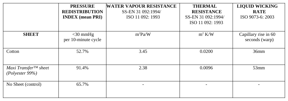
The sheets were draped loosely over an ‘active’ (alternating) pressure-redistributing (PR) mattress, which is designed to periodically off-load the tissue through the cyclic inflation and deflation of a series of transverse air cells [Phillips, 2012]. Using standardised methodology [Phillips, 2007] and an anatomically weighted test mannequin, the pressure redistribution index (PRI) of the mattress was measured to determine whether the sheets were flexible enough to avoid interference with the important off-loading properties of the mattress. This technique records the pressure at the interface between a single cell of the mattress and an anatomical point of the test mannequin; the pressures are traced as the cell inflates and deflates. Data is reported for the time over each 10-minute inflation and deflation cycle that the pressure is below a nominal threshold, in this case, 30 mmHg. Further standardised tests (Table 1) compared the ability of both textiles to potentially influence tissue microclimate at the body-mattress interface by determining the liquid wicking rate, water vapour transfer rate and thermal resistance.
Results
Table 1: Bed sheet performance characteristics WATER VAPOUR RESISTANCE
Discussion
Compared to the cotton sheet, the synthetic sheet appears more breathable, has lower heat retention properties, superior wicking and better synergy with the mattress. In addition, PRI data produced a surprising result, showing the synthetic sheet to be some 37% better than no sheet at all! This might be explained by a lower coefficient of friction allowing the sensor to move across the cells during inflation-deflation; a characteristic yet to be formally tested. The synthetic sheet has a number of other properties that are considered advantageous for tissue viability such as an ability to reduce ‘hammocking’ across the deflated mattress cells which might otherwise interfere with the performance of the underlying support surface [Iuchi, 2014]. Synthetic textiles, when used for bedding and clothing, have already been shown to reduce the development and duration of pressure ulcers in a number of ways [Smith and Ingram, 2010; Colandonato, 2012]. Reducing the build up of heat in the tissue lowers metabolism and reduces sweating, keeping the skin dry reduces maceration and a low-friction surface reduces the impact of tissue distortion. Other studies indicate that polyester slings used for the care of seated individuals may actually reduce, rather than increase, interface pressure, as previously feared [Mellson and Richardson, 2012]. The design of a lift sheet, which is soft enough to be comfortable, yet flexible enough to work sympathetically with a therapeutic mattresses, is likely to be clinically advantageous compared to the most commonly encountered cotton bed sheet.
CONCLUSION AND CLINICAL RELEVANCE Regular repositioning, the cornerstone of PU prevention, is most likely to occur when clinicians have immediate access to lifting equipment. A pragmatic, and refreshingly simple solution, to address the lack of access to lift equipment, would be sheets that are designed to work in synergy with therapeutic support surfaces, to provide an environment conducive to tissue viability when retained in situ and also be a useful tool for repositioning the patient. Replacing the standard bed sheet with a lifting device, may positively impact concordance with repositioning protocols and so improve patient outcomes and, importantly, caregiver safety.
Fortunately, the development of contemporary textiles has eradicated many of the drawbacks associated with traditional repositioning equipment and so, today, lifting sheets may be similar in feel, appearance and quality to the standard cotton or polyester-cotton hospital bed sheet. Synthetic textiles, such as that used to in the low-friction Maxi Transfer™ sheet, may drape in the same way as cotton, can be seam free, quick to dry and have equitable or superior moisture-vapour transfer rates, as well as being pleasant to lie upon. These characteristics call for a reconsideration of pressure ulcer prevention guidelines so that lift equipment, provided it has been shown to have suitable characteristics, may be left in situ.
Preventing pressure ulcers and caregiver MSD represents a significant challenge to healthcare providers; both conditions are common, costly and can prove difficult to control. It is important to recognise that the very act that protects the patient also places the caregiver at greatest risk and so the solution must bring together expertise and collaboration from the fields of both the tissue viability and safe patient handling.
REFERENCES
Bansal C, Scott R, Stewart D et al (2005), “Decubitus ulcers: A review of the literature”. Int J Derm. Volume 44 No.10 pp. 805-810 Bennett G, Dealey C, & Posnett J (2004), “The cost of pressure ulcers in the UK. Age and Aging.” Volume 3 No. 33 pp. 230–235. Brem H, Maggi J, Nierman D et al (2010), “High cost of stage IV pressure ulcers”. Am J Surg. Volume 200 No. 4 pp. 473-477 Brienza DM, & Geyer MJ (2005) “Using support surfaces to manage tissue integrity”. Adv Skin Wound Care. Volume 18 No.3 pp.151-157 Buerhaus, PI. Auerbach, DI. & Staiger, DO. (2009) “The Recent Surge In Nurse Employment: Causes and Implications”, Health Affairs. Volume 28 No. 4 pp. w657-w668. Bureau of Labor Statistics, U.S. Department of Labor (2013), “Non-fatal occupational injuries and illnesses requiring days away from work - 2012”. USDL-13-2257. http://www.bls.gov/news.release/pdf/osh2.pdf Buttery J., & Phillips L. (2009), “Pressure ulcer audit highlights important gaps in the delivery of preventative care in England and Wales 2005-
2008” EWMA Journal. Volume 9 No. 3 pp. 27-31 Coladonato J, Smith A, Watson N et al (2012). “Prospective, nonrandomized controlled trials to compare the effects of a silk-like fabric to standard hospital linens on the rate of hospital acquired pressure ulcers”. Ostomy Wound Management. Volume 58 No. 10 pp. 14-31 Dawson, A. P., McLennan, S. N., Schiller, S. D., Jull, G. A., Hodges, P. W., & Stewart, S. (2007). “Interventions to prevent back pain and back injury in nurses: a systematic review”. Occupational and Environmental Medicine Volume 64 No. 10 pp. 642-650. Dealey C, Posnett J, &Walker A. (2012) “The cost of pressure ulcers in the United Kingdom” J Wound Care Volume 21 No. 6 pp 264-266 Gallagher P, Barry P, Hartigan I et al (2008) “Prevalence of pressure ulcers in three university teaching hospitals in Ireland”. J TissueViability.
Volume 17 No. 4 pp. 103–109. Graves N, Birrell F, Whitby M et al. (2005). “Effect of Pressure Ulcers on Length of Hospital Stay”. Infection Control and Hospital Epidemiology. Volume 26 No. 3 pp. 293-297 Gulácsi L. (2001).“Hungarian health care in transition; Studies on the improvement of the effectiveness of health care in Hungary by implementing quality assurance”. PhD Thesis. University of Amsterdam (ISBN 90-71433-64-1) Iuchi T, Nakajima Y, Fukuda M et al (2014).“Using an extreme bony prominence anatomical model to examine the influence of bed sheet
materials and bed making methods on the distribution of pressure on the support surface”. J Tissue Viability (in press) http://dx.doi.org/10.1016/j.jtv.2014.01.003 Keir, P. J., & MacDonell, C. W. (2004).“Muscle activity during patient transfers: a preliminary study on the influence of lift assists and experience”. Ergonomics, Volume 47 No 3 pp. 296-306. McCoskey, K. L. (2007). “Ergonomics and patient handling”. AAOHN Journal Volume 55 No 11 pp. 454-462. Mellson J. & Richardson N. (2012). “The impact of hoist sling fabrics on gluteal interface pressures while sitting in healthy individuals: a
controlled pre-post test study” Am J SPHM Volume 3 No. 3 pp.79086 Moore Z. & Cowman S. (2012) “Pressure ulcer prevalence and prevention practices in care of the older person in the Republic of Ireland”. Journ Clin Nurs Volume 21 No. 3-4 pp. 362-371 National Institute for Health and Clinical Excellence (2005). “Clinical Guideline CG29: Pressure Ulcer Management”.
www.nice.org.uk/guidance/CG29 National Pressure Ulcer Advisory Panel and European Pressure Ulcer Advisory Panel. (2009), “Prevention and treatment of pressure ulcers: clinical practice guideline”. Washington DC. Phillips L. (2007) “Interface pressure measurement: appropriate interpretation of this simple laboratory technique used in the design and assessment of pressure ulcer management devices. Primary Intention. Volume 15 No 3 pp106-113 Phillips L, Goossens R, Takahashi M. & Clark M, (2012) Defining active pressure redistribution. Wounds International. Volume 3 No 3 pp.52-56 Posnett, J., Gottrup, F., Lundgren, H., & Saal, G. (2009) “The resource impact of wounds on health-care providers in Europe”. J Wound Care.
Volume18 No 4 pp. 154–161 Severens JL, Habraken JM, Duivenvoorden S, & Frederiks CM. (2002). “The cost of illness of pressure ulcers in The Netherlands”. Adv Skin Wound Care. Volume 15 No 2 pp. 72–77. Skotte, J., & Fallentin, N. (2008). “Low back injury risk during repositioning of patients in bed: the influence of handling technique, patient weight and disability”. Ergonomics Volume 51 No. 7 pp. 1042-1052. Shreve J, Van Den Bos J, Gray T, Halford M, Rustagi K, & Ziemkiewicz E (2010) The Economic Measurement of Medical Errors. Society of
Actuaries. www.healthlawyers.org Smith G, & Ingram A. (2010) “Clinical and cost effectiveness evaluation of low friction and shear garments”. Journal Wound Care. Volume 19 No. 12 pp. 535-542 Stevens GA, Singh GM, Lu Y et al. (2012). “National, regional, and global trends in adult overweight and obesity prevalences”. Population Health Metrics. http://www.pophealthmetrics.com/content/10/1/22 Thomas, D. R., Thomas, Y-L., Borner, H., Etherington, M., McMahon, A., Polaczuk, C., & Wallaart, J. (2009). Patient Handling Guidelines:
Literature Review. Wellington: ACC. Vanderwee K, Clark M. & Dealey C et al. (2007) “Pressure ulcer prevalence in Europe: a pilot study”. J Evaluation in Clinical Practice. Volume 13 No. 2 pp. 227–235. Van Gilder C, Macfarlane GD. & Meyer S. (2008) “Results of nine international pressure ulcer prevalence surveys: 1989–2005” Ostomy & Wound Management. Volume 54 No. 2 pp. 40–54 Woodburg MG. & Houghton PE. (2004). “Prevalence of pressure ulcers in Canadian healthcare settings”. Ostomy & Wound Management.
Volume 50 No. 10 pp. 22–38. Zulkowski K, Langemo D, & Posthauer ME. (2005). “Coming to consensus on deep tissue injury”. Advances in Skin & Wound Care. Volume 18 No. 1 pp. 28–29.
LISTED IN THE AHFE DIGITAL BOOKSTORE CATALOGUE
Explore the complete digital volume
Advances in Human Aspects of Healthcare
Read this individual research paper free through the original publisher, or explore the full digital proceedings book.
ISBN 978-1-4951-2093-0
Explore this ISBN in the AHFE Bookstore ↗Listed in the July 2026 bookstore catalogue; search by ISBN to confirm current availability · Digital book purchase optional