Sarah De_Cristofaro, Salvatore Parrulli, Bernardo Parrulli, Luca Rizzi (2026). Leveraging AI Tools for Emotion-Safe Dental Imaging: Enhancing Patient Communication While Preserving Anatomical Accuracy in Surgical Visualizations. In: Healthcare and Medical Devices. AHFE Open Access, vol. 211, pp. 282–290. https://doi.org/10.54941/ahfe1007491
This paper presents a preliminary study developed within the SMART DENT project, in collaboration with Studio Parrulli, investigating how AI-supported visual redesign strategies can improve dental diagnostic communication while preserving clinically relevant anatomical information. The study examines patients’ emotional and cognitive responses to different types of dental diagnostic images, with a focus on visual realism and representation style. An initial exploratory phase assessed reactions to 39 real clinical images using AI-based facial expression analysis, showing that highly realistic images, particularly intraoral scans depicting damaged dentition or implant-related conditions, tend to elicit discomfort. Due to methodological limitations in micro-emotion detection, a questionnaire-based evaluation was adopted as the primary assessment tool. The questionnaire, completed by 35 adult patients, indicates that simplified images are preferred for emotionally impactful content, while remaining sufficiently clear to support understanding of the treatment. Familiar radiographic images such as panoramic X-rays and CBCT were generally perceived as emotionally neutral, reducing the need for visual simplification. Overall, the results suggest that AI-supported image simplification is most effective for photorealistic intraoral scans, supporting more accessible and emotionally considerate communication in digital dentistry.
Dental anxiety is a well-documented barrier to timely and effective oral healthcare, often leading patients to delay or avoid treatment. Fear, emotional discomfort, and perceived loss of control significantly influence patient behaviour, contributing to worsening oral health outcomes (Appukuttan, 2016). One key factor underlying this phenomenon is difficulty in understanding diagnostic findings and treatment plans, especially when Received February 20, 2026; Revised March 27, 2026; Accepted April 14, 2026; Available online July 20, 2026
Leveraging AI Tools for Emotion-Safe Dental Imaging 283 information is conveyed through technical language or visually complex clinical imagery. Limited comprehension can increase emotional distress and reduced acceptance of care, particularly among anxious or vulnerable patients (Schulz-Weidner et al., 2024).
Effective communication strategies, supported by visual aids, have been shown to improve understanding, recall, and patient involvement in decision-making (Ho et al., 2024). In dentistry, tools such as photographs, radiographs, study models, and digital simulations are nowadays routinely used to explain treatment plans. A randomized clinical trial demonstrated that the use of intraoral scans (IOS) significantly enhanced comprehension compared to verbal explanation alone. However, even if personalized visual representations can increase clarity and acceptance of dental care, their effectiveness varies depending on the level of realism, abstraction, and emotional impact of the images presented. (Schulz- Weidner et al., 2024). Indeed, highly realistic clinical images may provoke discomfort, fear, or disgust, particularly when showing damaged teeth, exposed tissues, or invasive procedures (Appukuttan, 2016) (Schulz-Weidner et al., 2024). Broader psychological research similarly associates emotionally intense medical imagery with increased avoidance behaviour and heightened anxiety (Orellana et al., 2014; Nilchian et al., 2017).
From a Design for Inclusion perspective, these findings highlight a central challenge in digital dentistry: balancing informational accuracy with emotional comfort. Inclusive communication strategies should accommodate different levels of emotional sensitivity without compromising the clarity needed for informed decision-making.
Emerging digital technologies and free AI-based image processing tools offer new opportunities to address this challenge by generating alternative visual representations, such as simplified, stylized, or more emotionally neutral versions of diagnostic images, aimed at reducing emotional load while preserving clinically meaningful anatomical information. This approach is consistent with evidence suggesting that lowering visual complexity and realism can reduce anxiety without undermining comprehension, particularly in patients with elevated dental fear.
The present study, developed within the SMART DENT project funded by Regione PUGLIA, investigates how AI-supported simplification and stylistic transformation of dental diagnostic images can support a more inclusive, emotionally safe, and comprehensible patient communication. By combining questionnaire-based evaluation with AI-driven emotion analysis, the research preliminary assesses patient preferences, perceived clarity and emotional responses to different visualization strategies.
MATERIALS AND METHODS
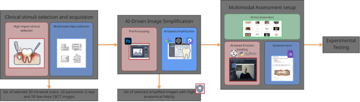
The methodological framework of the SMART DENT project was structured in four integrated phases to assess how AI-driven image simplification can enhance patient communication while preserving anatomical accuracy, as shown in Figure 1.
Figure 1: Methodological framework of the SMART DENT project.
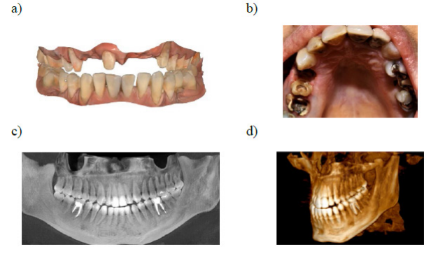
Clinical Stimuli Selection and Acquisition In the first phase, a representative set of anonymized diagnostic images was selected in collaboration with clinical staff to reflect real-world clinical practice while targeting varying degrees of emotional impact. The assets categorized for testing included clinical photographs (n = 4), intraoral 3D scans (n = 7), 2D panoramic X-rays (n = 1), and 3D low-dose CBCT images (Cone Beam Computed Tomography) (n = 1). A total of 13 anonymized images were chosen, depicting various conditions such as gingival inflammation, bleeding, damaged dentition, implant-related cases, and pre/post-treatment scenarios (Figure 2). The images were selected to balance emotional impact and clinical relevance, ensuring they were suitable for both the questionnaire and AI-driven emotion analysis in subsequent phases.
Figure 2: Example of clinical images selected for stimuli: a) intraoral 3D scan; b) photograph; c) 2D panoramic X-ray; d) 3D low-dose CBCT image.
AI-Driven Image Simplification The transformation of the selected clinical stimuli into simplified illustrations followed a systematic two-stage process:
Pre-processing with Adobe Photoshop (Adobe Photoshop, 2025): The original images were manually vectorized to normalize shapes, contours, and volumes. This step allowed for the creation of a stable visual structure that could be consistently used across different AI tools.
AI-Based Simplification: The pre-processed images were then simplified using AI-driven tools to generate visually reassuring illustrations while preserving anatomical accuracy. Three free online tools were employed for this process: Gemini Nano Banana (Gemini, 2025), ChatGPT (OpenAI, 2025) and PromeAI (PromeAI, 2025). A structured prompt was created to direct the transformation process, specifying the need for precise anatomical preservation, the removal of realistic and emotionally unsettling features, the application of soft pastel rendering, and the maintenance of clear, educational visuals. The generated simplified images were characterized by soft contours, pastel colour palettes (ivory for teeth, soft pink for gums), and minimal shading, ensuring a clean, non-clinical visual that would be emotionally neutral yet informative.
Multimodal Assessment Setup The third phase of the methodology involved the development of a multimodal assessment framework to evaluate emotional and cognitive responses to the clinical images presented. This phase combined AI-based emotion tracking and subjective questionnaire responses to collect a comprehensive analysis of patient reactions to visual stimuli.
To conduct the emotion tracking a custom Python script based on DeepFace, a Lightweight Face Recognition and Facial Attribute Analysis Library for Python (Serengil and Ozpinar, 2021) was developed. The script analyses video recordings of participants’ face captured while viewing a set of specific visual stimuli by the laptop webcam, to detect participants’ micro-expressions.
A total of 39 visual stimuli were used in the emotion tracking analysis to assess patients’ emotional responses to different types of dental diagnostic images. These stimuli included both real and simplified versions of clinical images, carefully selected to represent various stages of dental treatment and a wide range of emotional impacts. The stimuli were divided into four main categories: Clinical Photographs (n = 4, each depicting a different stage of dental issues such as Inflammatory conditions, exposed implant structures, severely damaged dentition, gingival bleeding and infection-related visuals); Intraoral Scans (n = 7, showing pre-treatment and post-treatment images; various stages of implantology, including pre-implant, post-implant, and hygiene treatment phases); Panoramic Radiographs (n = 2; showing a normal dentition scenario; a case with devitalized teeth and structural anomalies); Low-Dose 3D CBCT (n = 2, showing a scan depicting normal dentition and dentition with devitalized teeth and implants). 17 simplified versions of the same images obtained using Gemini was included to reduce emotional intensity by eliminating harsh details. An additional set of images sourced online was used exclusively to establish a baseline for expressions of joy, surprise, and disgust.
Alongside the biometric emotion tracking, a structured questionnaire was prepared in Google Form. The primary goal was to measure both the emotional comfort and the clarity of the images presented, while also evaluating participants’ preferences for simplified images in relation to emotional impact and diagnostic understanding.
The questionnaire consisted of 14 questions divided in multiple sections, each addressing specific aspects of the visual stimuli:
Emotional Comfort: Participants rated their discomfort with each image on a Likert scale from “no discomfort” to “extremely uncomfortable”. This section aimed to capture the emotional response triggered by highly realistic or potentially distressing images, such as those depicting implants, exposed tissues, or dental decay.
Clarity and Understanding: Participants assessed whether simplified images retained enough clarity to support understanding of the dental issue, comparing real and simplified versions for diagnostic effectiveness.
Image Preference: Participants indicated their preference between real and simplified images for different diagnostic scenarios, evaluating whether emotional relief outweighed any loss of detail.
Experimental Testing and Validation Field validation involved 35 adult volunteers (20 males, 15 females; aged 25–55). Participants viewed 39 visual stimuli (both real and simplified versions) in a controlled setting, then completed an anonymous questionnaire. The experimental protocol synchronized stimulus presentation with video recording of facial expressions, enabling alignment of detected micro- expressions with the corresponding stimuli during analysis for further considerations.
RESULTS
AI-Driven Image Simplification The AI-driven image simplification process involved three different AI tools: Gemini Nano Banana, ChatGPT, and PromeAI. The simplification process was applied to different types of clinical images, including intraoral scans (depicting pre- and post-treatment conditions), 2D panoramic radiographs and 3D low-dose CBCT scans. For each image type, the AI tools generated simplified versions that were softer, less detailed, and more visually neutral, which were perceived as more reassuring by the majority of participants. Despite some limitations in repeatability and anatomical accuracy, the overall outcome showed that simplified images could provide a useful tool for enhancing patient comfort during the communication of diagnostic and treatment plans.
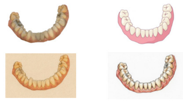
More in details, ChatGPT and PromeAI generated visually coherent images consistent with the requested style but struggled with maintaining anatomical fidelity. In particular, both tools produced simplified versions where some dental shapes were distorted or overly simplified, compromising
Leveraging AI Tools for Emotion-Safe Dental Imaging 287 diagnostic clarity (Figure 3). On the other side, Gemini Nano Banana, despite occasional variability in the output, demonstrated a stronger ability to preserve anatomical accuracy. It was better at maintaining the shapes and proportions of the teeth and gums while reducing emotional intensity. All of them exhibited low repeatability, meaning that the same prompt produced slightly different results each time, which posed challenges for consistent application Figure 4.
Figure 3: Simplification of an intraoral scan using ChatGPT. Notable variability is observed, along with substantial changes in anatomical features.
Gemini Nano Banana was ultimately selected as the most suitable tool for generating simplified images, as it offered the best balance between maintaining anatomical fidelity and reducing emotional impact. The simplified images produced by Gemini were used for subsequent testing and evaluation in the study to create the simplified images used in the multimodal emotion assessment.
Figure 4: Simplifications of an intraoral scan using Google Gemini generated repeating the same prompt.
Multimodal Emotion Assessment The analysis of the 35 video recordings of participants revealed important insights but also highlighted limitations in the ability to reliably capture micro-emotions. While the system was effective in tracking broad emotional categories (e.g., disgust, fear, happiness), the accuracy of detecting subtle emotions, particularly in response to images with varying emotional impact,
was compromised by factors such as lighting, facial obstructions (e.g., glasses, hands, beards), and variations in individual expressiveness.
Significant variability was observed in the classification of emotions like “anger” and “sadness,” where the DeepFace sometimes misclassified neutral or minor discomfort expressions as more intense emotions. This issue was particularly evident when viewing images of dental implants, exposed tissues, or severely damaged dentition, where discomfort was expected but the AI failed to consistently capture the intensity of the emotional response.
Despite these challenges, the emotional tracking system proved useful for identifying overall trends and provided some alignment with the subjective questionnaire results. For instance, images that were rated as highly discomforting (e.g., photographs of exposed implants) were often associated with the “disgust” category in the micro-emotion analysis, confirming findings from the questionnaire.
The questionnaire results extended the preliminary findings from the emotion tracking analysis, offering a comprehensive understanding of participants’ emotional and cognitive responses to the diagnostic images.
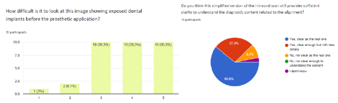
The images that elicited the most discomfort were those depicting particularly invasive clinical conditions, such as exposed dental implants before prosthetic application or severely damaged teeth with extensive decay. Over 60% of participants rated these images as very uncomfortable or extremely uncomfortable (Figure 5 a)), confirming that highly realistic images, especially those showing exposed tissues or compromised dentition, provoked a strong emotional reaction. This finding aligns with the results from the micro-expression analysis, which also identified significant negative emotional responses to these types of images.
Figure 5: Results of the questionnaire: a) Discomfort perceived with pre-implant images; b) Declared clarity for simplified version of intraoral scans to understand the proposed treatment.
In line with the emotional discomfort caused by the more realistic images, the majority of participants preferred the simplified versions. Specifically, more than 50% of participants favoured the simplified intraoral scans, particularly for restorative procedures and implantology, where the simplified images were perceived as more reassuring and emotionally neutral. This preference mirrors the emotional impact observed in the emotion tracking data, where simplified visuals were associated with “neutral” emotions. Most participants indicated that the simplified versions were clear enough to support understanding of the
Leveraging AI Tools for Emotion-Safe Dental Imaging 289 treatment plan, with 60% affirming that simplification did not compromise diagnostic clarity, demonstrating that emotional load can be reduced without sacrificing key information (Figure 5 b)).
However, the analysis also showed that for radiographic images (panoramic and CBCT), the emotional response was significantly lower, with over 70% of participants reporting minimal discomfort. This suggests that radiographs and CBCT models are perceived as standard clinical tools and emotionally neutral, thus not requiring visual simplification. Therefore, the preference for simplified versions was particularly relevant for photorealistic or highly detailed images, such as intraoral scans, where visual detail and texture can increase emotional discomfort.
These results confirm the findings from the emotion tracking analysis, highlighting that visual simplification can indeed reduce emotional discomfort without compromising comprehension of the treatment. Simplified images proved to be particularly effective in enhancing communication with sensitive patients, maintaining a balance between diagnostic clarity and emotional safety, especially for intraoral images and clinically complex scenarios.
CONCLUSION
The experiment conducted within the SMART DENT Project preliminarily evaluated the potential of free AI-tools for simplification of clinical visual contents to be used as a tool for supporting patient communication. The results confirm that transforming clinical images into stylized illustrations, featuring neutral colours, soft contours, and the removal of harsh details, effectively makes diagnostic content more accessible while simultaneously reducing the emotional load associated with more impactful images. This approach aligns with the preferences expressed by users involved in the tests, who positively rated the simplified versions, particularly for intraoral scans.
At the same time, the use of AI tools demonstrated significant potential for the automatic generation of such visual representations, though it also revealed certain challenges. Specifically, only a portion of the tested models adequately preserved anatomical fidelity, an essential element for maintaining diagnostic clarity. Furthermore, the low repeatability of results obtained from some free tools suggests the need for future development of specifically trained models for clinical image simplification, ensuring a stable and reproducible workflow.
Overall, the tests confirm that visual simplification is a valuable aid in explaining treatment plans. It facilitates the understanding of information and reduces the emotional impact of certain image types. However, the results also indicate that the dentist’s mediation remains an indispensable element for the correct interpretation of content and for managing patient anxiety. Simplified images do not replace the clinical relationship but serve as a useful and functional complement.
It is important to acknowledge that the limited number of images tested and the relatively small sample size of participants make this study a pilot experiment. While the results are consistent and indicative, larger-scale validation will be required to generalize the observed trends.
Finally, reflections from the data analysis suggest promising directions for future development. Specifically, the adoption of interactive interfaces capable of detecting not only micro-expressions but also spontaneous user behaviors, such as time spent on an image, zoom level, or tendency to avoid certain areas, could provide a more intuitive, less invasive, and potentially more reliable method to assess patients’ visual tolerance and personalize clinical communication in real time.
ACKNOWLEDGMENT
This work was funded under the “Programma Regionale 2021–2027-PR Puglia FERS FSE+ 2021–2027 AVVISO Pacchetti Integrati di Agevolazione per micro e piccole imprese – Mini PIA”.
REFERENCES
Adobe Photoshop. 2025. ‘Adobe Photoshop’. https://www.adobe.com/products/ photoshop.html. Appukuttan, Devapriya. 2016. ‘Strategies to Manage Patients with Dental Anxiety
and Dental Phobia: Literature Review’. Clinical, Cosmetic and Investigational Dentistry, March, 35. https://doi.org/10.2147/CCIDE.S63626. Gemini. 2025. ‘Google Gemini’. https://gemini.google.com. Ho, Jasmine, Hollis Chai, Edward Lo, Michelle Huang, and Chun Hung Chu. 2024.
‘Strategies for Effective Dentist-Patient Communication: A Literature Review’. Patient Preference and Adherence Volume 18 (July): 1385–94. https://doi.org/10.2147/PPA. S465221. Nilchian, Firoozeh, Fereshteh Shakibaei, and Zeinab Taghi Jarah. 2017. ‘Evaluation of Visual Pedagogy in Dental Check-Ups and Preventive Practices Among 6–12-Year-Old Children with Autism’. Journal of Autism and Developmental Disorders 47 (3): 858–64. https://doi.org/10.1007/s10803-016-2998-8. OpenAI. 2025. ‘Chat GPT’. https://chatgpt.com/. Orellana, Lorena M., Sonia Martínez-Sanchis, and Francisco J. Silvestre. 2014.
‘Training Adults and Children with an Autism Spectrum Disorder to Be Compliant with a Clinical Dental Assessment Using a TEACCH-Based Approach’. Journal of Autism and Developmental Disorders 44 (4): 776–85. https://doi.org/10.1007/ s10803-013-1930-8. PromeAI. 2025. ‘PromeAI’. https://www.promeai.pro/. Schulz-Weidner, Nelly, Marina Gruber, Eva May Schraml, Bernd Wöstmann, Norbert Krämer, and Maximiliane Amelie Schlenz. 2024. ‘Improving the Communication of Dental Findings in Pediatric Dentistry by Using Intraoral Scans as a Visual Aid: A Randomized Clinical Trial’. Dentistry Journal 12 (1): 15. https://doi. org/10.3390/dj12010015. Serengil, Sefik Ilkin, and Alper Ozpinar. (2020) 2026. ‘DeepFace’. In Gazi University
Journal of Science, Python, v. 1.0.0. no. Advanced Online Publication. February 8, released. https://doi.org/10.35378/gujs.1794891. Serengil, Sefik Ilkin, and Alper Ozpinar. 2021. ‘HyperExtended LightFace: A Facial Attribute Analysis Framework’. 2021 International Conference on Engineering and Emerging Technologies (ICEET), October 27, 1–4. https://doi.org/10.1109/ ICEET53442.2021.9659697.
LISTED IN THE AHFE DIGITAL BOOKSTORE CATALOGUE
Explore the complete digital volume
Healthcare and Medical Devices
Read this individual research paper free through the original publisher, or explore the full digital proceedings book.
ISBN 978-1-964867-87-8
Explore this ISBN in the AHFE Bookstore ↗Listed in the July 2026 bookstore catalogue; search by ISBN to confirm current availability · Digital book purchase optional