The bedroom thermal environment is a key determinant of sleep onset, yet most studies manipulate ambient temperature and evaluate sleep onset latency (SOL) between conditions. Continuous skin-temperature dynamics during the SOL, particularly across bedroom temperatures, remain poorly characterised. This study examined skin-temperature dynamics before objectively defined sleep onset across multiple bedroom temperatures. Twelve healthy adults (6 males, 6 females; 23–42 years; BMI 17.7–32.9 kg/m²) completed four overnight sessions at 22°C, 24°C, 26°C, and 28°C. From 23:00 lights-out to 07:00 lights-on, skin temperatures at nine sites were recorded every 30 seconds, while polysomnography was continuously monitored. Sleep stages were scored according to the American Academy of Sleep Medicine manual, and the first non-wake epoch defined sleep onset. Temperature series were aligned to this point, and distal (DST), proximal (PST), and mean skin temperatures (MST) were derived. The distal–proximal gradient (DPG) was calculated as the difference between distal and proximal temperatures. Linear mixed-effects models with ambient temperature and SOL segment (early: −30 to −15 min; late: −15 to 0 min) as fixed factors showed that ambient temperature strongly affected DST, MST, and DPG in the early period of SOL, but effects were markedly attenuated in the late period. Across all four bedroom temperatures, DST, MST, and DPG converged toward similar levels as sleep onset approached. These findings suggest that the body is not passively constrained by ambient temperature but actively adjusts skin temperature and heat dissipation through distal thermoregulation to reach a relatively stable sleep-conducive state.
Sleep is essential for health, cognitive performance, and well-being (McAlpine et al., 2019). Difficulties in initiating sleep are typically quantified using sleep onset latency (SOL), which is defined as the time from lights out or
bedtime to the first non-wakeful sleep epoch. Prolonged SOL is associated with impaired daytime functioning and increased risk of insomnia (Iijima et al., 2024). Therefore, understanding the physiological processes that precede sleep onset is important for both basic science and applied sleep engineering.
Thermoregulation plays a crucial role in sleep initiation. Prior research has shown that the transition to sleep is accompanied by a mild decline in core body temperature and an increase in distal skin temperature, often summarised by the distal–proximal gradient (DPG), which is defined as the temperature difference between distal and proximal sites. Higher DPG values, reflecting warmer extremities relative to the body, have been associated with shorter SOL (Kräuchi et al., 1999; Kräuchi et al., 2000; Krauchi et al., 2010). However, these studies examined the association between SOL and skin temperature features calculated at a single time point or over a brief predefined interval (e.g. at lights-out or a fixed delay after lights-out), and were typically conducted only under a single ambient temperature condition. For example, Minor et al. analysed wearable-derived sleep records and meteorological data from 68 countries and reported that higher nighttime ambient temperatures were associated with shorter sleep duration and longer SOL (Minor et al., 2020). Iijima et al. quantified ΔDPG as the difference between mean DPG during the 15 mins before lights-out and the 15 mins after lights-out, as an index of the reduction in heat loss from the pre-sleep preparation period to early sleep, and showed that changes in DPG around lights-out could predict subjective SOL (Iijima et al., 2024). Kräuchi et al., found that the mean DPG during the 1.5 hours before lights-out was a strong predictor of SOL, such that greater distal vasodilation was associated with faster sleep initiation (Kräuchi et al., 2000). In addition, studies by Tai et al., and Raymann et al., emphasised the role of distal temperature regulation and peripheral vasodilation in modulating SOL, suggesting that interventions such as hot-water bathing may facilitate sleep onset by enhancing heat dissipation (Raymann et al., 2007; Tai et al., 2021). In everyday bedroom environments, ambient temperature can vary markedly across seasons, building characteristics, and individual habits. At the same time, emerging smart mattresses and bedroom climate- control systems now permit relatively precise manipulation of bed and room temperatures. For the design of such systems, it is insufficient to simply assume that “warmer extremities are beneficial” for sleep onset. Therefore, it is necessary to characterise how skin temperatures evolve over time during the SOL, to determine whether there is a relatively stable SOL thermal state in the human body, and whether this thermal homeostasis is similar under different ambient temperature conditions.
The present study aims to address this question by examining continuous SOL skin-temperature changes in healthy adults under four bedroom temperatures (22°C, 24°C, 26°C, 28°C). We focus on the 30 minutes immediately preceding objectively defined sleep onset and, based on the first non-wakeful sleep epoch identified from polysomnography (PSG), divide this 30-minite SOL interval into an early SOL segment (−30 to −15 min) and a late SOL segment (−15 to 0 min). Our primary objective is to characterise how distal skin temperature (DST), proximal skin temperature (PST), mean skin temperature (MST), and DPG change with ambient temperature
Skin Temperature Dynamics During Sleep Onset Latency 199 in both phases, and whether these measures tend to approach a sleep- conducive thermal state as sleep onset draws near under different bedroom temperatures. Identifying such a thermal state would provide a physiological basis for specifying bedroom temperature settings and for designing adaptive control strategies in smart sleep environments.
METHODS
Participants Twelve healthy adults (6 males, 6 females; age 23–42 years; BMI 17.7–32.9 kg/m²) participated in the study. All participants reported regular sleep–wake schedules and no history of diagnosed sleep disorders, cardiovascular disease, or major psychiatric or metabolic conditions. Participants were asked to avoid caffeine and alcohol on experimental days and to maintain consistent bedtimes for at least one week before the first laboratory night. The study protocol was approved by the Ethics Committee of the School of Biological Science and Medical Engineering, Beihang University, and written informed consent was obtained from all participants.
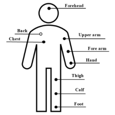
Figure 1: Distribution of skin temperature sensor sites on the body surface.
Experimental Design and Bedroom Conditions Each participant completed four non-consecutive overnight sleep sessions in the same controlled bedroom environment. The nominal bedroom air temperature was set to 22°C, 24°C, 26°C, or 28°C and assigned to nights according to a Latin square design, such that each temperature occurred equally often in each ordinal position across participants. The same standardised bed, mattress, bedding set, and summer sleepwear were used in all sessions. Room air temperature and relative humidity were continuously monitored throughout the night to ensure that the target temperature was maintained within a narrow range. The relative humidity in the room was approximately 65% ± 5%. Lights remained off throughout the night, and no external noise disturbances were present.
Measurements Skin temperature was measured at nine body sites (forehead, chest, back, upper arm, forearm, hand, thigh, calf, and foot) using miniature temperature– humidity loggers (iButton DS1923, Maxim Integrated, USA) as shown in
Figure 1. Sensors were affixed directly to the skin with hypoallergenic medical tape and configured to sample at 30-second intervals from 23:00 lights-out to 07:00 lights-on.
Figure available in the original PDF paper.
MST was calculated as a weighted average of the nine local skin temperatures using predefined regional weighting coefficients, as shown in Eq. (1) (Liu et al. 2011). In addition to MST, distal and proximal skin temperatures and their DPG are widely used indices in studies of thermoregulation during sleep (Xu et al., 2023). Accordingly, we derived DST, PST, and DPG for each epoch, as defined in Eqs. (2)–(4) (Zhang et al., 2019).
DPG = DST − PST (4) PSG signals were recorded using a portable PSG system (Alice PDx, Philips Respironics, USA) with a standard clinical montage. Sleep stages were scored in 30-second epochs by trained technologists according to the American Academy of Sleep Medicine criteria. Sleep onset was defined as the first non- wake epoch (N1, N2, N3, or REM) that was followed by predominantly sleep epochs, consistent with standard definitions of SOL. This epoch was taken as time 0. All skin-temperature time series were then re-aligned such that negative times represented minutes before this PSG-defined sleep onset.
Procedure On each experimental night, participants arrived at the laboratory at approximately 22:20. They changed into standardised lightweight sleepwear, after which skin-temperature sensors and PSG electrodes were attached. Participants then moved into the bedroom and sat quietly to adapt to the room conditions. At 23:00, lights were turned off and participants were instructed to attempt to fall asleep while freely adjusting their bedding according to comfort. PSG and skin-temperature recordings continued until final awakening at approximately 07:00.
Statistical Analysis All analyses were based on the sleep-onset–referenced skin-temperature time series, with time 0 corresponding to PSG-defined sleep onset and negative values indicating minutes before onset. For each night, we focused on the 30-min interval preceding sleep onset (−30 to 0 min). Within this window, an early SOL segment (−30 to −15 min) and a late SOL segment (−15 to 0 min) were defined. For each 30-second epoch in this interval, DST, PST, MST and DPG, were computed as defined in the Measurements section.
Skin Temperature Dynamics During Sleep Onset Latency 201 To examine the influence of bedroom temperature and SOL segment on skin temperature indices, linear mixed-effects models were fitted separately for DST, PST, MST and DPG. In each model, bedroom temperature was treated as a four-level factor with levels 22°C, 24°C, 26°C and 28°C, and SOL segment was treated as a two-level factor. The interaction between bedroom temperature and SOL segment was also included. Participant was entered as a random intercept, and sex, age and BMI were included as covariates. Wald chi-square tests were used to assess the significance of fixed effects. When a main effect or interaction involving temperature or segment was significant, trends were qualitatively assessed by examining the SOL temperature trajectories. Early and late segments within each temperature condition, as well as temperature conditions within each segment, were visually compared to determine whether the dependence of skin temperature indices on bedroom temperature differed between the early and late SOL segments.
RESULTS AND DISCUSSION
Linear Mixed-Effects Models of Skin Temperature Indices Mixed-effects models of the four skin-temperature indices are summarised in Table 1. Across outcomes, the SOL segment factor (Seg) showed a consistent effect on DST, PST and MST. Relative to the early SOL segment (−30 to −15 min), the late segment (−15 to 0 min) was associated with higher DST, PST and MST with model-estimated increases of 0.73°C, 0.47°C and 0.56°C, respectively. For DPG, Seg also shifted values upward by about 0.52°C, so that distal–proximal differences became more positive in the late segment. Taken together, these segment effects suggest that, as sleep onset is approached, the body shows a moderate overall rise in skin temperature that is driven disproportionately by distal warming, leading to a more positive DPG and enhanced distal heat dissipation.
Table 1: Wald chi-square tests for fixed effects in mixed- effects models of DST, PST, MST and DPG. Seg denotes the SOL segment factor (early and late), and Env denotes the bedroom temperature factor (22°C, 24°C, 26°C and 28°C).
Table available in the original PDF paper.
Outcome Effect df χ² p-value DST Seg 1 6.33 0.012
Env 3 21.98 <0.001 Seg × Env 3 3.59 0.310 PST Seg 1 4.09 0.043
Env 3 8.66 0.034 Seg × Env 3 0.68 0.878 MST Seg 1 5.87 0.015
Env 3 12.59 0.006 Seg × Env 3 1.97 0.578
DPG Seg 1 3.52 0.061 Env 3 9.74 0.021
Seg × Env 3 3.65 0.301
Bedroom temperature (Env) exerted systematic effects on all four indices. As bedroom temperature increased from 22°C to 28°C, DST, PST, MST, and DPG, all shifted upwards, with the smallest changes at 24°C, intermediate changes at 26°C and the largest increases at 28°C relative to 22°C (Table 1). Thus, within the tested range, warmer bedrooms were associated with higher DST, PST and MST and with more positive DPG values, showing that SOL skin-temperature patterns are strongly shaped by the bedroom thermal environment. The interaction between Seg and Env was small for all outcomes, indicating that the transition from the early to the late segment occurred in a similar way at all bedroom temperatures and that the overall pattern of SOL skin-temperature change was broadly comparable across thermal conditions.
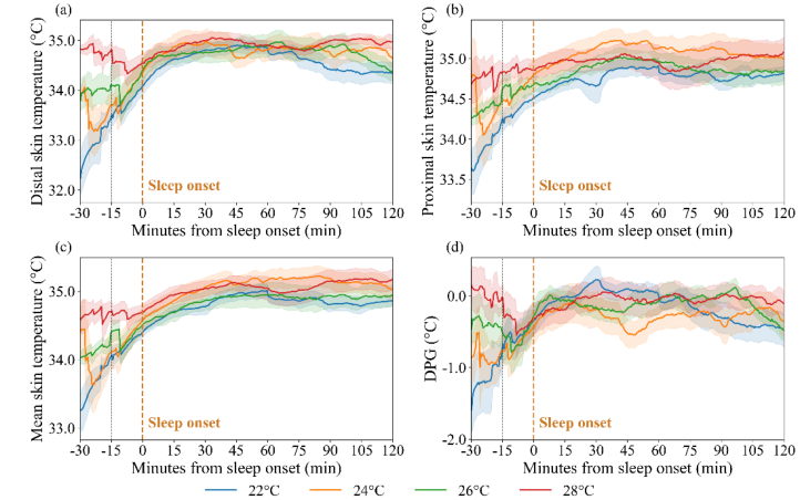
Figure 2: Sleep-onset–aligned trajectories of DST (a), PST (b) and MST (c) and DPG (d) across bedroom temperatures (22°C, 24°C, 26°C and 28°C). Solid lines denote means averaged across participants and nights, and shaded bands represent ±1 standard error of the mean (SEM). Vertical dashed lines mark −15 min and 0 min relative to PSG- defined sleep onset, separating the early (−30 to −15 min) and late (−15 to 0 min) SOL segments. Negative DPG values indicate cooler distal than proximal skin. Between- temperature differences are larger in the early segment and converge toward more similar levels as sleep onset is approached.
SOL Temperature Trajectories and Convergence Across Bedroom Temperatures Sleep-onset–aligned trajectories of DST, PST, MST and DPG across bedroom temperatures are shown in Figure 2. In the early SOL segment (−30 to −15 min), mean levels differed markedly between bedroom temperatures. Cooler rooms were associated with lower distal and mean skin temperatures and more
Skin Temperature Dynamics During Sleep Onset Latency 203 negative DPG, with the 22°C condition showing the lowest trajectories and the 28°C condition the highest. PST also varied with bedroom temperature, but the separation between conditions was less pronounced than for DST and MST. This pattern suggests that proximal regions, which more closely reflect core thermal status, were less influenced by the bedroom environment, and that SOL thermoregulatory adjustments were expressed mainly at distal sites. By contrast, during the late segment (−15 to 0 min), the curves for the four bedroom temperatures lay much closer together for all indices, and their shaded SEM bands showed substantial overlap. Visually, the between- temperature spread in DST, MST and DPG during this late SOL segment was much smaller than in the early segment, indicating that skin temperatures and DPG converged toward a common range as sleep onset was approached.
In summary, the mixed-effects results and sleep-onset–aligned trajectories together suggest that bedroom temperature mainly determines the initial SOL thermal level. As sleep onset approaches, DST and PST tend to converge towards a relatively common, sleep-conducive range across different ambient conditions. This convergence appears to be driven predominantly by SOL thermoregulatory adjustments at distal sites.
CONCLUSION
This study examined SOL skin-temperature dynamics across four ambient temperatures by aligning multi-site skin temperature to PSG-defined sleep onset and focusing on the 30 minutes that preceded it. Mixed-effects analyses indicated that higher bedroom temperatures were associated with higher distal, proximal and mean skin temperatures, as well as more positive DPG. These temperature parameters in the late SOL segment were also higher than in the early segment. The non-significant interaction between segment and bedroom temperature suggests that this early-to-late change in skin temperature did not depend strongly on bedroom temperature. The segment effect was larger for DST than for PST, indicating that these changes were driven mainly by distal warming and that SOL thermoregulation was expressed predominantly at distal sites. The trajectories aligned to sleep onset further showed that, in the early SOL segment, differences in DST, PST and DPG between bedroom temperatures were more pronounced, whereas these differences were clearly reduced in the late segment. Within the 22–28°C range, distal and proximal skin temperatures and DPG tended to fall into a relatively narrow range immediately before sleep onset. This convergence suggests that the bedroom environment determines the initial thermal state. SOL thermoregulatory processes then actively adjust skin temperatures so that they approach a broadly similar, sleep-conducive state across different ambient conditions.
These findings provide a sleep-onset–aligned, multi-bedroom temperature view of SOL thermoregulation that complements previous work based on single time points or single ambient settings. From an applied perspective, the convergent SOL temperature range identified in this study offers a physiological basis for defining target bedroom temperature settings and for developing adaptive control strategies in smart bedding and bedroom
climate-control systems. Future work with larger and more diverse samples and with different bedding and clothing configurations will be valuable to test the generalisability of these patterns to real-world sleeping environments.
ACKNOWLEDGEMENT This work was supported by the Digital Human and Intelligent Sleep Innovation Consortium Project under the Dongguan Key R&D Program for Priority Areas (No. 20261201500022).
REFERENCES
Iijima, R., Kadooka, A., Sugawara, K., Fushimi, M., Hosoe, M. and Aritake-Okada, S. (2024). Subjective sleep onset latency is influenced by sleep structure and body heat loss in human subjects. Journal of Sleep Research, Volume 33, No. 5, e14122. Kräuchi, K., Cajochen, C., Werth, E. and Wirz-Justice, A. (1999). Warm feet promote
the rapid onset of sleep. Nature, Volume 401, No. 6748, pp. 36–37. Kräuchi, K., Cajochen, C., Werth, E. and Wirz-Justice, A. (2000). Functional link between distal vasodilation and sleep-onset latency?. American Journal Of Physiology – Regulatory, Integrative and Comparative Physiology, Volume 278, No. 3, pp. R741–R748. Krauchi, K. and Deboer, T. (2010). The interrelationship between sleep regulation
and thermoregulation. FRONT BIOSCI, Volume 15, No. 2, pp. 604–625. Liu, W., Lian, Z., Deng, Q. and Liu, Y. (2011). Evaluation of calculation methods of mean skin temperature for use in thermal comfort study. Building and Environment, Volume 46, No. 2, pp. 478–488. McAlpine, C.S., Kiss, M.G., Rattik, S., He, S., Vassalli, A., Valet, C., Anzai, A.,
Chan, C.T., Mindur, J.E., Kahles, F. and Poller, W.C. (2019). Sleep modulates haematopoiesis and protects against atherosclerosis. Nature, Volume 566, No. 7744, pp. 383–387. Minor, K., Bjerre-Nielsen, A., Jonasdottir, S.S. et al. (2020). Ambient heat and human sleep. Arxiv Preprint, arXiv:2011.07161. Raymann, R.J., Swaab, D.F. and Van Someren, E.J. (2007). Skin temperature and
sleep-onset latency: changes with age and insomnia. Physiology & Behavior, Volume 90, No. 2–3, pp. 257–266. Tai, Y., Obayashi, K., Yamagami, Y. et al. (2021). Hot-water bathing before bedtime and shorter sleep onset latency are accompanied by a higher distal-proximal skin temperature gradient in older adults. Journal of Clinical Sleep Medicine, Volume 17, No. 6, pp. 1257–1266. Xu, X., Li, S., Yang, Y. and Lian, Z. (2023). Effects of thermal environment on body
temperature rhythm and thermal sensation before and after getting into bed: A laboratory study in Shanghai, China. Energy and Buildings, Volume 301, 113748. Zhang, N., Cao, B. and Zhu, Y. (2019). Effects of pre-sleep thermal environment on human thermal state and sleep quality. Building and Environment, Volume 148, pp. 600–608.
LISTED IN THE AHFE DIGITAL BOOKSTORE CATALOGUE
Explore the complete digital volume
Healthcare and Medical Devices
Read this individual research paper free through the original publisher, or explore the full digital proceedings book.
ISBN 978-1-964867-87-8
Explore this ISBN in the AHFE Bookstore ↗Listed in the July 2026 bookstore catalogue; search by ISBN to confirm current availability · Digital book purchase optional