Gloves and masks make fingerprint and face recognition difficult in hospitals and factories. In this research, as a precondition for developing an authentication system based on geometrical features inside the ear canal using a camera, we will verify whether it is possible to identify individuals from images inside the ear canal using machine learning. The method is to acquire image data of the ear canal using a camera and identify individuals using the image data and a model that has been learned in advance. As a preliminary step to machine learning, we conducted a questionnaire to investigate whether it is possible to identify the inside of the ear canal using human recognition ability. The questionnaire results showed that the correct response rate was approximately 64%. We also found that the correct response rate decreased for questions such as "select the image of the left ear from the image of the right ear," even for the same person. In this study, the VGG16 model was re-trained by transfer learning because of the small amount of training data we had prepared. The number of classes was 26 since the data we prepared was for 13 people and the number of left and right ears, and we used approximately 400 pieces of data per class. The experimental results showed that discrimination was possible with high accuracy. Accuracy, Recall, Precision, and F-measure were used as evaluation indices, and both Accuracy and F-measure were highly evaluated at 0.989. These results also indicate that the left and right ears can be discriminated against even if they are different during registration and evaluation. In the future, we are planning to study the imaging method for implementation and conduct experiments with third-party data.
The number of workers in Japan’s health and welfare industry gradually increases. In addition, more healthcare workers were expected to be needed due to the coronavirus. In line with this, there is a need to develop a medi- cal environment in which many healthcare workers can work efficiently and smoothly. In recent years, there have been many moves in the medical field to reduce various burdens on medical personnel through IT, such as the storage of patient information in electronic media like electronic medical records and the digitization of medical questionnaires. This has led to the development of devices that use biometric authentication, such as fingerprint recognition, to enhance security to protect personal information such as electronic medi- cal records.A biometric authentication system was a system that identifies an individual using physical characteristics such as human eyes and ears and behavioral characteristics. Because biometric authentication systems used the unique characteristics of each individual as a pass, they were complicated to impersonate or forge and were used in many fields as a robust security mea- sure.The types of biometrics currently in use were as follows. Fingerprint authentication was an authentication method that identifies and compares fingerprints engraved on a finger. Facial recognition was an authentication method that compares facial features. Otoacoustic authentication was an
authentication method that sends a sound into the ear and compares the fea- tures of the echo sound. In addition, “multimodal biometric authentication” was attracting attention as superior to conventional authentication systems. This multimodal biometric authentication was a revolutionary authentica- tion system that combines two or more biometric methods and was expected to improve performance significantly. However, people wear masks and glo- ves when working in hospitals and factories, making fingerprint and face recognition challenging to match. Therefore, we investigated an authentica- tion system that uses the ear as in otoacoustic authentication. The following was an overview of the system. In the current Japanese medical field, infor- mation technology was advancing, and biometric authentication was being considered to strengthen security to protect that information. However, there was a situation in which conventional biometric authentication was too cum- bersome due to masks, hats, and gloves in hospitals. Therefore, developing a personal authentication system based on a different approach other than acoustic features used in the ear was expected to complement multiple auth- entication methods and provide high authentication accuracy. The main goal of our research was to develop a new ear-based authentication system. We proposed a biometric authentication system that used geometrical (image) features within the ear canal.
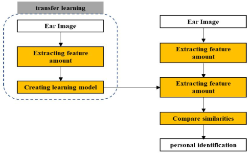
PROPOSED METHOD The flow of the proposed method is shown in Figure 1. In this study, a camera was used to capture the inside of a human ear and acquire image data. Fea- tures were extracted from the acquired image data, and the features were compared with a training model that was previously trained using images of the human ear canal. Personal identification was performed based on the results. The final goal was to develop a device that can be easily combined with other authentication systems by using a camera built into the earphone, thereby improving authentication accuracy without increasing the burden on the user. Since the amount of training data we prepared on our own was small, we used a VGG16 model that has already been trained and re-trained by
Personal Authentication Method Using Geometrical Features of External Auditory Canal 621
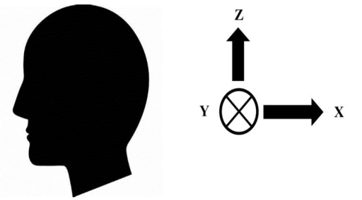
Figure 2: Coordinate system.
transfer learning (Simonyan et al., 2014), (Yusuke et al., 2013). The VGG16 model was a convolutional neural network consisting of 16 layers with para- meters pre-trained on the ImageNet dataset (Ciresan et al., 2011), (Deng et al., 2009). The advantage of transfer learning is that high discrimination accuracy can be achieved even with a small amount of data prepared by the researcher because it was based on a learned model. In addition, it can be applied to a wide range of domains and tasks that can be transferred.
EXPERIMENTAL METHOD Shooting Method
Figure 2 showed the coordinate system at the time of insertion. As shown in Figure 2, in this study, the camera angle was adjusted to capture images of a human profile with the direction from the nose to the back of the head as the X-axis, the direction from the front ear to the opposite ear as the Y-axis, and the direction from the neck to the top of the head as the Z-axis. The camera was used to take pictures with a device called EARPICK.
Thirteen subjects attempted to take pictures from five different angles with their left and right ears. First, the case in which the camera was inserted into the ear parallel to the Y-axis, with the ear hole position as the origin, was defined as the “normal shooting” case. Next, the subjects were asked to take photographs with the ear tilted 10 to 20 degrees in a positive direction along the X-axis and similarly tilted in a negative direction on the same axis. Finally, they were asked to take pictures with the ear tilted 10 to 20 degrees positively along the Z-axis and similarly tilted in a negative direction on the same axis. The reason for the ambiguity of 10 to 20 degrees is that the width of the ear can be moved depending on the size of the individual’s ear canal, so this time the camera was moved to the very edge of focus to take pictures from a variety of angles. There is a difference.
Questionnaire by Experiment As a preliminary step to machine learning, a questionnaire was used to verify how human cognitive abilities can identify images of the inner ear canal. The
Table 1. Questionnaire results.
Table available in the original PDF paper.
Combination (when registered: when answered correctly)
Percentage of correct answers (max: min) (%) (Right side: Right side) 72.0(86.7: 53.3) (Left side: Left side) 78.7(100: 53.3) (Right side: Left side) 42.7(73.3: 20.0) (Left side: Right side) 61.3(86.7: 20.0) Average of 4 patterns 63.7(80.0: 55.0)
questionnaire consisted of a question. Participants were asked to select one image of the external auditory canal and ten alternatives that they thought were identical to the image. The patterns of questions are shown below.
Pattern 1: Select the image of the right ear from the image of the right ear. Pattern 2: Select the image of the left ear from the image of the right ear. Pattern 3: Select the left ear image from the left ear image. Pattern 4: Select the right ear image from the left ear image.
Experiment by Machine Learning In the transfer learning, the number of classes was classified into 26 classes with left and right ears for 13 people, and 600 to 800 image data per class were randomly assigned to training and validation at a ratio of 4:1. The num- ber of epochs was performed ten times, and the training model was used to evaluate the individuality of the geometrical features within the ear canal.
EXPERIMENTAL RESULTS Questionnaire by Experiment
Table 1 showed the tabulation of the questionnaire results. The results sho- wed that when the ear at registration and the ear at evaluation was the same, the discrimination performance ranged from 72-78% as the average response rate for all respondents. On the other hand, when the left and right ears at the time of registration and at the time of evaluation were different, the discri- mination performance was significantly lower. However, the correct response rate exceeding 50% suggests that some parts are similar, so it will be essential to determine how much this affects the machine learning process.
Experiment by Machine Learning
Table 2 showed the results of the machine learning evaluation. The evaluation metrics were Accuracy, Recall, Precision, and F-measure. The odd-numbered classes were the image data of the left ear of each person, and the even- numbered classes were classified as the data of the right ear. They were also sorted one by one from the top (left and right ears of the same person in class 1 and class 2). Table 2 showed that the prediction results were high, with a 98.9% correct response rate. This confirmed a high degree of individuality in the shape (image) characteristics of an individual’s ears. In addition, the same person’s left and right ears also showed highly accurate discrimination
Personal Authentication Method Using Geometrical Features of External Auditory Canal 623
Table 2. Shows the results of the machine learning evaluation.
results for each. From these results, it could be said that a personal authen- tication system using both ears can be used for personal identification with higher accuracy than using only one ear. However, some images were judged to be similar, even though they were considered to be completely different by human recognition ability. This is thought to be due to dark images or white-outs.
CONCLUSION
In this study, we proposed a personal authentication system based on machine learning used image data of the ear canal captured by a camera, and evalua- ted the system through questionnaires and experiments. The results showed that the questionnaire indicated that human recognition ability was suffici- ent to recognize the ear canal and eardrum. In addition, a high discrimination rate of approximately 98.9% was achieved in learning, confirming that the human ear canal’s image characteristics vary among individuals. It was also confirmed that the left and right ears of the same person had different cha- racteristics. On the other hand, some of the images were recognized as not
similar by human recognition ability. In the future, the insertion angle and length of the camera should be strictly considered. In addition, experiments that used photographs and third-party data under actual usage conditions were also necessary.
REFERENCES
AVNET Corporation (November 12, 2018). Biometric identification, overview of six major technologies. AVNET Corporation Website: https://www.avnet.com/wps/ portal/japan?locale=ja-JP > D Ciresan, U Meier, J Masci, L Gambardella, and J Schmidhuber (2011). Flexi-
ble, High Performance Convolutional Neural Networks for Image Classification. IJCAI. J Deng, W Dong, R Socher, L Li, K Li, and F Li (2009). ImageNet: A large-scale hierarchical image database. CVPR. K Simonyan, and A Zisserman (2014) .Two-stream convolutional networks for
action recognition in videos. NIPS. Ministry of Health (October 21, 2020). Labour and Welfare, Trends in the diffusion of electronic medical record systems, Ministry of Health Website: https://www. mhlw.go.jp/index.html Mynavi (2019) “Deep Learning and VGG16”, in:Learn while you make! Develo-
pment Deep Learning by Pytorch, O Yutaro. pp. 6–17 NEC. biometric identification. NEC Website: https://jpn.nec.com/ Statistics Bureau (2021). Ministry of Internal Affairs and Communications, Japan, Labor Force Survey, Statistics Bureau Website: https://www.stat.go.jp/data/roud ou/sokuhou/tsuki/index.html U Yusuke, and Y Takayoshi (2017). Research Trend of Convolutional Neural
Networks. Information and Communication Engineers.
Read this individual research paper free through the original publisher, or explore the full digital proceedings book.
ISBN 978-1-958651-44-5
Explore this ISBN in the AHFE Bookstore ↗Listed in the July 2026 bookstore catalogue; search by ISBN to confirm current availability · Digital book purchase optional