Ramnaath M, Mounika D, Murali Subramaniyam, Seung Nam Min (2022). Measurement of Spine Curvature using Flexicurve Integrated with Machine Vision. In: Human Factors and Wearable Technologies. AHFE Open Access, vol. 29, pp. 82–86. https://doi.org/10.54941/ahfe1001477
Many Spinal disorders require medication and prolonged rehabilitation, which consist of physical exercise and posture control. During restoration, frequent monitoring of spine curvature is necessary. The professionals do not suggest the radiography method more frequently since it has harmful effects on our bodies. Flexicurve has been widely used in much research to measure spine curvature in the sagittal plane. However, the data extraction from Flexicurve are not standardized or the current methodology adopted is difficult and time-consuming. Researchers use a graph (by hand) based method to extract curvature data from Flexicurve. Changes in spine curvature measurements can lead to the wrong methodology for correcting it. So, it is necessary to automate the data collection while measuring spine curvature with Flexicurve. This study proposes a method to automatically record the spine curvature data using the machine vision technique.
The spine, often known as the backbone or vertebral column, consists of small bones (vertebrae) piled on top of each other and discs. The most signifi- cant part of the axial skeleton system is the spine. A healthy spine has smooth bends or curves, including cervical, thoracic, lumbar, and scrum when viewed from the side. These bends in the spine absorb stress caused by bodily move- ment and gravity. A spine deformity is when a spine curvature shows more than 10
of variation than normal spine curvature (Good C.R et al., 2011). The significant deformities in the spine are Scoliosis, kyphosis, Flatback syn- drome, Lordosis, Ankylosing spondylitis, Spondylolisthesis (Clément et al., 2013). Most medical practitioners, including doctors and physiotherapi- sts, decide their medication and treatment methodology based on the spine curvature. During rehabilitation, regular monitoring of spine curvature is mandatory (Horng et al., 2019). A slight deviation in the measurement of spine curvature can go wrong in the treatment methods.
Measurement of Spine Curvature using Flexicurve Integrated with Machine Vision 83
The methods of measuring spine curvature are X-ray, inclinometer, Arcometer, spinal mouse, Flexicurve, etc. The gold standard technique of measuring spine curvature is X-ray radiography; various medical experts still utilize the conventional radiography technique, even though this method is not easy to perform and requires a shielding room to protect others from radi- ation exposure. Hence, the researchers have used a non-radiographic method for measuring the spine curvature. The inclinometer or clinometer is cheap and affordable but not accurate. The electronics inclinometer (Adams et al., 1986) works on the optoelectronic circuit, giving the output a voltage propor- tional to angles between the flat face and vertical. This device only measures the lumbar curvature. Two electronic inclinometers were used to measure the lumbar curvature angles. An Arcometer (Chaise et al., 2011) is an instrument that measures the distance between fixed points. It used a metal rule that can be fixed to the base and then manoeuvres in a single plane to determine the length of different points in the spine from the selected reference.
Spine positioning point measuring instrument (Hou Jianjun et al., 2012) describes a measuring device, which aims to provide a measuring instru- ment with the advantages of simple structure and low cost. The size of the spine positioning point on an X-axis, a Y-axis and a Z-axis can be artifici- ally read or electronically displayed. The human spinal column measurement and display system (Eiichi Ichikawa and Morio Ichikawa, 2011) include a probe pinched between fingers and moved from the first thoracic vertebra to the fifth lumbar vertebra to get X, Y, and Z position. After the measu- rement, the three-dimensional image of the spinal column is displayed on the display screen by integrating the measured data and image of the spinal column. The integration of (Inertial Measurement Unit) IMU sensors with microcontroller is attached to the shirt to measure the spine curvature in the sagittal plane. Then the spine curvature data is sent to AutoCAD to get the curvature (Voinea et al., 2017). Another study aimed to create a digital goni- ometer using machine vision that allows instantaneous measurement of the elbow and knee joint angles through pictures. Statistical analysis shows a 98.25% and 98.09% accuracy for the elbow and knee joints, respectively, resulting from closely related values between the actual goniometer and the device created from the study (Bumacod et al., 2020). The author measu- res the thoracic kyphosis angle with motion capture and flexicurve in the survey. The systematic review shows that flexicurve provides high reliability and validity by considering instruments other than radiography (Itoi, E. and Mori, Y, 2017).
Flexicurve is one of the most frequently and widely used equipment by various researchers for spine curvature measurement. The flexicurve com- prises flexible lead enclosed within a plastic casing to replicate the patient’s spine surface shape by moulding it to the body. The flexicurve forms the spine’s shape, and it is reflected on paper to work out the points (de Oli- verira et al., 2012). This technique requires the manual calculation of spine curvature angles, which is time-consuming (Horng et al., 2019). The flexi- curve instrument is valid and reliable to measure spine curvature for persons with BMI normal and underweight in the sagittal plane (Raupp et al., 2017). The spine curvature was measured using Flexicurve and then compared with
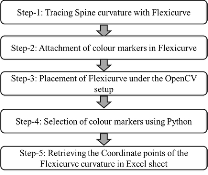
Figure 1: Steps followed in coordinates retrieval of Flexicurve.
radiography. The results show that Flexicurve produces Moderate to Good results as radiography and has certain limitations like a person’s BMI (Bar- rett et al., 2014). But the problem involved in the manual measurement of the Flexicurve technique is it takes more time, and it is difficult to follow this methodology if we have a more significant number of participants.
Moreover, there are some chances of getting errors in spine curvature from flexicurve due to improper handling. For flexicurve measurement, the author uses a formula to find the kyphosis angle concerning length and depth of the curvature obtained from flexicurve θ = 2sin−1 (4dL/ 4d2 + L2) (Grindle et al., 2020). This present study proposes a method to overcome the draw- backs of the flexicurve technique and improve the measurement accuracy. This study aims to measure spine curvature using flexicurve by integrating machine vision techniques.
INTEGRATION OF MACHINE VISION AND FLEXICURVE This study proposes a new methodology to measure the spine curvature angles using a flexicurve instrument with a machine vision system. This study is constrained to measuring spine curvature in the thoracic and lumbar regions—T1 to T12 and L1 to L5. Colour markers are added with the traced Flexicurve, and a camera identifies these markers in OpenCV, and it gives the coordinate points between T1 and T12 and L1 and L5 using python pro- gramming. The spine curvature data is stored automatically in a drive or excel sheet. OpenCV based Python program is developed to solve computer vision- related difficulties with Python bindings library. OpenCV is capable of image analysis and processing. It allows you to analyze the image through different patterns. Image Normalization, Edge Detection and several other exceptional features are utilized in OpenCV. It takes frames from the video or two fra- mes from a stereoscopic camera and performs algorithms and mathematical tools to extract information and retrieve coordinate points. A high-definition webcam has been utilized to capture the image.
Measurement of Spine Curvature using Flexicurve Integrated with Machine Vision 85
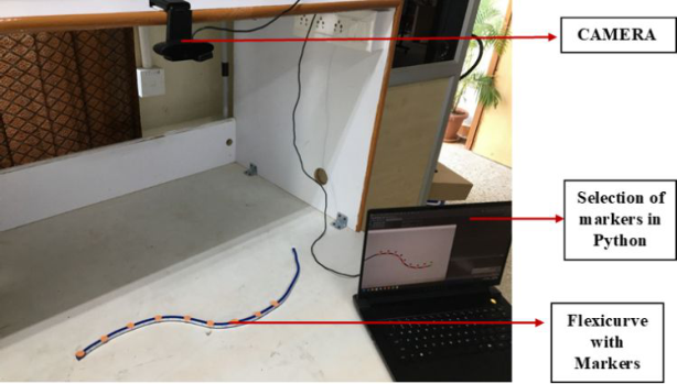
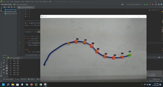
Figure 2: Flexicurve placed under OpenCV setup.Figure 3: Selection of colour markers in python.
The spine curvature is traced using flexicurve, and then colour markers are attached on the side of the flexicurve. The flexicurve with colour markers is placed under the OpenCV setup (see Figure 2). The python program is coded to track the through an interface. While selecting each marker, the video frame will freeze, and then a marker selection is made (see Figure 3). After choosing the last marker, the coordinate points are automatically stored in an excel sheet.
CONCLUSION
Numerous researchers have been utilizing the Flexicurve instrument to under- stand the spine curvature for research purposes. The proposed methodology of measuring spine curvature is expected to be precise; cost-efficient requires less time and less computational power. In future, the proposed technique has to be validated with standard procedures for reliability.
REFERENCES
Adams, M. A., Dolan, P., Marx, C., & Hutton, W. C. (1986). An electronic inclinometer technique for measuring lumbar curvature. Clinical Biomechanics, 1(3), 130–134. Barrett, E., McCreesh, K. and Lewis, J., (2014). Reliability and validity of non-
radiographic methods of thoracic kyphosis measurement: a systematic review. Manual therapy, 19(1), pp. 10–17. Bumacod, D.S.F., Delfin, J.V., Linsangan, N. and Angelia, R.E., (2020), December.
Image-Processing-based Digital Goniometer using OpenCV. In 2020 IEEE 12th International Conference on Humanoid, Nanotechnology, Information Techno- logy, Communication and Control, Environment, and Management (HNICEM) (pp. 1–6). IEEE. Chaise, F. O., Candotti, C. T., Torre, M. L., Furlanetto, T. S., Pelinson, P., & Loss, J. F.
(2011). Validation, repeatability and reproducibility of a noninvasive instrument for measuring thoracic and lumbar curvature of the spine in the sagittal plane. Brazilian Journal of Physical Therapy, 15(6), 511–517. Clément, J.L., Geoffray, A., Yagoubi, F., Chau, E., Solla, F., Oborocianu, I. and Rampal, V., (2013). Relationship between thoracic hypokyphosis, lumbar lordosis and sagittal pelvic parameters in adolescent idiopathic Scoliosis. European spine journal, 22(11), pp. 2414–2420. de Oliveira, T. S., Candotti, C. T., La Torre, M., Pelinson, P. P. T., Furlanetto, T. S.,
Kutchak, F. M., & Loss, J. F. (2012). Validity and reproducibility of the measure- ments obtained using the flexicurve instrument to evaluate the angles of thoracic and lumbar curvatures of the spine in the sagittal plane. Rehabilitation research and practice, 2012. Eiichi Ichikawa and Morio Ichikawa, (2011). Human body backbone measuring/displaying system, United States US7883477B2. Good, C.R., Auerbach, J.D., O’Leary, P.T. and Schuler, T.C., (2011). Adult spine
deformity. Current reviews in musculoskeletal medicine, 4(4), pp. 159–167. Grindle, D.M., Mousavi, S.J., Allaire, B.T., White, A.P. and Anderson, D.E., (2020).
Validity of flexicurve and motion capture for measurements of thoracic kyphosis vs standing radiographic measurements. JOR spine, 3(3), p.e1120. Horng, M.H., Kuok, C.P., Fu, M.J., Lin, C.J. and Sun, Y.N., (2019). Cobb angle measurement of spine from X-ray images using convolutional neural network. Computational and mathematical methods in medicine. Hou Jianjun, Yoon Wu, Shen Liming., (2012). Spine Positioning Point Measuring
Instrument. China patent CN103070689A, 2012–12-14. Itoi, E. and Mori, Y., (2017). Posture Analysis in Patients with Spinal Osteoporosis. In Non-Pharmacological Management of Osteoporosis (pp. 31-43). Springer, Cham. Raupp, E.G., Candotti, C.T., Marchetti, B.V., Vieira, A., Medeiros, F.S. and Loss,
J.F., (2017). The validity and reproducibility of the Flexicurve in the evaluation of cervical spine lordosis. Journal of manipulative and physiological therapeutics, 40(7), pp. 501–510. Voinea, G.D., Butnariu, S. and Mogan, G., (2017). Measurement and geometric modelling of human spine posture for medical rehabilitation purposes using a wearable monitoring system based on inertial sensors. Sensors, 17(1), p. 3.
LISTED IN THE AHFE DIGITAL BOOKSTORE CATALOGUE
Explore the complete digital volume
Human Factors and Wearable Technologies
Read this individual research paper free through the original publisher, or explore the full digital proceedings book.
ISBN 978-1-958651-05-6
Explore this ISBN in the AHFE Bookstore ↗Listed in the July 2026 bookstore catalogue; search by ISBN to confirm current availability · Digital book purchase optional