Refractory patients who fail standard therapies have limited options, and NK92-, CD16+NK92-, and CAR-NK92–based approaches are being explored to prolong survival and serve as bridging therapy to transplantation. Because NK92 cells are infused after irradiation, their in vivo persistence and activity are short-lived. We will generate CD16-expressing NK92 cells to enhance cytotoxic potency and broaden applicability by enabling tumor-associated antigen (TAA)–directed, monoclonal antibody–dependent cellular cytotoxicity (ADCC). In parallel, we will engineer “ASSASSIN” cells (CD16+IL12+NK92) and introduce BCMA-specific CARs to create CAR ASSASSIN (CAR+CD16+IL12+NK92) cells capable of dual/bispecific killing via CAR recognition and CD16-driven ADCC. This combinatorial targeting is designed to reduce antigen-escape–mediated loss of efficacy, a key limitation of conventional CAR therapies. To promote more durable immune control despite the transient lifespan of irradiated NK92 cells, IL-12 secretion is incorporated to stimulate the host immune system, aiming to sustain antitumor activity after infused cells disappear. Overall, this study proposes a ready-to-use, IL-12–secreting NK92 platform that can be redirected to multiple tumor antigens within a bridging-therapy framework and may support the development of standardized, flexible, and clinically actionable treatment approaches for time-sensitive therapeutic settings.
Standard anticancer therapeutic approaches have led to measurable survival benefits across many tumor types. However, despite gains in overall survival, the inability to consistently improve progression-free survival and, more importantly, the failure of combined immunotherapy protocols to achieve durable cures have generated disappointment in the field (Bilusic and Gulley, 2012). These limitations highlight key biological barriers such as tumor immune evasion, antigen heterogeneity, and treatment-driven selection, underscoring the need for next-generation immune-based strategies.
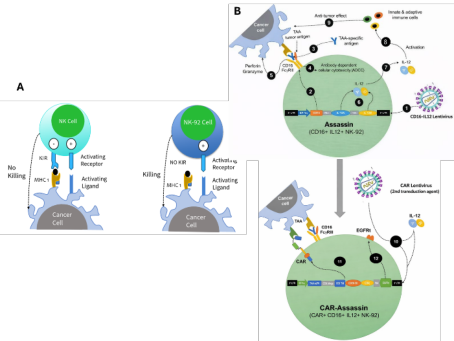
Advances in molecular biology and the increasing accessibility of genetic engineering have accelerated a shift from chemotherapy-dominated regimens toward targeted and cellular therapies. The overarching goals of these innovative approaches are to increase response rates and quality of life, improve overall survival, and prolong disease-free intervals (Qian et al., 2025; Min and Lee, 2022). In this context, chimeric antigen receptor (CAR)–engineered NK platforms offer an additional tumor-recognition mechanism, particularly in patient settings where ligands required for endogenous NK receptor activation are downregulated (Tang and Chen, 2025). Moreover, CAR signaling following tumor engagement can promote NK-cell proliferation and contribute to improved persistence (Acharya et al., 2024; Marin et al., 2024; Balkhi et al., 2025). In the present study, we aim to enhance cytotoxic efficacy and enable a broadly applicable, tumor-type–agnostic (“universal targeting”) strategy by generating CD16-expressing NK92 cells. CD16 expression will allow antibody-dependent cellular cytotoxicity (ADCC) to be triggered using tumor-associated antigen (TAA)–specific monoclonal antibodies, thereby diversifying and strengthening antitumor activity. In parallel, we define “ASSASSIN” cells as antibody-activated CD16+IL12+NK92 cells, and we will further engineer these cells to express TAA-specific CAR receptors to create BCMA CAR ASSASSIN (CAR+CD16+IL12+NK92) cells (Figure 1). This design is intended to produce bispecific (“double-hit”) cytotoxicity by combining CAR-mediated recognition with CD16-driven ADCC. Such combinatorial targeting is expected to mitigate a major limitation of conventional CAR therapies—loss of efficacy due to target antigen escape.
In addition to its biological novelty, this strategy has practical translational relevance for time-sensitive clinical settings. Because refractory patients often face narrow treatment windows, bridging approaches must combine antitumor potency with rapid deployability, safe implementation, and manufacturing consistency. A ready-to-use NK92-based platform with flexible retargeting capacity may therefore provide practical value by supporting standardization and facilitating integration into real-world treatment workflows.
A further challenge for NK92-based products is their limited lifespan, which may restrict durable immune control. To address this, IL-12 expression in ASSASSIN and CAR ASSASSIN cells is incorporated to stimulate the host innate immune system and promote sustained antitumor immunity. We hypothesize that, even after the infused ASSASSIN cells are eliminated, an activated host immune response will continue to exert pressure on the tumor and contribute to prolonged disease control.
Figure 1: NK92 vs ASSASSIN vs BCMA CAR ASSASSIN (biorender.com).
MATERIALS AND METHODS
Synthesis of CAR Construct and Lentivirus (LV) Production A BCMA-specific CAR–encoding lentiviral vector (CAR-LV; anti-BCMA scFv h(CD28–CD3ζ)–EGFRt) was designed and synthesized by Creative Biolabs. The pCMV-VSV-G envelope plasmid (gift from Bob Weinberg; Addgene #8454; RRID: Addgene_8454) and the psPAX2 packaging plasmid (gift from Didier Trono; Addgene #12260; RRID: Addgene_12260) were obtained from Addgene. The CD16-IL12 construct was designed in our laboratory and purchased from GenScript. Except for the updated plasmid constructs, all procedures—including lentiviral production, LV titration and calculation of number of infection units per milliliter (IFU/mL) and quality control tests (appearance, viral titer, sterility, mycoplasma, identity, host cell protein, replication-competent lentivirus)—were carried out without further modification according to our previously published protocol (Taştan et al., 2020).
NK-92 Cell Transduction and Culture Conditions NK-92 cells were maintained in suspension at 37 °C in a humidified 5% CO2 incubator in TexMACS medium supplemented with 20% heat-inactivated fetal bovine serum, 2 mM L-glutamine, and recombinant human IL-2 (5 ng/ mL, Miltenyi Biotech). Cultures were gently resuspended to break up large
Improving Outcomes in Patients’ Refractory to Standard Therapies: Dual-Mechanism 157 aggregates and were passaged by dilution with fresh medium every 2–3 days, maintaining cell density at approximately 0,1–0,4 × 10⁶ cells/mL; IL-2 was kept in the medium throughout, as NK-92 proliferation is IL-2 dependent. Transduction was performed sequentially—first with the CD16-IL12 LV (5 MOI) and then with the CAR BCMA LV (5 MOI)-using the concentrated (100×) lentiviral preparation in the presence of Vectofusin-1 (10 μg/mL; Miltenyi MACS), followed by centrifugation (400×g for 60 min). To enhance transduction efficiency in NK-92 cells, BX-795 (Sigma-Aldrich) was added to a final concentration of 6 μM for 6 h during lentiviral exposure, as previously reported for several cell types (Sutlu et al., 2012). CD16-IL12+ or CAR+ cells were isolated using a streptavidin-based selection approach and subsequently expanded under cGMP-compliant conditions. Following transduction, the engineered NK-92 cells were subsequently cultured, expanded and sorted via flow cytometry. Transduced cells were sent to Acıbadem Altunizade Hospital for irradiation. Cells were irradiated at 1,000 cGy, returned to the laboratory immediately, washed twice with DPBS, and counted prior to downstream experiments. Alternatively, cells were cryopreserved and stored as an off-the- shelf product until use. Quality control assessment was done according to previously published protocol (Taştan et al., 2020).
In Vitro Efficacy Analysis Cytotoxicity assays were performed by co-culturing target tumor cell lines BCMA+ H929 cells with ASSASSIN or BCMA CAR-ASSASSIN effector cells for 24h (effector:target; 1:1, 5:1, 10:1). H929 cells were transduced with an fLuciferase–mCherry plasmid for tumor cell tracking. Flow cytometry–based in vitro cytotoxicity assays were performed by co-culturing mCherry+ H929 target cells with effector cells at effector-to-target (E:T) ratios of 1:1, 5:1, and 10:1, and tumor burden was quantified based on mCherry expression to assess cytotoxic activity.
Mice Xenograft Model and Histological Analysis All animal experiments were conducted in accordance with protocols approved by the Acıbadem Mehmet Ali Aydınlar University Laboratory Animal Application and Research Center (ACUDEHAM; ACU-HADYEK approval no. 2019/56), where all procedures were performed. The 5x10⁵ fLuciferase-mCherry-positive H929 cells were then injected intraperitoneally (IP) into each NOD/SCID mouse in all groups at day 0. Bioluminescent positive tumors were determined within 3 days after the injection. Tumor challenge was established by i.p. injection of 5×10^5 Fluc-mCherry– expressing (85,76%) H929 cells. For intraperitoneal (i.p.) treatment, BCMA
CAR-ASSASSIN (or ASSASSIN) cells were administered i.p. at 4×10^7 cells per NOD/SCID mouse. Each experimental group included 5 mice (n = 5). Tumor growth and development were monitored by bioluminescence with Luciferin (1 mg/mL) injection. The emitting bioluminescence signal of tumors was measured with the IVIS in vivo imaging system. Liver, spleen, and heart tissue samples were collected at the end of the experimental period and fixed in 10% neutral buffered formalin for histopathological examination. Following fixation, tissues were trimmed routinely, dehydrated through graded alcohol series, cleared in xylene, and embedded in paraffin blocks. Paraffin-embedded tissue blocks were sectioned at a thickness of 5 µm using a rotary microtome. Tissue sections were mounted on glass slides and stained with hematoxylin and eosin (H&E) according to standard histological procedures. All stained sections were examined under a light microscope at magnifications of ×40 and ×400. Histological evaluation focused on: Liver: hepatocyte morphology, sinusoidal structure, vascular congestion, and Kupffer cell distribution Spleen: organization of white pulp, red pulp, presence of primary and secondary follicles, and macrophage infiltration Heart: myocardial fiber organization, cardiomyocyte nuclei, and structural integrity Representative micrographs were captured for each experimental group.
Statistics Analysis All data are presented as mean ± standard error of the mean (SEM). One-way analysis of variance (ANOVA) and two-way ANOVA were used to evaluate the parameters, as appropriate. A P value<0.05 was considered statistically significant. Statistical analyses were performed using GraphPad Prism (version 8).
RESULTS AND DISCUSSION
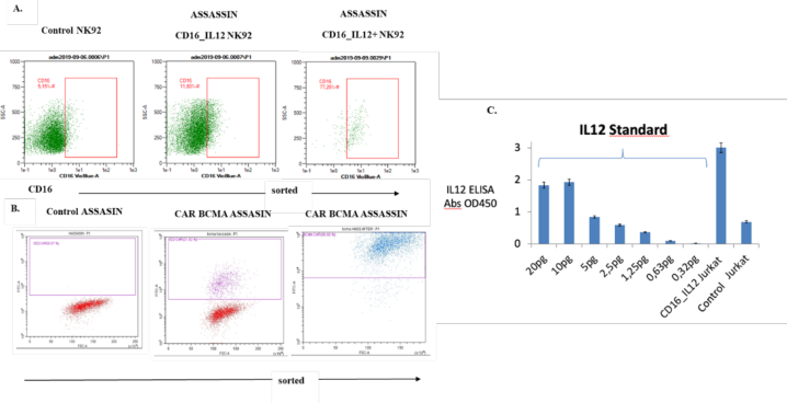
ASSASSIN and BCMA CAR-ASSASSIN Cell Process Outputs: Production Yield, QC Metrics, and Performance An NK-92 cell line expressing CD16 and IL-12 was generated by lentiviral transduction, establishing a universal cell line. To increase the transduction efficiency, CD16-based FACS selection was performed. Expression levels were analyzed by flow cytometry, showing 77,25% positive cells after sorting. On the other hand, CAR BCMA ASSASSIN cells, the BCMA-specific CAR expression level was determined by flow cytometry to be 96,66% (Figure 2). Upon repeating the flow cytometry analysis two months later, the BCMA CAR expression profile was found to be approximately 82% (Data not shown). Using Jurkat cells as a control transduction model, ELISA analysis demonstrated that the construct successfully mediates and significantly increases IL-12 expression (Figure 2).
Figure 2: Production of ASSASSIN and BCMA CAR–ASSASSIN cells. Representative flow cytometry plots showing (A) NK-92 cells, either untransduced or transduced with CD16–IL-12, were stained with an anti-CD16 antibody to generate ASSASSIN cells, followed by post-sort purity assessment. (B) ASSASSIN cells, either untransduced or transduced with a BCMA-specific CAR, were stained with an αEGFR-A488 antibody, followed by post-sort data acquisition to assess population purity. (C) ELISA-based validation of IL-12 expression using a Jurkat control transduction model.
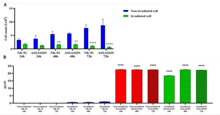
To prevent secondary tumorigenesis in in vivo applications (Tonn and Schwabe, 2016; Klingemann, 2023; Klingemann, 2025), NK-92 and ASSASSIN cells intended for use in this study were irradiated at 1,000 cGy, and both preclinical in vitro and in vivo workflows were planned accordingly. Therefore, we evaluated whether irradiation affected post-irradiation cell viability. Irradiated and non-irradiated NK-92 and ASSASSIN cells were seeded into well plates, and viability was compared at 24, 48, and 72 hours. In addition, IL-12 expression levels in irradiated (+) and non-irradiated (−) NK-92 and ASSASSIN cells were measured using a commercial ELISA kit (Thermo Fisher Scientific) according to the manufacturer’s instructions. As expected, although the viability of irradiated (+) NK-92 and ASSASSIN cells decreased compared with non-irradiated (−) cells, the irradiated cells were able to maintain viability for up to 48 hours. Due to the proliferation observed in non-irradiated (−) cells, a significant difference in cell number was detected at 48 hours compared with irradiated (+) cells (p < 0.001), and this difference further increased at 72 hours (p < 0.0001) (Figure 3, Çilingir, 2024).
Figure 3: A. Post-irradiation cell viability in irradiated and non-irradiated NK-92 and ASSASSIN cells B. IL-12 expression levels in irradiated (+) and non-irradiated (−) NK- 92 and ASSASSIN.
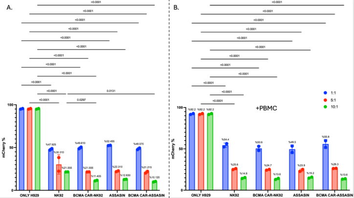
Each cell product was tested by Acıbadem Labmed Laboratory for mycoplasma, endotoxin, VDRL (Venereal Disease Research Laboratory) test, fungal culture, aerobic and anaerobic bacterial cultures, hepatitis B, hepatitis C, human immunodeficiency virus (HIV), and Epstein–Barr virus (EBV) and found to be negative (Data not shown, Çilingir, 2024). To assess the activity of irradiated effector cells under human-simulating conditions, in vitro cytotoxicity assays were performed in the presence and absence of PBMCs. H929 tumore cells were transduced with an fLuciferase–mCherry plasmid using a lentiviral vector at an MOI of 10 for tumor cell tracking, followed by fluorescence-activated cell sorting to enrich mCherry-positive cells, resulting in a final purity of 97% (Data not shown). In vitro cytotoxicity assays against H929 multiple myeloma cells indicated that BCMA CAR–engineered ASSASSIN cells mediated the highest antitumor activity, yielding the lowest residual H929 viability at an effector-to-target ratio of 10:1 (10.12% without PBMCs and 13.57% in the presence of PBMCs, Gafarlı İ, 2022). Although PBMC co-culture did not further increase killing, the sustained activity observed in long-term culture suggests that BCMA CAR–ASSASSIN cells may retain efficacy under preclinical in vivo conditions (Figure 4).
Figure 4: Flow cytometry–based cytotoxicity assessment of ASSASSIN and BCMA CAR–ASSASSIN cells against mCherry⁺ H929 tumor cells. mCherry⁺ H929 cells were co-cultured with NK-92, BCMA CAR–NK92, ASSASSIN, or BCMA CAR–ASSASSIN effector cells at effector-to-target (E:T) ratios of 1:1, 5:1, and 10:1. Tumor cell burden was quantified by flow cytometric analysis of mCherry expression. (A) Cytotoxicity assay performed in the absence of PBMCs. (B) Cytotoxicity assay performed in the presence of PBMCs (+PBMC). Data are presented as mean ± SEM. Statistical significance was determined using multiple comparisons, with indicated p-values.
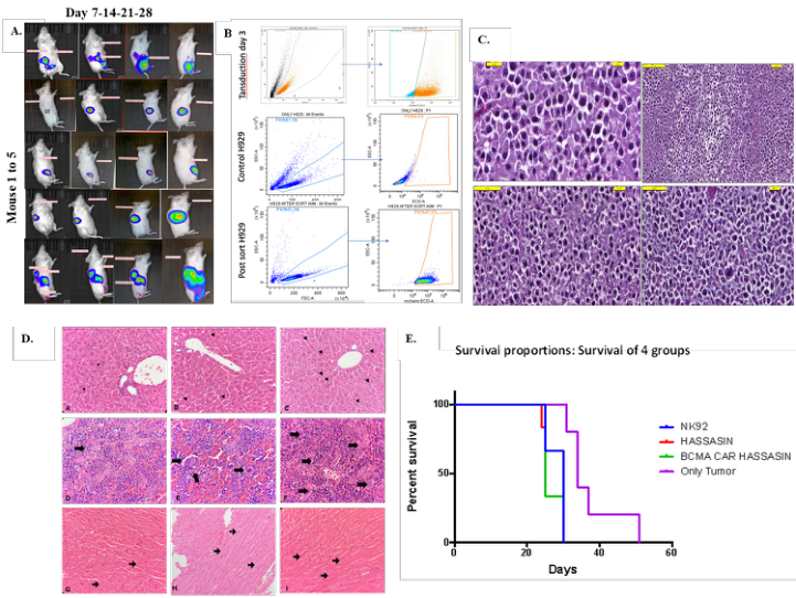
In Vivo Validation of ASSASSIN and BCMA CAR–ASSASSIN Cell Antitumor Efficacy With H929 Cancer Model Preclinically efficient ASSASSIN and BCMA CAR ASSASSIN cells were next tested in an in vivo cancer model with NOD/SCID mice. Figure 4 shows the in vivo experimental approach, where fLuciferase-mCherry-positive H929 cells, control ASSASSIN and CAR BCMA ASSASSIN cells were transferred to NOD/SCID mice. The growth rates of the fLuc mCherry-expressing H929 cells (98%, Figure 5) in the mice were tracked using luciferin for in vivo bioluminescence screening for approximately 50 days. Tumors were established by inoculating 5 × 10⁵ tumor cells. ASSASSIN and BCMA CAR–ASSASSIN effector cells were administered at a dose of 4 × 10⁷ cells (number of transduced (marker-positive) cells) per treatment on days 5, 7, and 9, followed by longitudinal monitoring of the animals. Imaging analyses on day 5 confirmed tumor establishment. In the between-group analysis of toxicological evaluation, the BCMA CAR–ASSASSIN group showed a significant change in heart and spleen weights compared with the control group (p < 0.05). In addition, one mouse died in this group. These findings may indicate treatment-associated toxicity, potentially related to heightened activation in the BCMA CAR–ASSASSIN group. In survival analysis, no significant survival advantage was observed in the treatment groups compared with the tumor group. Specimens consistent with multiple myeloma tumor tissue were submitted, and histopathological examination of the received material demonstrated a plasma cell infiltration exceeding 10% (Figure 5).
Histopathological analysis revealed organ-specific and treatment- dependent responses across experimental groups. ASSASSIN administration resulted in mild hepatic vascular congestion without hepatocellular damage, accompanied by pronounced immune activation in splenic tissue, as evidenced by increased primary and secondary follicle formation. In contrast, BCMA CAR ASSASSIN treatment maintained normal hepatic and cardiac morphology while inducing prominence Kupffer cell visibility in the liver and a marked expansion of primary splenic follicles, suggesting a shift toward macrophage-mediated immune modulation rather than overt inflammatory activation. Notably, no histopathological alterations were detected in myocardial tissue across any treatment group, indicating the absence of cardiotoxic effects (Figure 5).
Figure 5: A. Bioluminescence radiance of all mice in the only tumour groups (mouse 1 to 5). B. Flow cytometric gating strategy and enrichment of mCherry⁺ H929 tumor cells. C. Histopathologic evaluation of multiple myeloma tumour tissues (NK-92 Group, ASSASSIN Group, CAR-BCMA ASSASSIN Group, Tumor Group, respectively). D. Histopathological evaluation of liver, spleen, and heart tissues. Representative hematoxylin and eosin–stained sections of liver (A–C), spleen (D–F), and heart (G–I) tissues from experimental groups are shown. Panels A, D, and G represent the control group; panels B, E, and H represent the ASSASSIN-treated group; and panels C, F, and I represent the BCMA CAR– ASSASSIN -treated group. In liver sections (A–C), normal hepatic architecture is preserved across groups, with prominence of Kupffer cells indicated by arrowheads. Spleen sections (D–F) demonstrate differences in follicular organization, with arrows indicating primary follicle formation and immune cell aggregation. Heart sections (G–I) display normal myocardial fiber organization in all groups, with arrows indicating cardiomyocyte nuclei. Arrows and arrowheads indicate relevant histological features as described. H&E×400. E. Mean of survival probability rates of the groups based on screening for 50 days.
CONCLUSION
In conclusion, donor-independent, cryopreservable ASSASSIN and BCMA CAR–ASSASSIN cell lines were developed as therapeutic candidates with faster manufacturing timelines and lower production costs. These cells demonstrated effective cytotoxic activity against multiple myeloma and modulated the host immune response. Notably, BCMA CAR–ASSASSIN cells demonstrated superior antitumor activity. Their efficacy could be further enhanced through combination strategies, particularly with anti-BCMA monoclonal antibodies, given the CD16 expression on ASSASSIN cells and the potential to augment antibody-dependent cellular cytotoxicity (ADCC). While these findings are promising, additional follow-up studies are required to comprehensively assess safety and efficacy, and evaluating combination approaches with anti-BCMA monoclonal antibodies represents a key focus of future work. Taken together, these findings highlight both the therapeutic promise of the platform and its translational potential as a standardized and readily deployable strategy for clinically time-sensitive settings, while underscoring the need for careful evaluation of safety, dosing, and regulatory feasibility prior to clinical implementation.
In contrast, in a rapidly progressing systemic tumor model, administration of irradiated NK-92–derived products did not confer sustained antitumor benefit and may have imposed additional physiological stress. The lack of in vivo persistence and proliferative capacity of NK-92 cells following 1000 Gy irradiation, together with potential acute inflammatory or cytotoxic effects immediately after cell infusion, may have contributed to the reduced survival observed in treated groups. These findings indicate that, while target recognition and cytotoxic function are effective in vitro, durable disease control in aggressive systemic malignancies will likely require effector cells with enhanced persistence and expansion capacity. Accordingly, future studies will focus on evaluating the ASSASSIN platform in non-irradiated, persistence-competent effector formats, such as primary NK cells, to determine whether improved in vivo expansion and persistence can enhance efficacy in systemic models.
ACKNOWLEDGEMENT
The author(s) received no financial support for the research. The authors thank Acibadem Labcell Cellular Therapy Center for their support of the project. We thank Dr. Tuğba Bağcı Önder from Koç University, who gave us the fLuciferase-mCherry encoding lentiviral plasmid, and Dr. Dilek Telci from Yeditepe University, who gave us the HEK293T cells. The research in this study, supervised by Dr. Ercüment Ovalı, was supported in Turkey by Technology and Innovation Funding Programs Directorate (TEYDEB) grant 3191231.
REFERENCES
Acharya, S., Basar, R., Daher, M. et al. (2024) ‘CD28 Costimulation Augments CAR Signaling in NK Cells via the LCK/CD3ζ/ZAP70 Signaling Axis’, Cancer Discovery, 14(10), pp. 1879–1900. Balkhi, S., Zuccolotto, G., Di Spirito, A., Rosato, A. and Mortara, L. (2025) ‘CAR-NK
cell therapy: promise and challenges in solid tumors’, Frontiers in Immunology, 16, 1574742. Bilusic, M. and Gulley, J.L. (2012) ‘Endpoints, patient selection, and biomarkers in the design of clinical trials for cancer vaccines’, Cancer Immunology, Immunotherapy, 61, pp. 109–117.Jiang et al. 2014 Çilingir, S, et al. (2024) HAŞŞAŞİN (IL-12 CD16+ NK-92) ve Muc-1 spesifik CAR
HAŞŞAŞİN’in(Muc-1 CAR IL-12 CD16+ NK-92) In Vitro ve In Vivo pankreas kanseri modellerinde etkinliği. [Acıbadem Mehmet Ali Aydınlar Üniversitesi, Sağlık Bilimleri Enstitüsü, Fizyoloji ABD], [İstanbul]. Gafarli, I. (2022) Multipl Myelom Hastalığında BCMA CAR, CD16 ve IL-12 Eksprese Eden NK-92 Hücrelerinin (BCMA CAR HAŞHAŞİN) Preklinik Etkinlik Çalışması. Tıpta Uzmanlık Tezi. [Acıbadem Altunizade Hastanesi, İç Hastalıklar ABD], [İstanbul]. https://hdl.handle.net/11443/ 2994 Klingemann, H. (2023) ‘The NK-92 cell line—30 years later: its impact on natural
killer cell research and treatment of cancer’, Cytotherapy, 25(5), pp. 451–457. Klingemann, H. (2025) ‘The Natural Killer Cell Line NK-92 and Its Genetic Variants: Impact on NK Cell Research and Cancer Immunotherapy’, Cancers, 17(12), 1968. Marin, D., Li, Y., Basar, R. et al. (2024) ‘Safety, efficacy and determinants of response
of allogeneic CD19-specific CAR-NK cells in CD19+ B cell tumors: a phase 1/2 trial’, Nature Medicine, 30, pp. 772–784. Min, H.-Y. and Lee, H.-Y. (2022) ‘Molecular targeted therapy for anticancer treatment’, Experimental & Molecular Medicine, 54(10), pp. 1670–1694. doi:10.1038/s12276-022-00864-3. Qian, Y., Ma, W. and Xu, X.N. (2025) ‘Breakthrough for Anticancer Immunotherapy:
Current Advances in Manufacturing Protocols of Chimeric Antigen Receptor- Based Therapies’, Antibodies, 14(4), p. 105. Sutlu, T., Nyström, S., Gilljam, M., Stellan, B., Applequist, S.E. and Alici, E. (2012) ‘Inhibition of intracellular antiviral defense mechanisms augments lentiviral transduction of human natural killer cells: implications for gene therapy’, Human Gene Therapy, 23, pp. 1090–1100. Tang, E. and Chen, J.Y. (2025) ‘Shaping the glioblastoma microenvironment to
enhance CAR-NK immunotherapy’, Frontiers in Immunology, 16, 1681966. Taştan, C., Kançağı, D.D., Turan, R.D. et al. (2020) ‘Preclinical Assessment of Efficacy and Safety Analysis of CAR-T Cells (ISIKOK-19) Targeting CD19-Expressing B-Cells for the First Turkish Academic Clinical Trial with Relapsed/Refractory ALL and NHL Patients’, Turkish Journal of Hematology, 37(4), pp. 234–247. Tonn, T. and Schwabe, D. (2016) ‘Natural Killer Cells for Immunotherapy –
Advantages of the NK-92 Cell Line’, Frontiers in Immunology, 7, 91.
LISTED IN THE AHFE DIGITAL BOOKSTORE CATALOGUE
Explore the complete digital volume
Healthcare and Medical Devices
Read this individual research paper free through the original publisher, or explore the full digital proceedings book.
ISBN 978-1-964867-87-8
Explore this ISBN in the AHFE Bookstore ↗Listed in the July 2026 bookstore catalogue; search by ISBN to confirm current availability · Digital book purchase optional