Lucas Paletta, Michael Schneeberger, Martin Pszeida, Dietmar Maurer (2026). Rapid Military Triage of Traumatic Brain Injury Using Eye Tracking and Pupillometry. In: Cognitive Computing and Internet of Things. AHFE Open Access, vol. 205, pp. 249–265. https://doi.org/10.54941/ahfe1008068
Rapid triage in military operations must reliably identify “hidden” neurological impairment among soldiers who appear only slightly wounded. Mild to moderate traumatic brain injury (TBI), concussion, and neurotoxic exposures can present with subtle early signs that are easily missed under stress, time pressure, and resource constraints. Eye tracking and pupillometry are promising for early triage because they probe brainstem and oculomotor function, can be captured quickly with deployable devices, and provide quantitative, repeatable measures suitable for decision support. This paper proposes a research program to develop an eye tracking–based, explainable rule base with operational thresholds to support early military triage. The goal is a field-ready decision logic that estimates the likelihood of clinically relevant impairment (e.g., concussion/moderate TBI, severe TBI warning signs, or neurotoxic syndromes) in “walking wounded” soldiers and outputs a transparent triage recommendation (e.g., high / medium / low risk), aligned with medic workflows. Methodology. We will implement a rapid assessment protocol combining (1) pupillary light reflex metrics, (2) basic oculomotor screening (e.g., gaze stability, smooth pursuit, saccadic control), and (3) short mobile cognitive-oculomotor tasks that can be completed in minutes. Candidate rules will be derived from a structured synthesis of prior clinical and applied evidence and formalized into interpretable “if–then” statements with thresholds and confidence flags. The rule base will be designed with explicit handling of confounds relevant to deployment (illumination variability, fatigue, stress, motion, and partial occlusion), including quality checks and fallback logic when signals are unreliable. Expected results. The project will deliver (i) a prototype triage tool integrating eye tracking data capture and automated rule evaluation, (ii) an initial threshold library for the most informative pupil and oculomotor features, and (iii) a validation plan and performance targets emphasizing sensitivity to clinically relevant TBI risk while maintaining practical specificity for operational use. We will prioritize explainability (rules that medics can interpret) and actionability (clear next steps such as observation, repeat testing, or expedited evacuation).Future work will include controlled studies for normative baselines, simulated field trials, and prospective evaluations in training environments, with iterative refinement toward robust deployment and integration with other physiological measures where beneficial.
Military medics often have to decide within minutes whether a soldier who is able to walk and communicate can remain under observation, should be removed from duty, or requires urgent evacuation. The most difficult cases are not the visibly severe casualties, but the hidden neurological injuries after blast, blunt head impact, acceleration-deceleration trauma or chemical
exposure. These cases can present with headache, dizziness, confusion, light sensitivity, balance disturbance, nausea, abnormal eye movements or subtle pupil abnormalities while the casualty still appears operationally functional.
Current concussion screening and neurological examination remain essential, but field use is constrained by noise, darkness, time pressure, stress, protective equipment, mixed injury mechanisms and the need for rapid prioritization. Military concussion tools such as MACE 2 were designed to support deployed assessment, yet research on usability shows that even accepted tools require training and can be perceived as complex in operational settings (Khokhar et al., 2021). A deployable digital biomarker system should therefore not replace the medic. It should add quantitative, repeatable observations that can be interpreted quickly and transparently together with history, symptoms, vital signs and tactical constraints.
(a)
(b) (c)
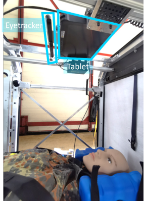
Figure 1: Functional demonstrator for an eye-tracking-enabled ‘patient box’ for rapid military triage: (a) patient box with integrated casualty position and sensor infrastructure, (b) unmanned ground vehicle (UGV1) for mobile deployment1, and (c) close-up view of the internal eye-tracking (Tobii Pro Fusion/Spark) and tablet- based assessment setup for pupillometry and oculomotor screening. (Credits: M. Schneeberger, ChatGPT for image postprocessing; patient box by courtesy of Autoflug GmbH).
In the envisaged operational concept, the eye-tracking triage module is integrated into a protected patient box that can be transported by an unmanned ground vehicle (UGV; see Figure 1). The patient box provides a controlled and repeatable measurement environment for the injured soldier, including a fixed head position, defined viewing geometry, a tablet display and an eye tracker positioned close to the casualty’s face. This setup is 1Credit: Milrem Robotics, NATO Centre of Excellence for Military Medicine.
Rapid Military Triage of Traumatic Brain Injury Using Eye Tracking and Pupillometry 251 intended to support rapid pupillometry and oculomotor screening during evacuation or at a casualty collection point, while reducing variability caused by hand-held operation, unstable posture, ambient light and movement. The UGV-mounted patient box therefore links autonomous casualty transport with objective neurological triage: while the soldier is physically protected and moved out of the danger zone, eye-tracking-based biomarkers can be collected to support early detection of traumatic brain injury, concussion or other neurological impairment. This concept complements the paper’s aim of developing deployable, explainable decision support for walking-wounded or evacuated soldiers under military field conditions.
The eye provides a particularly attractive measurement window because the pupil and oculomotor system are influenced by brainstem circuits, cranial nerves, autonomic state, cortical attention systems and vestibular-ocular integration. Quantitative pupillometry and eye tracking can measure pupil size, pupillary light reflex (PLR), saccades, smooth pursuit, gaze stability, nystagmus, convergence and task-linked visual-cognitive performance. This creates a compact measurement set that is relevant for TBI and concussion, but also for cholinergic neurotoxic exposure, where miosis and impaired vision are prominent early signs.
The contribution of this paper is a structured concept for an explainable, rule-based triage system. The overall system is designed within the European EDF project iMEDCAP2 as an explorative research component first, not for immediate clinical deployment. The central design principle is conservative decision support: the rule base should never make a definitive diagnosis of TBI or toxic exposure. Instead, it should flag risk levels, report the features that triggered the flag, document signal quality and medication confounds and recommend operational next steps such as repeat testing, observation, CBRN decontamination workflow or expedited evacuation.
RELATED WORK AND CONSIDERATIONS Mild TBI (mTBI; Eapen et al., 2022) is difficult to diagnose because symptoms can be transient, delayed or masked by adrenaline, pain, fatigue and mission demands. The Military Acute Concussion Evaluation was created to help deployed medics and corpsmen identify concussion, and the MACE 2 update has been evaluated for usability, utility and provider confidence in military health-care settings (Khokhar et al., 2021). However, deployed assessment remains vulnerable to self-report, environmental disruption and examiner variability. These limitations motivate additional objective signals that are rapid, quantitative and compatible with a medic workflow.
Eye tracking in mTBI. A structured review by Stuart et al. (2020) concluded that eye-movement assessment is promising in mTBI because saccades, smooth pursuit, fixations and nystagmus can reflect distributed neural systems, but also emphasized that protocols, instrumentation, processing algorithms and confound reporting are not yet standardized. This is directly relevant for military triage: a field system should avoid a black-box score and should document the precise task, sampling rate, 2https://defence-industry-space.ec.europa.eu/system/files/2023-06/iMEDCAP-Factsheet_EDF22.pdf
signal quality, luminance conditions and medication status behind each recommendation.
Visual and ocular biomarkers. Acute military mTBI studies have found that PLR velocity measures, near point of convergence and rapid number- naming or visual tracking tasks can discriminate injured from control participants. Capo-Aponte et al. (2018) reported that average constriction velocity, average dilation velocity and re-dilation time were affected in acute mTBI, and that dilation velocity and near point of convergence showed comparatively high predictive value. Maruta et al. (2010) proposed visual tracking synchronization as a concussion screening metric, while Murray et al. (2020) demonstrated deficits in smooth pursuit and saccadic performance after sport-related concussion. Vestibular/Ocular Motor Screening (VOMS) research supports brief screening of smooth pursuit, saccades, convergence, vestibular-ocular reflex and visual motion sensitivity, but symptom- provocation scores alone remain partly subjective (Mucha et al., 2014).
Quantitative pupillometry. Automated infrared pupillometry has advanced from intensive care monitoring to potential triage and mTBI applications. In severe TBI, Neurological Pupil index (NPi), constriction velocity, dilation velocity and latency have been associated with intracranial hypertension, neurological deterioration or outcome (Jahns et al., 2019; El Ahmadieh et al., 2021; Boulter et al., 2021). Recent mTBI studies support the potential of quantitative pupillometry in subtle impairment detection and clinical evolution tracking (Dengler et al., 2025; Mitschang et al., 2025). A key translational problem is that hospital thresholds cannot simply be copied into the field; they must be re-estimated under device-specific, illumination- controlled and medication-aware conditions.
Neurotoxic exposure. Organophosphate nerve agents inhibit acetylcholinesterase and can produce a cholinergic syndrome involving miosis, secretions, bronchospasm, fasciculations, seizures and respiratory failure. Reviews of nerve-agent medicine emphasize that ocular findings, especially miosis and impaired vision, may occur early and may persist after exposure (Candiotti, 2017; Figueiredo et al., 2018). For example, VX is a highly toxic organophosphate nerve agent. It belongs to the class of chemical warfare agents that inhibit acetylcholinesterase, an enzyme needed to stop nerve signalling at synapses and neuromuscular junctions. VX is operationally important because it is persistent and can produce delayed systemic toxicity after dermal exposure. Case literature and experimental ocular exposure studies demonstrate that VX and sarin can produce ocular injury, long-lasting miosis and impaired visual function (Nozaki et al., 1995; Egoz et al., 2017; Gore et al., 2019). In a triage system, miosis alone is therefore insufficient: the rule base must distinguish cholinergic toxicity patterns from opioid analgesia, bright light, ocular trauma and prior antidote administration.
Medication and physiological confounds. Pupil and oculomotor signals are strongly influenced by immediate field medication and physiological status. Opioids such as fentanyl or morphine can produce miosis and alter PLR, especially in hypoxia or hypercarbia (Rollins et al., 2014). Sedatives and antihistamines can modify arousal and pupil dynamics, while diazepam may not produce the simple miosis expected from sedation alone (Hou et al., 2006).
Rapid Military Triage of Traumatic Brain Injury Using Eye Tracking and Pupillometry 253 Atropine, used in nerve-agent treatment, is antimuscarinic and can create mydriasis or a nonreactive pupil, thereby masking the original cholinergic signal. Ketamine is useful for combat analgesia but may create nystagmus, dissociation and unreliable cognitive-oculomotor task performance. A robust triage system must therefore log all administered drugs and interpret the ocular result as medication-adjusted risk, not as an isolated biomarker.
RAPID EYE-TRACKING TRIAGE PROTOCOL The proposed assessment is designed for walking-wounded soldiers who are hemodynamically stable enough for a short measurement but may have hidden neurological impairment. The workflow is deliberately short. It can be performed after immediate life threats have been addressed, during observation, or at a casualty collection point. The intended sequence is: confirm safety and basic responsiveness; record mechanism, symptoms and medication; measure ambient illumination; conduct PLR; run oculomotor tasks; perform a 30-60 s cognitive-oculomotor task; and output an explainable risk flag with recommended next action (Table 1). An NPi-equivalent pupillary responsiveness index (NEPRI) is calculated from pupil diameter, latency, constriction velocity, dilation velocity and inter-eye asymmetry. The metric is conceptually inspired by the Neurological Pupil Index but does not reproduce the proprietary NPi algorithm.”
Table 1: Candidate eye-tracking and pupillometry feature set for rapid military triage.
Table available in the original PDF paper.
Module Primary Features Clinical/Operational Meaning
Quality and Confound Controls
PLR / pupil size
Brainstem/cranial nerve integrity, autonomic balance, miosis/mydriasis, severe neurological warning signs.
Controlled or measured luminance; repeat both eyes; exclude direct ocular trauma; log opioids, atropine, anticholinergics and chemical-antidote status.
Static and dynamic anisocoria
Difference in baseline diameter, difference in constriction amplitude or NEPRI between eyes, unilateral fixed or sluggish pupil.
Operationally relevant attention, scanning, executive control and situational-awareness proxy.
Keep task language- independent and less than one minute; use individual baseline when available.
The protocol should be implemented with either a handheld quantitative pupillometer, a mobile eye-tracking headset, eye-tracking glasses or a rugged tablet/smartphone configuration with a controlled light stimulus.
For field use, the system should include an illuminance sensor, a short calibration check, blink and occlusion detection, motion quality monitoring and an explicit “repeat/invalid” state. If the measurement is unreliable, the correct output is not a risk score but a quality-controlled instruction to repeat under improved conditions or fall back to manual neurological assessment.
The triage protocol (Table 1) should maintain a separation between three layers: raw observations, derived features and rule decisions. Raw observations include pupil videos, gaze samples and illuminance. Derived features include latencies, velocities and asymmetry indices. Rule decisions only use derived features after signal-quality gates are passed. This separation is crucial for scientific validation and medico-legal traceability.
EXPLAINABLE RULE BASE FOR FIELD TRIAGE The initial rule base is intentionally transparent (Table 2). Each rule has four fields: condition, evidence strength, confound handling and recommended action. The thresholds in Table 3 are candidate implementation thresholds for a proof-of-system study. They are not final diagnostic cut-offs and must be calibrated by device, task, illumination and population. In operational use, the rule base should prioritize sensitivity for severe risk while using repeated testing and symptom context to preserve specificity.
A key design decision is that the system should output both a risk level and a rationale. For example, a high-risk result might read: “High neurological risk: left NEPRI below threshold; anisocoria increased on repeat measurement; valid signal quality; no atropine or opioid documented.” A toxicology result might read: “High CBRN-compatible ocular pattern: bilateral miosis under measured low luminance plus secretions and exposure cluster; pupil score treatment-confounded after atropine.” This wording supports medic interpretation and prevents overclaiming.
Table 2: Explainable rule base for high-, medium- and low-risk triage outputs.
Table available in the original PDF paper.
Rule Family Candidate If-Condition Risk Output Recommended Operational Action Severe TBI warning High Expedited evacuation / urgent clinician review. Do not wait for cognitive task completion.
Any unilateral fixed/ dilated pupil; absent PLR in one eye; NEPRI <3.0 in one or both eyes; rapidly worsening anisocoria; repeated vomiting, seizure, deteriorating consciousness or focal neurological deficit.
Medium to high
Concussion / moderate TBI suspected
Remove from duty, observe, repeat test after short interval, apply MACE 2/ neurological exam and escalate if symptoms evolve.
Reliable measurement plus abnormal PLR velocity or latency relative to device baseline; abnormal smooth pursuit or saccade control; symptom provocation; blast/head impact history; cognitive-oculomotor task below threshold.
High if systemic signs present
Cholinergic neurotoxic syndrome suspected
Activate CBRN/ decontamination and medical nerve-agent workflow. After atropine/antidote, classify pupil metrics as treatment- confounded.
Bilateral miosis or pinpoint pupils under non-bright light; sluggish PLR; lacrimation/ rhinorrhea/salivation/ sweating/bronchospasm/ fasciculations; multiple similarly affected casualties or CBRN context.
Localized ocular exposure possible
Marked unilateral miosis or ocular pain/blurred vision after suspected chemical splash/vapor exposure without clear TBI mechanism.
Medium Protect responder, decontaminate according to protocol, repeat ocular assessment, monitor for systemic signs.
Medium / confounded
Medication- confounded miosis
Miosis after opioid/fentanyl/ morphine or hypoxia/ hypercarbia without cholinergic secretions and without exposure cluster.
Log medication and respiratory status; do not classify as nerve-agent exposure based on pupil size alone.
Confounded Use history and systemic signs; repeat later; do not use pupil thresholds as primary neurological evidence.
Mydriasis or weak PLR after atropine, anticholinergic exposure, sympathomimetic, ocular drops or local eye trauma.
Low apparent ocular risk
Low Document result; return to observation pathway only according to medic judgement and local policy.
Reliable bilateral PLR and oculomotor screen within expected range, no red flags, no symptoms, no concerning mechanism and no deterioration.
The rule base also requires negative logic. An isolated abnormal pupil finding should not be automatically treated as TBI or VX exposure. It should trigger a structured check for illumination, ocular injury, contact lenses, dust or smoke irritation, medications, hypoxia, intoxication, migraine, pre- existing anisocoria and prior eye surgery. Only the combination of ocular features, mechanism, systemic signs and signal quality should determine the final triage flag (Table 3).
Table 3: Candidate operational threshold library for proof-of-system implementation.
Table available in the original PDF paper.
Interpretation Field Limitation Evidence Anchor
Candidate Threshold
Shoyombo et al., 2018; Boulter et al., 2021
NEPRI <3.0 Abnormal or sluggish PLR in many automated pupillometry systems.
Do not transfer uncritically between devices; use as high- salience warning only until locally validated.
Constriction velocity <0.8-1.0 mm/s
Affected by baseline pupil size, light stimulus, opioids and sedation.
Slowed response, possible brainstem/ autonomic impairment or medication effect.
Shoyombo et al., 2018; Capo- Aponte et al., 2018 Repeat and compare with history; physiologic anisocoria exists.
El Ahmadieh et al., 2021; Boulter et al., 2021
Equation reproduced from the original PDF.
Potential focal neurological warning, ocular trauma or unilateral exposure.
Requires illuminance and medication context; not diagnostic alone.
Figueiredo et al., 2018; Rollins et al., 2014
Bilateral diameter <2.0 mm outside bright photopic light
Miosis; compatible with cholinergic exposure or opioid effect.
Candiotti, 2017; Hou et al., 2006
Diameter >6.0 mm in room light or >8.0 mm extreme
Atropine and ocular drops can invalidate toxicology/TBI interpretation.
Mydriasis; may indicate severe neurological process, anticholinergic drug or sympathetic arousal.
Smooth pursuit gain/phase error outside baseline
Concussion-related oculomotor control deficit.
Murray et al., 2020; Stuart et al., 2020
Needs calibration and stable target; exclude ketamine/nystagmus as primary cause.
Saccade latency/ error increase or antisaccade failure
Cognitive-oculomotor impairment, fatigue or frontal-executive load.
Interpret with sleep deprivation, pain and stress covariates.
Maruta et al., 2010; Stuart et al., 2020
IMMEDIATE FIELD MEDICATION AND MEASUREMENT IMPACT Military casualties may receive analgesics, sedatives, antiemetics, antibiotics, tranexamic acid or CBRN antidotes before or between eye measurements. The triage system must therefore record medication time stamps and classify each feature as valid, confounded or uninterpretable. The central principle is simple: medication affects the eye and central nervous system- based performance, but the effect is itself useful contextual information if documented (see Table 4).
Rapid Military Triage of Traumatic Brain Injury Using Eye Tracking and Pupillometry 257 ENVISAGED PROOF-OF-SYSTEM
The envisioned proof-of-system consists of a rugged measurement module, a local rule engine and a medic-facing user interface. The measurement module captures bilateral pupil videos, gaze samples, device motion, illuminance and timestamps for clinical events. The rule engine performs signal quality checks, extracts features, evaluates rules and produces a transparent triage card. The user interface shows risk level, evidence features, confound flags, recommended next action and a countdown for repeat measurement if needed.
The proof-of-system should work offline and store all raw and derived data for later audit. It should use secure case identifiers, minimal manual input and an event log for injury mechanism, symptoms, medication and CBRN context. A typical triage card should fit on one screen: patient ID, time since event, measurement quality, high-risk triggers, medication confounds, recommended action and whether a repeat test is advised. For research use, the same data structure supports blinded validation against clinical diagnosis, MACE 2, neuroimaging, symptom evolution and return-to-duty decisions.
Table 4: Field medication and physiological confound handling for ocular triage.
Table available in the original PDF paper.
Rule-Base Handling
Immediate Factor Expected Influence on Eye/Cognition
Risk for Screening Logic
Opioids: fentanyl, morphine, related analgesics
Can mimic nerve- agent miosis and reduce PLR-based sensitivity.
Record vital signs and context; use multimodal convergence rather than a single feature.
Non-drug physiological confounds: pain, shock, hypoxia, hypercarbia, fatigue, stress The technical architecture should remain modular. A clinical-grade pupillometer can be used as a reference standard in laboratory and clinical studies, while deployable devices can be evaluated against it. Eye-tracking glasses or headset-integrated eye tracking can add gaze and oculomotor tasks when conditions allow. A tablet-only mode can run cognitive-oculomotor tasks and provide a lower-confidence fallback when direct PLR hardware is unavailable.
Human-factors design is central. The medic must not be required to interpret raw curves. The system should display simple statements such as “repeat because blink/illumination quality was poor,” “high-risk pupil asymmetry,” or “miosis present but opioid documented.” The rationale should be visible enough for trust but brief enough for stress and time pressure. In training and debriefing settings, a detailed view can show pupil curves, gaze traces and rule evaluation history.
Table 5: Proposed research roadmap and measurable KPIs.
Table available in the original PDF paper.
Phase Core Activity Deliverable Primary KPIs Phase 1: Evidence and rule ontology Structured review, expert workshops, mapping of ocular signs to TBI, concussion and neurotoxic syndromes.
Prospective blinded evaluation in training/ deployment-adjacent settings with outcome adjudication.
High-risk sensitivity maintained; specificity acceptable for evacuation burden; no undocu- mented black-box overrides.
RESEARCH ROADMAP AND KPI FRAMEWORK The research roadmap (Table 5) follows a staged translational pathway from evidence synthesis to normative baselines, technical validation, simulated field deployment and prospective operational evaluation. Because the final use case is high-stakes triage, development should emphasize safety, repeatability, explainability and integration with existing military medical workflows. The proposed key performance indicators (KPIs) were selected to capture complementary aspects of technical feasibility, diagnostic validity and operational usability under realistic military field conditions.
The KPI framework distinguishes technical KPIs from clinical and operational KPIs. Technical KPIs include valid sample percentage, calibration success, illuminance logging, blink and occlusion rates, repeatability, device agreement and time to result. Clinical KPIs include sensitivity, specificity, negative predictive value for severe warning signs, AUC for mTBI risk and accuracy of toxicology pattern classification. Operational KPIs include medic workload, trust, interpretability, repeat-test feasibility, battery life, cleaning time, glove compatibility, offline functionality and integration into casualty documentation.
Thresholds should be derived conservatively and refined iteratively. The initial rule library should use expert-defined thresholds and confidence flags and be refined using Bayesian or mixed-effects models accounting for baseline, device, luminance, medication, fatigue and injury class. A final deployable rule set should be locked only after prospective validation. Until then, all thresholds should be labelled “research candidate” in the software and manuscript.
(a)
(b)
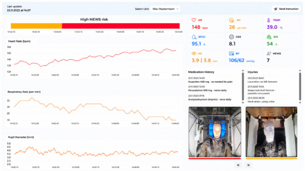
Figure 2: Sketch of telemedical eye-tracking triage interface for military casualty assessment: (a) audio-visual communication between a remote medical expert and an injured soldier, including integrated pupil-diameter readout from Tobii Pro Fusion/ Spark eye-tracker; (b) medic-facing monitoring dashboard showing MEWS risk, vital parameters, pupil-diameter trends and live patient-box camera views. (Credits: a. D. Maurer, b. D. Tomašević & F. Haid; ChatGPT for image post-processing).
Figure 2 illustrates the intended user interface concept for remote, eye- tracking-supported triage in military medical evacuation.
The audio-visual interface in Figure 2a enables a medical expert to communicate with the casualty or the local medic while simultaneously inspecting the soldier’s condition and basic pupillometry values.
Rapid Military Triage of Traumatic Brain Injury Using Eye Tracking and Pupillometry 261 The dashboard in Figure 2b extends this concept by combining vital signs, MEWS-based risk indication, medication and injury history, pupil-diameter trends and live views from the patient box. Together, these views support a telemedical workflow in which ocular biomarkers are not interpreted in isolation but are embedded into a broader clinical and operational decision- support environment.
DISCUSSION
The proposed system addresses a practical gap between traditional neurological screening and the need for rapid, data-supported decisions in austere military settings. Eye tracking and pupillometry are attractive because they can be fast, non-invasive and repeatable, and because the eye is linked to brainstem reflexes, cortical attention and autonomic state. Yet these same strengths create a risk of overinterpretation. Pupil diameter is not a disease-specific marker; it is a physiological signal shaped by light, arousal, medication, pain, hypoxia and ocular factors. The rule base must therefore be explainable and context-aware.
The most promising near-term application is not a definitive diagnosis of concussion or nerve-agent poisoning, but risk stratification of walking- wounded soldiers. A low-risk output can support observation and documentation but should not authorize return to duty without policy-based medical judgement. A medium-risk output can trigger repeat assessment, MACE 2, VOMS or observation. A high-risk output should prioritize evacuation, urgent clinician review or CBRN workflow when systemic signs and exposure context support it.
The proposed rule-based approach has advantages over a purely machine- learning classifier in early development. It allows domain experts to inspect the logic, enables medics to understand why a flag appeared and supports incremental validation. Machine learning may still be valuable for feature extraction, signal-quality assessment and threshold refinement, but final triage output should remain interpretable, auditable and overridable by the medic.
A further advantage is compatibility with multimodal sensing. Future versions can include heart rate, HRV, electrodermal activity, SpO2, respiration, inertial data and speech features. These signals should not be added for complexity alone. They should be integrated only when they reduce ambiguity: for example, SpO2 and respiratory rate can help separate opioid-related miosis with respiratory compromise from cholinergic toxicity; HR/EDA can help contextualize stress; and inertial data can identify motion- corrupted eye tracking.
LIMITATIONS, ETHICS AND OPERATIONAL SAFETY Several limitations must be addressed before deployment. First, ocular metrics in mTBI are promising but not yet standardized across devices and protocols. Second, thresholds derived from hospital or sports studies
may not generalize to soldiers, night conditions, blast environments or protective equipment. Third, medication can directly alter pupil physiology and cognitive-oculomotor performance. Fourth, neurotoxic exposure cannot ethically be validated through harmful human exposure; validation must rely on safe simulation, historical data, animal/experimental evidence and clinical toxicology cases.
Operational safety requires that the system be framed as decision support. It must not delay life-saving interventions, decontamination, airway management, haemorrhage control or evacuation. It must not recommend antidote administration, doses or battlefield treatment steps. Instead, it should identify patterns compatible with TBI or cholinergic toxicity and prompt the appropriate established medical or CBRN pathway. The medic remains responsible for triage decisions, supported by clear evidence, quality flags and documented uncertainty.
Privacy and governance are also important. Eye videos and gaze traces are biometric and potentially sensitive. Data storage should be minimized, encrypted and separated from personal identifiers when possible. Research use should require ethics approval, informed consent where feasible, and clear handling of incidental clinical findings. For operational settings, governance must define who can view raw data, how long it is retained and how algorithm changes are controlled.
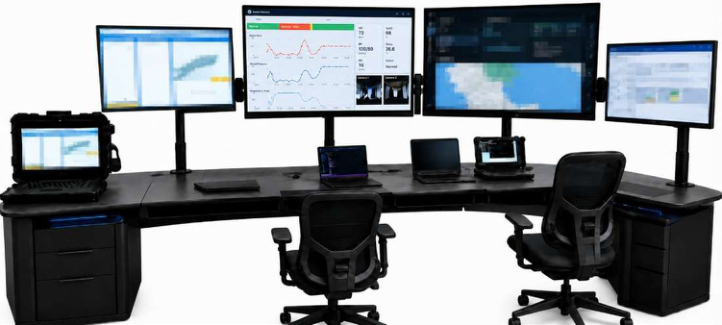
Figure 3: Patient Evacuation Control Centre (PECC) concept for integrated resource management, military triage, continuous patient care via bidirectional audio-video communication and patient monitoring, with human-in-the-loop medical robot intervention control (Developed by the iMEDCAP consortium; Credit: M. Schneeberger, ChatGPT for image post-processing).
CONCLUSIONS AND OUTLOOK This paper proposes a research program for rapid military triage using eye tracking and pupillometry as explainable digital biomarkers for hidden neurological impairment. The concept focuses on walking-wounded soldiers
Rapid Military Triage of Traumatic Brain Injury Using Eye Tracking and Pupillometry 263 after blast, head impact or suspected neurotoxic exposure, where subtle signs can be missed under operational pressure. The proposed system combines PLR, pupil size, anisocoria, gaze stability, smooth pursuit, saccades and short cognitive-oculomotor tasks with a transparent rule base and explicit medication/confound handling.
The main expected outcome is not a single diagnostic score, but a defensible triage recommendation with an auditable rationale: high risk, medium risk, low risk or invalid/confounded. The research roadmap emphasizes staged validation, normative baselines, simulated field trials and prospective evaluation. If validated, the approach could improve early recognition of TBI warning signs, support CBRN-compatible ocular screening, reduce missed impairment among walking wounded and provide a foundation for integration with broader physiological monitoring in military medicine.
Figure 3 illustrates the envisioned Patient Evacuation Control Centre (PECC) as an integrated command environment for resource management, military triage, continuous patient care, patient monitoring and human-in- the-loop medical robot intervention control. By fusing patient, platform, reconnaissance and mission information into a common operational picture, the PECC enables simultaneous management of multiple casualties, telemedical supervision and evidence-based control of distributed autonomous medical assets.
ACKNOWLEDGMENT
The project iMEDCAP is funded by the European Union under Grant Agreement No: 101121421. Views and opinions expressed are however those of the author(s) only and do not necessarily reflect those of the European Union or the European Commission. Neither the European Union nor the granting authority can be held responsible for them.
REFERENCES
Boulter, J. H., Shields, M. M., Meister, M. R., Murtha, G., Curry, B. P., and Dengler, B. A. (2021). The expanding role of quantitative pupillometry in the evaluation and management of traumatic brain injury. Frontiers in Neurology, 12, 685313. https://doi.org/10.3389/fneur.2021.685313. Candiotti, K. (2017). A primer on nerve agents: What the emergency responder,
anesthesiologist, and intensivist needs to know. Canadian Journal of Anesthesia/ Journal canadien d’anesthesie, 64, 1059–1070. https://doi.org/10.1007/s12630- 017-0920-2. Capo-Aponte, J. E., Beltran, T. A., Walsh, D. V., Cole, W. R., and Dumayas, J. Y.
(2018). Validation of visual objective biomarkers for acute concussion. Military Medicine, 183(suppl_1), 9-17. https://doi.org/10.1093/milmed/usx166. Ciuffreda, K. J., Joshi, N. R., and Truong, J. Q. (2017). Understanding the effects of mild traumatic brain injury on the pupillary light reflex. Concussion, 2(3), CNC36. https://doi.org/10.2217/cnc-2016-0029.
Dengler, B. A., Meister, M., Aderman, M., Malvasi, S. R., Ross, J. D., Fu, A., Haight, T., Bartanusz, V., Kambe, C., Boulter, J. H., and Cameron, K. L. (2025). Quantitative pupillometry predicts return to play and tracks the clinical evolution of mild traumatic brain injury in US military academy cadets: A Military Traumatic Brain Injury Initiative study. Neurosurgery, 96(1), 142-151. https://doi.org/10.1227/ neu.0000000000003032. Eapen, B. C., Bowles, A. O., Sall, J., Heriseanu, R. E., O’Neil, M. E., Blackman, J.
A., & Cifu, D. X. (2022). The management and rehabilitation of post-acute mild traumatic brain injury. Brain Injury, 36(5), 693–702. https://doi.org/10.1080/02 699052.2022.2033848 Egoz, I., Nili, U., Grauer, E., and Gore, A. (2017). Optimization of the ocular treatment following organophosphate nerve agent insult. Toxicological Sciences, 159(1), 50-63. https://doi.org/10.1093/toxsci/kfx119. El Ahmadieh, T. Y., Bedros, N. M., Stutzman, S. E., Nyancho, D., Venkatachalam,
A. M., MacAllister, M., et al. (2021). Automated pupillometry as a triage and assessment tool in patients with traumatic brain injury. World Neurosurgery, 145, e163-e169. https://doi.org/10.1016/j.wneu.2020.09.152. Figueiredo, T. H., Apland, J. P., Braga, M. F. M., and Marini, A. M. (2018). Acute and long-term consequences of exposure to organophosphate nerve agents in humans. Epilepsia, 59(Suppl. 2), 92-99. https://doi.org/10.1111/epi.14500. Fisher, A. D., DesRosiers, T. T., Papalski, W., Remley, M. A., Schauer, S. G., April,
M. D., Blackman, V., Brown, J., Butler, F. K., Cunningham, C. W., Gurney, J. M., Holcomb, J. B., Montgomery, H. R., Morgan, M. M., Motov, S. M., Shackelford, S. A., Sprunger, T., and Drew, B. G. (2022). Analgesia and sedation for Tactical Combat Casualty Care: TCCC proposed change 21-02. Journal of Special Operations Medicine, 22(2), 154-165. https://doi.org/10.55460/8CBI-GAOD. Gore, A., Lazar, S., Yacov, G., Kadar, T., et al. (2019). Ocular surface histopathological insult following sarin and VX exposure and potential treatments in the rat model. Toxicology Letters, 314, 153-163. https://doi.org/10.1016/j.toxlet.2019.08.002. Hou, R. H., Scaife, J., Freeman, C., Langley, R. W., Szabadi, E., and Bradshaw, C.
M. (2006). Relationship between sedation and pupillary function: comparison of diazepam and diphenhydramine. British Journal of Clinical Pharmacology, 61(6), 752-760. https://doi.org/10.1111/j.1365-2125.2006.02632.x. Jahns, F. P., Miroz, J. P., Messerer, M., Daniel, R. T., Taccone, F. S., Eckert, P., and Oddo, M. (2019). Quantitative pupillometry for the monitoring of intracranial hypertension in patients with severe traumatic brain injury. Critical Care, 23, 155. https://doi.org/10.1186/s13054-019-2436-3. Khokhar, B., Jorgensen-Wagers, K., Marion, D., and Kiser, S. (2021). Military Acute
Concussion Evaluation: A report on clinical usability, utility, and user’s perceived confidence. Journal of Neurotrauma, 38(2), 246-250. https://doi.org/10.1089/ neu.2020.7176. Maruta, J., Suh, M., Niogi, S. N., Mukherjee, P., and Ghajar, J. (2010). Visual tracking synchronization as a metric for concussion screening. Journal of Head Trauma Rehabilitation, 25(4), 293-305. https://doi.org/10.1097/ HTR.0b013e3181e67936. Mitschang, D., Sydorenko, V., Kuehlwein, D., Beynon, C., Younsi, A., Krieg, S. M.,
et al. (2025). Quantitative pupillometry as a sensitive biomarker for detecting subtle neurological impairment in mild traumatic brain injury - a pilot study. Neurological Sciences, 46, 5243-5251. https://doi.org/10.1007/s10072-025- 08405-2.
Rapid Military Triage of Traumatic Brain Injury Using Eye Tracking and Pupillometry 265 Mucha, A., Collins, M. W., Elbin, R. J., Furman, J. M., Troutman-Enseki, C., DeWolf,
R. M., Marchetti, G. F., and Kontos, A. P. (2014). A brief Vestibular/Ocular Motor Screening (VOMS) assessment to evaluate concussions: preliminary findings. American Journal of Sports Medicine, 42(10), 2479-2486. https://doi. org/10.1177/0363546514543775. Murray, N. G., Szekely, B., Islas, A., Munkasy, B., Gore, R., Berryhill, M., Reed-Jones, R. J., and Peterson, M. (2020). Smooth pursuit and saccades after sport-related concussion. Journal of Neurotrauma, 37(2), 340-346. https://doi.org/10.1089/ neu.2019.6595. Nozaki, H., Aikawa, N., Fujishima, S., Suzuki, M., Shinozawa, Y., Hori, S., and
Nogawa, S. (1995). A case of VX poisoning and the difference from sarin. The Lancet, 346(8976), 698-699. https://doi.org/10.1016/S0140-6736(95)92306-3. Oddo, M., Sandroni, C., Citerio, G., et al. (2023). The Neurological Pupil index for outcome prognostication in people with acute brain injury (ORANGE): A prospective, observational, multicentre cohort study. The Lancet Neurology, 22(10), 925-936. https://doi.org/10.1016/S1474-4422(23)00271-5. Rollins, M. D., Feiner, J. R., Lee, J. M., Shah, S., and Larson, M. (2014). Pupillary
effects of high-dose opioid quantified with infrared pupillometry. Anesthesiology, 121(5), 1037-1044. https://doi.org/10.1097/ALN.0000000000000384. Shoyombo, I., Aiyagari, V., Stutzman, S. E., Atem, F., Hill, M., Figueroa, S. A., Miller, C., Howard, A., and Olson, D. M. (2018). Understanding the relationship between the Neurologic Pupil Index and constriction velocity values. Scientific Reports, 8, 6992. https://doi.org/10.1038/s41598-018-25477-7. Stuart, S., Parrington, L., Martini, D., Peterka, R., Chesnutt, J., and King, L. (2020).
The measurement of eye movements in mild traumatic brain injury: a structured review of an emerging area. Frontiers in Sports and Active Living, 2, 5. https:// doi.org/10.3389/fspor.2020.00005. Vrettou, C. S., Fragkou, P. C., Mallios, I., Barba, C., Giannopoulos, C., Gavrielatou, E., and Dimopoulou, I. (2024). The role of automated infrared pupillometry in traumatic brain injury: a narrative review. Journal of Clinical Medicine, 13(2), 614. https://doi.org/10.3390/jcm13020614.
LISTED IN THE AHFE DIGITAL BOOKSTORE CATALOGUE
Explore the complete digital volume
Cognitive Computing and Internet of Things
Read this individual research paper free through the original publisher, or explore the full digital proceedings book.
ISBN 978-1-964867-81-6
Explore this ISBN in the AHFE Bookstore ↗Listed in the July 2026 bookstore catalogue; search by ISBN to confirm current availability · Digital book purchase optional