Matteo Iurato, Ronny Stanzani, Mirko Job, Andres Gaggero, Igor Ingegnosi, Marco Testa (2025). Assessment of Upper Limb Functional Workspace through Inertial Measurement Units: a Pilot Study. In: Human Interaction and Emerging Technologies (IHIET-FS 2025): Future Systems and Artificial Intelligence Applications. AHFE Open Access, vol. 196, pp. 163–172. https://doi.org/10.54941/ahfe1005965
The assessment of the upper limb functional workspace in an ecological environment is important for the evaluation of clinical progress in persons suffering from musculoskeletal disorders or neurological impairments. Inertial Measurement Units (IMUs) represent a very effective technology for the assessment of human movement in ecological settings. This work presents a preliminary validation of a methodology for reconstructing and assessing the upper limb functional workspace explored during the daily routine in ecological setting through IMUs. Participants in the study were involved in 7 hours data acquisition with IMUs performing two different protocols simulating an active and a non-active arm, respectively. For each of the two protocols, a workspace for each limb segment and each participant was reconstructed by evaluating the estimated spatial position of the sensors over time. A density and clusterization assessment was performed on each workspace through the application of a Gaussian kernel and k-means algorithm. Next, workspaces from the non-active and the active protocols were compared by performing statistical tests on the distributions of points in the respective workspaces along the the three spatial coordinates. Results showed significant difference between the two protocols (active and non-active) on every spatial coordinate and every of the three segments in the upper limb (arm, forearm, hand) and different clusterization of the workspaces. The findings represent a preliminary confirmation of the applicability of IMUs to the assessment of changes in the functional workspace of the upper limb. Further developments may involve enlarging the sample size, testing on impaired persons, and assessing in more realistic scenarios.
Musculoskeletal disorders (MSDs) and neurological impairments can significantly affect the mobility of the upper limb (Almomani et al., 2019; Urwin et al., 1998). This results in compensative motor strategies that reduce the dimension of the explored space during the execution of the activities of daily living (ADL) (Almomani et al., 2019; Magermans et al., 2005).
Therefore, when assessing the clinical progress of patients recovering from a motor disorder involving the upper limb, it is important to evaluate the changes in their functional workspace (Kurillo et al., 2013; Matthew et al., 2020). This assessment should preferably be performed in an ecological setting, to foster the investigation of the upper limb mobility during the everyday life of the patient (Davis and Burton, 1991).
Among the available technologies for movement analysis, the most suitable for kinematic monitoring in ecological settings are Inertial Measurement Units (IMUs) (Grip et al., 2022; Hindle et al., 2021; Poitras et al., 2019). These sensors can indeed be easily worn and attached to a person, measuring joint angles and accelerations without the need for any additional devices or supervision (Grip et al., 2022).
In this work, we present a pilot attempt to reconstruct and assess the functional workspace from IMU data related to the orientation of all segments in the upper limb kinematic chain.
RELATED WORKS Because of the clinical importance of this metric (Matthew et al., 2020), literature about the use of technologies for movement analysis to reconstruct the functional reachable workspace of the upper limb has grown over time. The majority of studies focused on evaluating this metric relying on datasets acquired using Microsoft Kinect (Kurillo et al., 2013; Lee et al., 2020; Matthew et al., 2020).
Notably, the work by Carmona-Ortiz et al. (Carmona-Ortiz et al., 2020) presented an attempt at using IMUs to reconstruct the upper limb functional workspace: they performed a pilot study comparing the functional workspace of one healthy and one impaired participant, focusing on the space explored by the wrist joint.
In this work, we present a preliminary evaluation of a methodology for the reconstruction through IMUs of the functional workspace of all the three segments of the upper limb (arm, forearm, hand) and the detection of changes in the workspace between an active and a non-active upper limb.
METHODS
Measurement System Orientation data were acquired using four BNO080 IMUs, connected to a central unit that synchronizes signals and saves data on a SD card. Previous works in the literature demonstrated the accuracy of this type of sensors and their reliability for the study of the upper limb (Stanzani et al., 2020). The output of the sensors are unitary quaternions representing their orientation in the Earth’s reference frame.
Experimental Protocol The four IMUs were placed on the sternum, arm, forearm and hand respectively. Participants wore these sensors by attaching them to the limbs using elastic bands.
Assessment of Upper Limb Functional Workspace Through Inertial Measurement Units 165
Each participant performed two different data acquisition sessions on two different days: on the first day the reduced mobility protocol (RMP) was performed, while on the second day the high mobility protocol (HMP) was performed. Participants were all right-handed and the dominant right arm was monitored through IMUs during both the RMP and HMP sessions.
RMP involved a 7 hours acquisition session, during which participants were asked to perform only the tasks of a routine office working day which mainly involved sitting at their desk, trying to limit wide-range movements (e.g. to stretch oneselves).
HMP involved a 7 hours acquisition session, during which the regular office activity at the desk was alternated with a set of tasks simulating activities performed at home during a daily routine and involving a high amount of mobility of the upper limb. Detailed instructions for these tasks and frequency of execution are reported in Table 1.
Table 1: Tasks performed during the high mobility protocol. Repetitions were performed throughout the 7h acquisition.
Table available in the original PDF paper.
Activity Instructions Repetitions Move objects Open a wardrobe, pick up
2 times the whole sequence, each time moving 10 objects
objects from a high shelf, move them onto a low shelf, close the wardrobe
2 times the entire sequence
Take the rubbish bag, carry it
Throw garbage
to the bin, open the bin, throw the bag, close the bin
in the dumpster Sweep the floor Take the broom, sweep the
2 times the entire sequence; the act of sweeping was performed for 1 minute
floor, take the dustpan, collect residual dust, throw in the bin, put broom and dustpan back in place Clean surfaces Open a wardrobe, take cleaning products, put cleaning product onto a rag, move the rag around the surface, put cleaning products back in place
2 times the entire sequence with the rag moving action lasting 30 seconds; the first time a horizontal surface (e.g. table) was cleaned, while the second a vertical surface (e.g. window) was cleaned Pour water into
Take a glass placed on a high a glass and drink shelf, put it on the table, open the cap of a bottle, pour water, close the bottle cap, drink the water, wash the glass, put the glass back in place
10 times the sequence opening the bottle cap, drinking and reclosing it followed by the washing action involving rubbing the entire glass surface 10 times to wash, and 10 additional times to dry Continued
Table 1: Continued Activity Instructions Repetitions
Table available in the original PDF paper.
Get dressed and
Take off the jacket, take off
10 times the entire sequence
undressed
the sweatshirt or jumper, take off the shirt, put the shirt back on, put the sweatshirt or jumper back on, put the jacket back on
Write on the
Write a sentence and draw a blackboard figure on the blackboard
10 times writing and 10 times drawing Unscrew a light
Raise the arm so that the hand
10 rotations
bulb
is pointing towards the ceiling, rotate the limb to unscrew
Draw the
Open and reclose the curtains 10 times curtains Stretch oneself Freely performed 10 stretching sequences lasting 20 seconds each
Workspace Reconstruction Hereafter the processing of the quaternions output by the IMUs to obtain the functional workspace is described.
Processing was performed in Python 3.11, with Numpy 1.26 and Scipy 1.11 for calculations and Matplotlib 3.8 for data plotting. Quaternions are below defined as q = w + ix + jy + kz with i, j, k denoting the imaginary units.
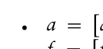
We first defined a unitary axis as a unit quaternion with a single imaginary part: v = i. At each time frame, for each of the four IMUs, the unitary axis v was then rotated according to the quaternion output q from the IMU, based on the following equation: l = qvq∗ (1)
where ∗indicates the conjugate of the quaternion.
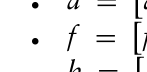
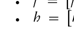
The unitary rotated axes l for each of the four IMUs were then multiplied by the anthropometric average length of the segment relative to each IMU, obtained from anthropometric tables [13]. Therefore, at the end of this process, four quaternions representing the orientated axes of each limb were obtained. Cartesian coordinates of those axes were directly obtained from the imaginary parts of the said quaternions. Consequently, after this process three axes for each limb segment were available:
Assessment of Upper Limb Functional Workspace Through Inertial Measurement Units 167
Equation reproduced from the original PDF.
for the arm
Equations and mathematical formulas are available in the original paper PDF.
Equation reproduced from the original PDF.
for the forearm
Equations and mathematical formulas are available in the original paper PDF.
Equation reproduced from the original PDF.
for the hand
hx, hy, hz The positions pa, pf, ph of the extremities of each of the three limb segments were therefore calculated based on the following kinematic chain:
pa =
Equations and mathematical formulas are available in the original paper PDF.
pf = +
+
ax, ay, az
fx, fy, fz
hx, hy, hz
ph = These positions represent one point at the considered time frame in the functional workspace for each of the three limbs. Repeating the process for each time frame in the dataset allowed to build the complete functional workspace for each limb segment explored during the acquisition.
Signals relative to the x, y, z of each limb were then downsampled to 10000 samples using linear interpolation for the purpose of data visualization.
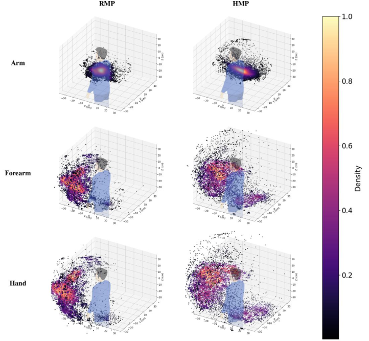
Based on the overall downsampled functional workspace, a density value for each point was then calculated through a kernel-density estimation using a Gaussian kernel (µ = 0, σ = 0.1).
Workspace Clustering On each of the reconstructed workspaces, a gradual thresholding based on the density values obtained through the Gaussian kernel was performed.
Specifically, given a density threshold t, all points with a density lower than t were removed from the workspace.
This process was repeated iteratively by variating t from 0 to 0.8 with steps of 0.1.
On each of the thresholded workspaces, the k-means algorithm was run and a curve for the inertia as a function of the number of selected clusters k was obtained.
Statistical Analysis The distributions of position data for each of the three limbs (arm, forearm, hand) from all subjects for every x, y, z spatial coordinate and for both the RMP and HMP protocol were computed.
Normality was evaluated for each distribution through a Shapiro-Wilk test.
For normal distributions, a two-sided t-test was performed, whereas for non-normal distributions a two-sided Mann Whitney U-test was applied, to test the hypothesis of dissimilarity between the corresponding distributions in RMP and HMP protocol.
A Bonferroni correction was applied to the tests performed on each of the three limbs (arm, forearm, hand) to account for multiple testing (three tests on each x, y, z spatial coordinate).
For each of the inertia curves obtained through density thresholding and k-means algorithm application as explained in the previous section, the average and standard deviation across participants were calculated.
RESULTS
Number of Participants We included 5 male healthy participants (average age ± standard deviation: 38.8 ± 13.73 years).
Workspace Reconstruction
Figure 1 shows examples of workspaces for one representative participant for the three limb segments in both the RMP and HMP protocol.
Figure 1: Functional workspaces for arm, forearm and hand in both the RMP and HMP protocol for one representative participant.
Assessment of Upper Limb Functional Workspace Through Inertial Measurement Units 169
Workspace Spatial Distribution
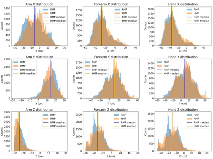
Figure 2 shows the distribution of points reached by all participants during both RMP and HMP protocols along the three spatial coordinates x, y, z for every of the three limb segments (arm, forearm, hand).
All Shapiro-Wilk tests performed on the distributions exhibited p < 0.05: therefore, the null hypothesis is rejected for every test and all distributions are assumed to not be normal. Consequently, results from Mann-Whitney tests comparing RMP and HMP distributions are presented.
All Mann-Whitney tests comparing RMP and HMP distributions showed p < 0.05. The Bonferroni correction on these tests led to a corrected alpha equal to 0.02 and showed all corrected p < 0.02. Therefore, the null hypothesis is always rejected and statistical differences between the RMP and HMP population are visible for every of the three limb segments (arm, forearm, hand) and for all the three spatial coordinates x, y, z.
Figure 2: Distribution of points reached by all participants during both RMP and HMP protocols along the three spatial coordinates x, y, z for every of the three limb segments (arm, forearm, hand).
Workspace Clustering
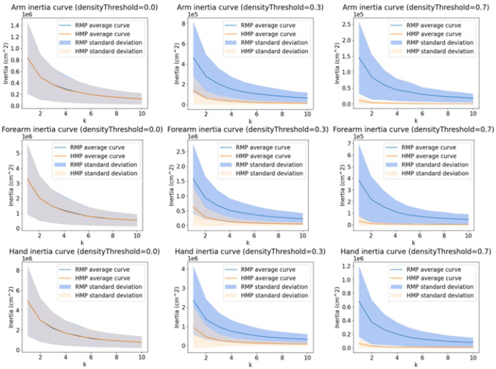
Figure 3 shows the average curves of inertia as a function of the number of clusters k for every of the three upper limb segments (arm, forearm, hand) obtained after density thresholding and k-means clustering for three representative values of the density threshold in both RMP and HMP.
It is noticeable that, while increasing the density threshold, for every of the three limb segments, the inertia of clusters detected in the HMP protocols decreases more rapidly than for the RMP protocols.
Figure 3: Average curves with standard deviation showing inertia as a function of the number of clusters k obtained after density thresholding of the three upper limb segments workspaces for three representative density thresholds (0, 0.3, 0.7) for the two RMP and HMP protocols.
DISCUSSION
Our findings support the potential applicability of the IMUs to the assessment of the changes in the functional workspace of the upper limb explored by a patient during his daily life.
The results we obtained strengthen findings from the previous pilot presented in (Carmona-Ortiz et al., 2020) by relying on statistical comparison thanks to a larger sample size and extending the analysis to all the segments of the upper limb (arm, forearm, hand).
The cluster analysis through Gaussian kernel and k-means revealed lower inertia in the HMP protocol while increasing the density threshold. This suggests the presence of more tight high-density clusters with respect to the RMP protocol, which can be interpreted as an effect of the additional performing of ADL outside the arm stationary area for a more prolonged time. This evidence supports the possibility of using this methodology for the clinical assessment of the amount of mobility of the upper limb.
Assessment of Upper Limb Functional Workspace Through Inertial Measurement Units 171
Nonetheless, this work represents only a preliminary exploration and has several limitations. First, the included sample size, even if larger than in the attempts we found in the literature, was composed of only five participants and no impaired persons were involved. Enlarging the sample size would strengthen the validity of the results.
Moreover, we just simulated a subset of possible movements that are performed by persons with an unimpaired healthy arm in an ecological setting, but we did not test in a real scenario. Performing further acquisition in a real-life setting (e.g. at home) would provide more realistic insights about the applicability of the approach. In this latter case, indeed, ADL would be performed for a higher amount of time: combined with the inclusion of impaired participants (which should exhibit even remarkably lower mobility than what we simulated in the RMP), this further development could lead to detect greater differences in the workspaces.
Also, improved results could arise from enlarging the duration of the observation (e.g. one-week monitoring) in order to evaluate long-term variations in the use of the upper limb.
However, even if our findings should just be considered very preliminary results, they provide promising indications in the direction of using IMUs as a tool to monitor patient progresses in an ecological setting. The assessment of the functional workspace indeed provides very important information about the mobility of the upper limb, which can be extremely helpful for a thorough clinical assessment (Kurillo et al., 2013).
The use of motion analysis technologies can help developing quantitative indicators that provide an objective evaluation of the variations in the functional workspace. However, the type of motion analysis technologies employed can strongly impact the usability of the system. IMUs represent a non-cumbersome and easy to wear type of sensors: therefore, validating their applicability in the assessment of functional workspace represents an important step that can enlarge their employment in objective clinical assessment in an ecological setting.
CONCLUSION
In this study we presented a preliminary evaluation for the assessment of the functional workspace explored by a person in an ecological environment through IMUs, comparing the workspace from an active arm with the one from a non-active arm.
The preliminary results that we obtained suggest a good applicability of the methodology for the reconstruction of the workspace from IMU data and for the assessment of such workspace. Further developments of this work include testing on a larger sample size, on impaired persons, and in a more realistic scenarios and for longer time.
Exploring these future directions could strengthen the validity of our results and validating the applicability of IMUs in the assessment of the functional workspace, fostering the application of this technology for an objective and quantitative assessment of the upper limb mobility in ecological environment.
REFERENCES
Almomani, F., Alghwiri, A. A., Alghadir, A. H., Al-momani, A., Iqbal, A. (2019).
Prevalence of upper limb pain and disability and its correlates with demographic and personal factors. J. Pain Res. 12, 2691–2700. Carmona-Ortiz, V. A., Lobo-Prat, J., Van Ruysevelt, J., Torras, C., Font- Llagunes, J. M. (2020). Development and Pilot Evaluation of the ArmTracker: A Wearable System to Monitor Arm Kinematics During Daily Life, in: 2020 8th IEEE RAS/EMBS International Conference for Biomedical Robotics and Biomechatronics (BioRob), pp. 759–764. Davis, W. E., Burton, A. W. (1991). Ecological Task Analysis: Translating Movement
Behavior Theory into Practice. Grip, H., Källströmer, A., Öhberg, F. (2022). Validity and Reliability of Wearable Motion Sensors for Clinical Assessment of Shoulder Function in Brachial Plexus Birth Injury. Sensors 22, 9557. Hindle, B. R., Keogh, J. W. L., Lorimer, A. V. (2021). Inertial-Based Human
Motion Capture: A Technical Summary of Current Processing Methodologies for Spatiotemporal and Kinematic Measures. Appl. Bionics Biomech. 2021, e6628320. Kurillo, G., Chen, A., Bajcsy, R., Han, J. J. (2013). Evaluation of upper extremity reachable workspace using Kinect camera. Technol. Health Care 21, 641–656. Lee, Y. M., Lee, S., Uhm, K. E., Kurillo, G., Han, J. J., Lee, J. (2020). Upper Limb
Three-Dimensional Reachable Workspace Analysis Using the Kinect Sensor in Hemiplegic Stroke Patients: A Cross-Sectional Observational Study. Am. J. Phys. Med. Rehabil. 99, 397. Magermans, D. J., Chadwick, E. K. J., Veeger, H. E. J., van der Helm, F. C. T. (2005).
Requirements for upper extremity motions during activities of daily living. Clin. Biomech. 20, 591–599. Matthew, R. P., Seko, S., Kurillo, G., Bajcsy, R., Cheng, L., Han, J. J., Lotz, J. (2020).
Reachable Workspace and Proximal Function Measures for Quantifying Upper Limb Motion. IEEE J. Biomed. Health Inform. 24, 3285–3294. Poitras, I., Dupuis, F., Bielmann, M., Campeau-Lecours, A., Mercier, C., Bouyer, L. J., Roy, J.-S. (2019). Validity and Reliability of Wearable Sensors for Joint Angle Estimation: A Systematic Review. Sensors 19, 1555. Stanzani, R., Dondero, P., Mantero, A., Testa, M. (2020). Measurement Accuracy
of an Upper Limb Tracking System Based on Two Hillcrest Labs BNO080 IMU Sensors: An Environmental Assessment. IEEE Sens. J. 20, 10267–10274. Urwin, M., Symmons, D., Allison, T., Brammah, T., Busby, H., Roxby, M., Simmons, A., Williams, G. (1998). Estimating the burden of musculoskeletal disorders in the community: the comparative prevalence of symptoms at different anatomical sites, and the relation to social deprivation. Ann. Rheum. Dis. 57, 649–655.
LISTED IN THE AHFE DIGITAL BOOKSTORE CATALOGUE
Explore the complete digital volume
Human Interaction and Emerging Technologies (IHIET-FS 2025): Future Systems and Artificial Intelligence Applications
Read this individual research paper free through the original publisher, or explore the full digital proceedings book.
ISBN 978-1-964867-72-4
Explore this ISBN in the AHFE Bookstore ↗Listed in the July 2026 bookstore catalogue; search by ISBN to confirm current availability · Digital book purchase optional