Tsai-chun Huang, Bingxue Li (2024). Back skin deformation during anaerobic exercises for ergonomics design application. In: Healthcare and Medical Devices. AHFE Open Access, vol. 130, pp. 161–171. https://doi.org/10.54941/ahfe1004849
The purpose of this study is to propose a muscular-based superficial skin deformation method to visualise the deformation of human back during resistance training, thus, to provide a reference for the back design of gym wear and wearable devices. Among the muscle groups, the back has numerous muscles and is a very difficult area to train, therefore, there is an urgent need for the study of the skin deformation of the back during anaerobic training. Several studies have been conducted on skin deformation during aerobic exercise, such as rowing and cycling using the division of anthropometric measurements, however, back muscle contractions in anaerobic exercise in which the skin deformation was caused by muscle movements need more exploration. In this study, eight regular trained males were instructed to perform three back training exercises: seated pulldown, seated row and standing pulldown. Their backs were marked points of 3 cm interval according to the surface division of the two major muscles: the trapezius and the latissimus dorsi. The statistics skin measurements of starting point and completion of the exercises were taken by 3D scanner Artec Eva. The mean, maximum values and standard deviation of the data were compared and analyzed according to muscle morphology and fibre bundle orientation. The results showed that the maximum deformation of trapezius, was 24.23% and 11.97%, 9.31% and 8.94%, 1.51% and 9.78%, for starting and completion in the horizontal, longitudinal and diagonal direction, respectively. For the latissimus dorsi, the greatest deformation manifested as 7.23% and 5.36 %, 14.59% and 8.52%,17.60% and 9.70% for starting and completion along the longitudinal and diagonal directions, respectively. These muscle-based deformation can be implemented in the design of prototypes in the corresponding regions to improve the dynamic fit during exercise.Both muscles showed different degrees of segmental deformation on the triangular shaped sides. The horizontal and longitudinal directions of the trapezius muscle show a state of compression in the whole segment during movement, but with the degree of compression ranging from 1.99% to 14.29% for each sub-segment. The oblique side of the trapezius showed a concentration of deformation in the lower half of the muscle, with a range of 10.83%-38.49%, while the latissimus dorsi shows a more uneven deformation dominated by the oblique upper third extrusion deformation. For the dramatic variable sub-regions, additional structures or strengthen materials should be taken into account to support the muscle, providing bracing forces for better muscle perception and avoid injuries.In this research, skin deformation characteristics during back anaerobic training were proposed and regional analysis based on muscle morphology and kinesiology are performed. These zoning values can be used as a reference for intelligent monitoring, so that the design of sensing accessories can be better matched. The visual mapping of results offered guidance for the cutting lines of tight fitness apparel prototype and the back structural design of sportswear, applying the deformation characteristics of anaerobic exercise to improve the dynamic comfort.
This research would like to understand the skin surface deformation caused by body movements during back resistant training, specifically, seated pull- down, standing lat pull-down, and seated row, as the foundation for the assistive training gears, or garments design in the future.
Gym exercise has become a growing trend for fitness people due to its sta- ble indoor environment and multiple choices of equipment from aerobic to anaerobic. Adding some anaerobic exercises using resistance equipment to the training session can be very effective in increasing the proportion of mus- cles in the body, in this way enhancing basal metabolism, physical endurance and immunity. With the arising awareness of anaerobics for these health ben- efits, more people are performing resistance training targeting a particular part of the muscles during their gym workouts. However, back could be one of the most difficult parts for sensing muscle activation during training.
Different movements during resistance training result in different levels of activation in each muscle (Lehman et al., 2004). For example, in pull-downs and rowing, which are often used in back resistance training, the latissimus dorsi muscle will have a higher level of activation and the highest level of variability among different movements relative to the other muscles. Among human muscles, the back muscles are the most involved muscle groups in the human body and are the focus of many people’s upper body training, including the latissimus dorsi, trapezius, teres major, teres minor, infraspina- tus, rhomboids, and erector spine (Hass et al., 2001). As the muscle and tissue actions directly affect superficial skin deformation (Carnevale et al., 2021), a large number of back muscle groups will cause complex skin defor- mations, resulting in the structural design at the back of the garment, which can greatly influence training effectiveness and comfort. At the same time, due to the invisibility of the back, there is an urgent need to improve human perception during training through wearable design. Therefore, skin defor- mation analysis of the back muscle is needed to inform the design of clothing and wearable designs for the back, to promote comfort level of movement when people are training their back muscles.
Many studies have investigated skin deformation in various exercises and applied it in sportswear design. The drastic motion of limbs leads to large skin deformation areas first drew some researchers’ attention. Choi and Ash- down (2011) measured changes in the lower limbs in dynamic postures using 3D scanning (Choi et al., 2011). The selection of functional seamlines for ordinary outdoor pants was also proposed through 3D scanning when peo- ple perform standing, 90-degree flexion and squatting (Lee et al., 2013). A gel rubbing method was adopted by Luo in 2017 to obtain the skin deforma- tion areas from the waist to the knee of subjects’ lower limbs in 12 cycling postures (Luo et al., 2017). These studies provide useful guidance for body measurement methods based on lower limb skin surface deformations.
A small number of studies also have used manual or 3D scanning meth- ods to investigate the skin deformation of the upper body during outdoor aerobic activities like cycling (Wang et al., 2021; Shi et al., 2023). Missing data on skin deformation distribution in various riding terrains was noted by Wang et al. and they used elastic bandages to measure the skin defor- mation on all road conditions (Wang et al., 2021). The 3D space capture was used by Shi et al. (2023) to further explore the skin deformation of the torso and thigh of cyclists during dynamic cycling, hence suggested the opti- mum ease amounts for the compression cycling skinsuit pattern to promote performance.
From Question to Approach Previous studies provide a fundamental database of skin deformation of the upper body during aerobic exercises, the method that they chose to estab- lish the divisions mainly from anthropometric reference, little of them have explored the relationship between the activation of particular muscle and skin deformation during anaerobic exercise.
Back Skin Deformation During Anaerobic Exercises for Ergonomics Application 163
Unlike aerobic exercises, anaerobic resistance training puts more empha- sis on activating a certain part of the muscle independently for the exercise. One of the fitness training plans is to target a certain muscle group on a par- ticular day to get a more focused training intensity and then 48-hour rest recommended by experts (Kraemer et al., 2004; Hass et al., 2001). Thus, the design of fitness garments and wearables should include the streamline series of muscle resistance training to consider the fit comprehensively and to enhance the workout outcome. It is essential to develop a new method for skin deformation measurement based on muscle division and movement orientation.
METHODS
Participants A total of eight male trainers with over three years of gym training expe- rience were employed in this study, and their anthropometric and exercise information are listed in Table 1. All participants were healthy adults and right-hand dominant, who practice two - four times per week including regu- lar training sessions on the pull-up and row equipment. Among them, subjects with a history of upper back pain or discomfort in the past six months were excluded. Before the experiment stage, the subjects will be informed about the purpose of this study and methods and written informed consent. Sub- jects were invited to familiarise themselves with the equipment the day before the experiment, in order to avoid errors caused by their unfamiliarity. The ethics approval was granted by The Hong Kong Polytechnic University Ethics Committee (HSEARS20230412008).
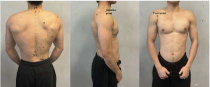
Surface Division In order to better focus on each muscle in resistance training, points were marked at the apex of the shape of each back muscle according to the struc- ture of the muscle and the direction of movements, which were then divided between the markers at a distance of 30 mm from each other.
A professional clinician from the rehabilitation discipline was invited to guide the palpation, and the palpation was conducted by the same investi- gator during the experiment. It is also important to note that the trapezius muscle referred to in this study is the middle and lower part of the fibers from the seventh cervical vertebra (C7) downwards. The marked points are shown in Figure 1.
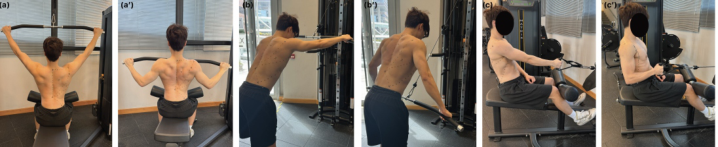
Motion In gym, back resistance training is mainly divided into two categories: pulling and rowing. Compared to the free weight exercise, the fixed apparatus is more capable of stabilizing the body of subjects to reduce the error. In this experiment, we used the pullover and seating row equipment and chose the movements according to the difference of torso and arm angles to activate dif- ferent muscles, including seated pull-down, standing pull-down, and seated row. The standard of these exercises is shown in Figure 2.
164 Huang and Li
Procedure The specific exercise programme was designed and adjusted according to expert recommendations for adult men with some training experience (Kraeme et al., 2004): one set of three repetitions of each training, with one minute of rest after each set, and five minutes of rest after each movement cycle. As the scanning was conducted during exercises, only 50% of their daily exercise weight was used, which allowed the subjects to maintain the posture.
The whole cycle of each movement was recorded at two key points: the start point and the end point. In order to scan the key movements, we asked the subjects to maintain each posture for 15 seconds.
Table 1. Information and body measurements of subjects.
Table available in the original PDF paper.
Subjects
1 2 3 4 5 6 7 8 Mean SD
2 - 4
Age 29 23 26 24 27 21 28 22 25.00 2.93 Height(cm) 172 177 173 173 164 173 168 175 171.88 4.09 Weight(kg) 75 73 68 69 66 69 70 69 69.88 2.85 Years of training Above 5 years - Anaerobic frequency(times/week)
1–2 2–4 1–2
Average duration(min) 40 min above 30 min - 40 min - Back training frequency (times/week)
After marking, the above back exercise movements were performed, and the back deformation was measured using 3D software. The deformation data measured under different back exercise movements was analysed in
Back Skin Deformation During Anaerobic Exercises for Ergonomics Application 165
terms of horizontal, longitudinal, grid. The data were visually presented on the human body.
Figure 2: Motion demonstration.
RESULTS
In terms of counting the skin deformation, the following equation was estab- lished, where L’ is the length of the skin segment during exercise, and L0 is the initial length: Skin Deformation(1%) = (L’−L0)/ L0 % The mean values and standard deviation were calculated based on the data of eight subjects in the state of three movements of six postures.
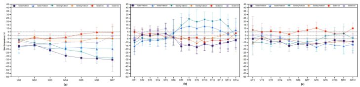
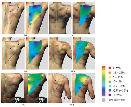
Trapezius Neck-Shoulder Direction The overall deformation of the neck-shoulder (NS) direction during seated pull-down is shown in Figure 3(a). The dark purple and blue lines show the segmental deformation of the trapezius muscle during the seated pull-down. The surface deformation of the NS direction was most significant in the seated pull-down, where the interval ranges between −35.34% ∼−13.27%. The closer the segments are to the shoulders, the higher degree of contracting is observed, reaching a maximum of contracting at NS7.
The standing pull-down showed the same trend of contracting through green and orange lines suggesting an overall compression, while the NS region in the seated row presented in Figure 3(a) was relatively stable, with a max- imum stretch of 5.01% only. Formal movement with the arm retracted, and scapular posterior rotation was accompanied by contraction of the trapezius muscle, reflected in contractions of 8.06% ∼12.89% in the NS segment. The degree of change in NS of three movements was maximal near the point of the shoulder.
Figure 3: Deformation of trapezius (a) neck-shoulder direction (b) shoulder-t10 direction (c) neck-t10 direction.
166 Huang and Li
Trapezius Shoulder-t10 Direction The overall variation in the direction of shoulder-t10 (ST) is presented in Figure 3(b), where three movements performed all show a contracting state and changed most when performing the seated pull-down, reaching a degree of 17.27%. The general variation in other movements ranged from −2.15% ∼7.87%.
As the longest obliquely orientated side of the trapezius structure, the shoulder-t10 direction covers a large area from the shoulder to the middle back. It has more pronounced segmental variations, such as in the seated pull-down, where it is changed from a 2.88% extension to the 17.27% deformation at the middle part of ST, more dramatic variation was also demonstrated in preparing standing pull-down ranging from −10.77% to 23.40%. However, due to the 30-degree forward angle of the torso during standing pull-down, the downward force was dispersed during the move- ment, and the maximum compression when performing the movement only reached 11.55% at ST9 in the middle section of ST.
During the seated row, the ST direction is shown to be squeezed to a certain extent, and the overall maximum deformation was −5.56% while perform- ing the movement. The segmental deformation presented during seated row was within −15.52% ∼9.9%.
Trapezius Neck-t10 Direction From the analysis of Figure 3 (c), it can be seen that the vertical region from the neck point to T10 (NT) presented a mean value of 5.23% stretch during the seated row preparatory movement and the stretch level is relatively stable in each segment.
The remaining five cases showed more contracted segments of the lower half of the NT, i.e., NT8-NT14, presenting remarkable deformations of 11.97% ∼18.13%, and reaches a maximum value of 18.13% at the lower segment of NT13, with an overall deformation of 17.10% during the seated pull-down. In comparison, the NT8-NT14 segments show slightly different segmental trends in standing pull-down, with a decreasing trend at the lower segment and a compression value of only 0.23% at NT13.
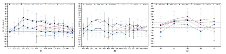
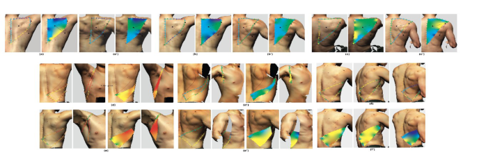
Figure 4: Deformation of Latissimus dorsi (a) front-t10 direction (b) front-abs direction (c) T10-abs direction.
Back Skin Deformation During Anaerobic Exercises for Ergonomics Application 167
Latissimus Dorsi Front-t10 Direction As shown in Figure 4 (a), the oblique edge deformation in the direction of the front point to t10 (FT) was most obvious in the seated pull-down preparation, showing a large peak where the FT4 and FT5 segments reach more than 30% stretching, whereas FT14 - FT15 have only less than 10% deformation and suggest a trend from extension to compression in the direction close to the spine.
The trend characteristics of the deformation segments during the seated pull-down were almost the same as preparation, however, the maximum stretching deformation was reduced to 12.31%, and a small contraction deformation was observed from FT8 onwards.
The deformation of FT9 - FT15 during standing pull-down only showed a remarkable tensile deformation with the maximum value of 32.9% during preparation, while the deformation did not show any significant trend char- acteristics which move up and down around the zero line when performing the movement.
The greatest extrusion of the FT segments among the three movements was in seated row, which showed a greater mean line than the other movements, with several points of FT7 - FT11 falling below the −15% value.
Latissimus Dorsi Front-abs Direction The segments of the front point to abs (FA) during the preparatory move- ments of seated pull-down and standing pull-down have similar skin defor- mation trends, both of the deformation gradually increased from the upper segments, and then showed steady decrease after reaching more than 30% deformation in the interval of FA4-FA6. FA7-FA16 part reached the mini- mum deformation values at FA15-16, which are 12.95% and 13.79%.
When performing the seated pull-down preparation, segments FA2-FA5 showed 3.53% ∼9.59% stretching, while the deformation of FA6-FA10 had an unstable tendency fluctuating between extension and compression in the range of 1.51%-7.07%, and the deformations of FA11 - FA16 all showed 2.98% ∼7.43% stretching. The arm position is pulled back, and the axilla is clamped during the stand- ing pull-down and seated rowing, the area of the anterior and the auxiliary region is obscured. Therefore, only the comparisons of the posterior segments of muscle can be carried, i.e. FA6 - FA16 (standing pull-down), FA11-FA16 (seated row). From the line of Fig. 4 (b), it can be seen that the lower half of the FA presents a relatively large stretch when performing the standing pull-down and reaches a maximum value of 13.72% at the bottom.
Latissimus Dorsi t10-abs Direction
Figure 4 (c) demonstrates that the deformations from t10 to abs (TA) are presented to a small extent in all movements, with each segment fluctuating between −11.68% and 15.41%. The maximum partial degree of stretch is reflected in TA5, reaching 11.49% when preparing seated row.
Only the whole segment characteristics of the seated pull-down differed between the preparatory and the performed segments, with the preparatory segment showing 6.78% stretching of the whole segment while the performed segment showed 3.89% compression.
168 Huang and Li
Regional Analysis of Skin Deformation Based on the above calculation of horizontal, vertical, and oblique segmental deformation measurements of trapezius (middle and lower) and latissimus dorsi muscles, the deformation was graded in steps of 10% as shown in Figure 5–6, where the ± 5% (± 1.5 mm) interval, i.e., the green area, was considered as insignificant deformation.
It can be seen that the horizontal neck-shoulder region is the most promi- nent region of extrusion in the three back exercises, in most cases above 15% and not showing any stretching manifestations. The stretching and squeez- ing characteristics in the vertical direction varied according to the different dynamics of the back, with squeezing predominating in the standing and seated pull-downs and partial stretching presented in the seated row.
The greatest variability was seen in the oblique side of the segment, with the middle and lower trapezius fibres connected to the scapula and divided by the scapular gonad, as can be seen from the regional maps, the horizontal fibres of the middle trapezius above the scapula were predominantly extruded by 5%–15% during the two pull-down exercises, with minor stretching only occurring in seated rowing preparation.
The lower region of the scapula has the most pronounced movement vari- ability and the largest drop in deformation. The entire segment was stretched in the seated pull-down and then squeezed in preparation for the standing pull-down.
For the regional deformation of the latissimus dorsi, the auxiliary region showed a very large, long segment deformation of 25% or more in prepa- ration for the two pull-down movements. The skin recovered during the formal seated pull-down and was squeezing the posterior part of the latis- simus dorsi muscle. Overall, the skin deformation induced by the direction of the latissimus dorsi fibres on was very significant in all three movements.
In this study, a muscular-based method was proposed based on the two main muscles of the back which are the middle and lower trapezius and the latissimus dorsi muscles, and then used the three-dimensional scanner to obtain images of the back during three anaerobic training sessions. The regional extremes, regional variation characteristics for the same movement, and regional variation characteristics during different training movements were demonstrated.
The muscular-based method was developed to make up for the previ- ous gap in the study of skin deformation in anaerobic exercise, taking into account the special feature of anaerobic exercise. A few previous studies involved skin deformation of the upper body, targeting the direction of the direction but mainly aerobic exercise, Shi et al. (2023) and Wang et al., (2021) both used the division of the human body circumference method to study the skin deformation of the upper body during cycling. However, surface division according to anthropometric is only applicable to aerobic exercises.
From the results of present study, it appears that the deformation of the muscle zones is regionally distributed along the oblique edges of the muscle contour and that their distribution is correlated with the state of movement. A significant difference exists in the pull-down where the deformation of the segments along the lateral oblique edge of the trapezius muscle reaches a difference from −5% ∼15%. Such significant regional variability should be taken into account in functional design, dividing the design area according to regional skin deformation differences, to improve the zoning comfort based on anaerobically induced deformations.
One previous study conducted by Carnevale (2021) changed the tradi- tional division method by placing tracking points built on the scapular structure to analyse its deformations. One of its findings is similar to the state presented by the two pull-downs in this study, showing that auxiliary stretching deformations were most pronounced in the three planes of the arm at 90 and maximal degrees, respectively. Skin deformations induced during anaerobic training of the back need to be further investigated than just the scapular region.
170 Huang and Li
This paper points out the extreme deformities to watch out for in the dor- sal region. The largest area of deformation in back training was found in the auxiliary region of the latissimus dorsi muscle, with 37.41% and 34.99% of deformation caused by both sides of the latissimus dorsi muscle when per- forming seated and standing pull-downs, and the largest compression was found in the NS region in preparation for seated pull-down, with an average of 27.34%.These identified regional extremes need to be incorporated into the design considerations to ensure the design’s dynamics and comfort.
However, there are still some limitations of this study, for example, the underarm region points are shaded and could not be scanned during seated row and standing pull-down, resulting in missing data, this region could be further investigated in future studies. In addition, even though the invited participants were all trainers with daily exercise habits, the data obtained could not cover the whole group due to the differences in the exercise habits of each individual.
CONCLUSION
In conclusion, this paper investigated the skin deformation of the back induced by anaerobic training using a muscle-based segmentation approach and calculated regional deformations, and analysed the regional deformation characteristics. The results show that the latissimus dorsi region presented the greatest stretch in the auxiliary region during back exercises. In contrast, there is significant compression of the neck-shoulder region while perform- ing the preparation of the pull-down, also the oblique side of the latissimus dorsi while training through seated row. These results and visual deformation mapping provide interesting insights for the modular division and regional development of functional garment and wearable monitoring designs.
ACKNOWLEDGMENT
The financial support of publishing this research article is funded by the sup- ported by the School of Fashion and Textiles, The Hong Kong Polytechnic University, project P0044073 & P0046961.
REFERENCES
Carnevale, A., Schena, E., Formica, D., Massaroni, C., Longo, U. G., & Denaro, V. (2021). Skin strain analysis of the scapular region and wearables design. Sensors, 21(17), 5761. Choi, S., & Ashdown, S. P. (2011). 3D body scan analysis of dimensional change
in lower body measurements for active body positions. Textile research jour- nal, 81(1), 81–93. Hass, C. J., Feigenbaum, M. S., & Franklin, B. A. (2001). Prescription of resistance training for healthy populations. Sports medicine, 31, 953–964. Kraemer, W. J., & Ratamess, N. A. (2004). Fundamentals of resistance training: pro-
gression and exercise prescription. Medicine & science in sports & exercise, 36(4), 674–688.
Back Skin Deformation During Anaerobic Exercises for Ergonomics Application 171
Lee, H., Hong, K., & Lee, Y. (2013). Ergonomic mapping of skin deformation in dynamic postures to provide fundamental data for functional design lines of outdoor pants. Fibers and Polymers, 14, 2197–2201. Lehman, G. J., Buchan, D. D., Lundy, A., Myers, N., & Nalborczyk, A. (2004).
Variations in muscle activation levels during traditional latissimus dorsi weight training exercises: An experimental study. Dynamic Medicine, 3, 1–5. Luo, S., Wang, J., Yao, X., & Zhang, L. (2017). A novel method for determining skin deformation of lower limb in cycling. The Journal of The Textile Institute, 108(9), 1600–1608. Neumann, D. A. (2016). Kinesiology of the Musculoskeletal System-E-Book: Kinesi-
ology of the Musculoskeletal System-E-Book. Elsevier Health Sciences. Shi, Q. Q., Jiao, J., Shin, K., Chow, H. K., & Lau, N. (2023). Study of cyclists’ skin deformation for compression skinsuit design. Textile Research Journal, 93(19–20), 4548–4561. Wang, Y., Li, H., & Li, J. (2021). The skin deformation during cycling on terrains
for practical application of compression sportswear. The Journal of The Textile Institute, 112(5), 691–699. Watkins, S. M. (1985). Clothing: The portable environment.
LISTED IN THE AHFE DIGITAL BOOKSTORE CATALOGUE
Explore the complete digital volume
Healthcare and Medical Devices
Read this individual research paper free through the original publisher, or explore the full digital proceedings book.
ISBN 978-1-964867-06-9
Explore this ISBN in the AHFE Bookstore ↗Listed in the July 2026 bookstore catalogue; search by ISBN to confirm current availability · Digital book purchase optional