Eduardo Pinos, Roberto Coronel, Maria Cordero (2022). Three-dimensional construction of the corneal geometry from tomographic images and biomechanical parameters of the cornea. In: Applied Human Factors Research. AHFE Open Access, vol. 67, pp. 140–146. https://doi.org/10.54941/ahfe1002692
2 GIIATA, Research Group on Artificial Intelligence and Assistive Technologies, GIBI Research Group of Bietics in Research Universidad Politécnica Salesiana, Cuenca 010105, Ecuador ↗
Currently, there are various diseases that affect the corneal structure, when not treated in a timely manner can cause damage to the corneal structure, which affects the correct vision of people, in these cases the patient goes to the ophthalmologist, who through clinical studies determines the pathology and the degree of impairment of the corneal structure, in many cases surgery is necessary to correct this damage, however, there are cases in which after surgery the cornea suffers further damage to its structure, further affecting the visual acuity.To avoid damage after surgery, more effective methods are searched to determine whether the patient requires surgery or not, by analyzing the corneal biomechanics and its properties, in addition, the morphological analysis of the corneal tissue that allows observing the physical changes produced by these diseases. With the purpose of studying the physical structure and the forces applied on the corneal tissue, this article presents a construction of the corneal surface, for this, different types of elastic, hyperelastic and visco-elastic materials for simulation have been analyzed, whose characteristics resemble the flexible behavior of the corneal tissue in conditions of tension and deformation, in addition, a three-dimensional corneal geometry has been obtained from images of corneal tomographies, which were extracted from ophthalmologic examinations. Consequently, the unification of the three-dimensional geometry of the cornea with the properties of elasticity, Poisson's ratio and density assigned to a simulation material, results in a corneal model that resembles the natural corneal tissue.
The cornea is an element of great importance in the visual system, it is a delicate tissue that is responsible for protecting the internal structures of the eyeball and at the same time, it provides two thirds of the refractive power
Three-Dimensional Construction of the Corneal Geometry 141
of the vision mechanism of our eyes (Ophthalmology (AAO), 2012), for this reason when the cornea loses transparency or changes shape due to diseases or accidents, the correct vision is deficient and hinders the perception of the images of the environment (Chen, 2022).
Different devices for professional use make it possible to analyze the state of the tissues and corneal geometry in order to facilitate professional diagno- sis and the determination of diseases (Cordero et al., 2020), as well as possible solutions to improve the poor vision caused by corneal damage. Some meth- ods of vision correction are refractive and laser eye surgery (Dogan, 2019) or implants, whose action is to restore the shape of the cornea or replace tissues or functions of the cornea, so that it can again focus light correctly (Dunkin, 2020). Like any surgical procedure in the body, there may be secondary effects such as undercorrection, overcorrection, infection, delayed healing (Barrera Meléndez, 2019), however, a greater problem arises when postoperative ecta- sia is generated, that is, when the corneal deterioration returns or worsens after the surgery has been performed (Jin et al., 2020).
For this reason, it is necessary to look for new alternatives for the analy- sis of the cornea through its biomechanics, by creating a three-dimensional cornea and identifying modeling materials that resemble the tension and deformation behavior of corneal tissue (Barreto & Alvarez, 2018).
MODELING MATERIALS There are different types of materials used to describe the mechanical beha- vior of tissues since the mechanical properties of tissues have wide interest in different medical applications (Moreno et al., 2018). From the studies per- formed by (Kim et al., 2012; Zisis et al., 2015), it can be highlighted that when a small load is applied to a living tissue and subsequently removed, it has a linear elastic behavior, but when the tissue has a behavior similar to rubber or is subjected to large deformations it shows a nonlinear behavior (hyperelasticity) that said, the most optimal model that resembles corneal tis- sue and given the characteristics required for the specific material the corneal structure is modeled with a hyperelastic material (Nguyen et al., 2020).
The Mooney-Rivlin model is a hyperelastic material model that can resem- ble materials with isotropic and isothermal mechanical properties, it makes stress-strain relation by means of a strain energy density function, a linear combination of two invariants of the Cauchy-Green strain tensor (Ngu- yen et al., 2019), meanwhile, the Neo-Hookean hyperelastic model is a model similar to Hooke’s law, it is applicable in structures subjected to large deformations, this model can be applied in materials where initially the stress- strain relationship is linear up to a certain point and then this relationship changes to a nonlinear behavior (Kim et al., 2012; Moreno et al., 2018).
Because of the necessity of the integration of corneal geometry with bio- mechanical characteristics, Preview software will be used for this process, for this, the ideal is to find a model that with few parameters manages to describe the behavior in different situations of deformation of the corneal structure.
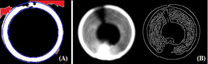
Figure 1: Problems when overriding errors. (A) By identification of the inner and outer circumference of the cornea, in the upper part of the figure, the blue lines do not follow the corneal contour due to a deformity, the areas marked in red are the features that do not belong to the cornea. (B) By edge identification, the edges do not follow a defined contour, but cross the cornea.
It is found that the Neo-Hookean model fits better taking into considera- tion that less data is needed for its input and at the same time these data are extracted from the cornea’s own biomechanics such as the modulus of ela- sticity, whose calculation can be found in the work done in a previous stage (Cordero-Mendieta et al., 2020).
THREE-DIMENSIONAL CORNEAL RECONSTRUCTION Two-dimensional corneal images are used to obtain a three-dimensional model of the cornea. The basis for obtaining these images are files in “dicom” format which come from corneal tomographies, however, in a tomography not only the images of the specific object are captured, but also the adjacent tissues and elements close to the cornea such as the iris and the pupil, addi- tionally, the images of the cornea can be altered by the reflection of light on this tissue, forming spots or clear areas that distort the shape of the cornea, this inconvenience can be corrected by eliminating these features foreign to the tissue to be studied.
Removal of Features that do not Belong to the Cornea There is a need to remove all stains and features that are not part of the cor- nea, for this, different methods have been tried to suppress these unnecessary features, in the first procedure that was tried to remove these noises, all the features that were outside the circles that form the inner and outer edge of the cornea for each slice of the tomography were annulled, based on (Kumar et al., 2015), however, when the cornea presents keratoconus or other path- ology that deforms its geometry , these edges are not uniform so they do not form a circumference which makes this method not viable since part of the corneal surface would be lost as seen in Figure 1(A). Another procedure with which we tried to eliminate the features that were not part of the cornea was by means of edge detection, during the test of this method it was evidenced that in those areas that presented dark spots the edges were also highlighted and in the areas where there were light spots edges that crossed the cornea
Three-Dimensional Construction of the Corneal Geometry 143
Figure 2: Non-solidified cornea from InVesalius.Figure 3: Anterior and posterior layer of the cornea.
were created as shown in figure 1(B), therefore, this method is less efficient than the one tested in the first instance.
A viable option to eliminate these errors is by means of the software “InVesalius”, this software makes it possible to segment the images manu- ally or automatically (by using filters), import the “dicom” format files, and the corneal images are manually modified using the software’s own tools, which allow drawing missing areas or deleting features that protrude from the edges of the cornea, preserving only those areas corresponding to the corneal geometry in each of the slices, either in the axial or sagittal plane.
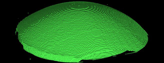
Subsequently, the information is re-rendered to create a first 3D object that approximates the three-dimensional geometry of the cornea to be created using the same software (See figure 2).
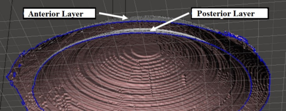
Solidification of the Three-Dimensional Geometry Since the three-dimensional reconstruction coming from the “InVesalius”sof- tware, the resulting object is not a solid, but only presents the anterior and posterior layers of the cornea as two objects separated by an air gap (see Figure 3), it is necessary that these objects form a single solid body similar to the corneal tissue.
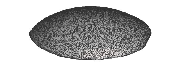
For this we made use of the Meshmixer software, with the use of the “Make Solid” tool, an approximation of the original shape is created using a large number of small cubes called “voxels” of modifiable size (Autodesk, 2018) that fill the empty space between the layers, thus creating a single solid. It is more suitable for the solidification of the object the “Accurate” mode, where the software takes the approximation of the cubes and calculates a
Figure 4: Solid cornea.
field of distances that approximate the original object, subsequently, you can modify the mesh density by increasing or reducing the size of the voxels and create a smoothing of the surface using the “Smooth” tool that smooths the surface as it is the cornea, finally obtaining the result seen in Figure 4.
As a last step, Poisson’s Ratio, density and corneal modulus of elasticity obtained from its biomechanical parameters are attached to the solidified three-dimensional cornea.
Finally, a structure containing the shape and characteristics that resemble as much as possible a specific cornea (healthy or pathological) is obtai- ned, thus allowing the use of the structure for the necessary analysis of the behavior of the corneal surface tissue.
CONCLUSION
The analysis of several models of hyperelastic materials has been carried out and it has been concluded that the Neo-Hookean material model, thanks to its hyperelastic characteristics, is ideal to analyze the stress-strain behavior of the corneal tissue, in addition, it allows obtaining its elastic modulus values through the biomechanical parameters extracted from the ophthalmologic examinations of each eye to which the three-dimensional model will be made.
In the construction process there are different methods to obtain the edges of the cornea in an axial tomography slice, however, when it presents path- ologies that deform its geometry it is not possible to determine its edges by locating circles because the characteristics of the cornea according to its path- ology do not form a homogeneous geometry so that when delimiting the edges through a circumference the information that remains outside the area is lost.
Due to the nature of information extraction and all the modifications made during the three-dimensional construction processes of the cornea, it is possi- ble that the final result that is presented loses some geometric characteristics of the initial product that the “dicom” files have, however, by combining it with the characteristics of the Neo-Hookean material, it is possible to have a model that has the shape and characteristics that resemble as much as possi- ble a specific natural cornea (healthy or pathological) thus allowing the use of the structure made for the necessary analysis on the behavior of the corneal surface tissue.
Three-Dimensional Construction of the Corneal Geometry 145
FUTURE WORK
The aim is to simulate the action of an external force similar to the air jet emitted by the tonometer applied on the corneal surface, in order to obtain pressure distribution maps and the tension exerted, being able to identify possible fragile areas in the tissue according to different corneal pathologies.
REFERENCES
Autodesk, H. (2018). Make Solid Tool | Search | Autodesk Knowledge Network.
https://knowledge.autodesk.com/search-result/caas/CloudHelp/cloudhelp/2019/ ENU/MSHMXR/files/GUID-F8C64203-AFB7-449C-A395-2C029A80BB6C-h tm.html Barrera Meléndez, A. (2019). Resultados a largo plazo de la cirugía refractiva corneal. https://uvadoc.uva.es/handle/10324/38356 Barreto, J. P. G., & Alvarez, J. C. O. (2018). Caracterización Y Diseño De Simulación
De Cornea Artificial. 74. Chen N. (2022). Research progress of natural biomaterials for construction of tissue engineering cornea. International Eye Science, 44–48. Cordero, M. I., Coronel, R., Pinos-Vélez, E., Ipanque, W., & Chacón, C. L. (2020).
Proposal for a Support Tool for the Study of Corneal Biomechanics and Its Influence in the Human Eye. Advances in Emerging Trends and Technologies, 219–228. Cordero-Mendieta, M. I., Pinos-Vélez, E., & Coronel-Berrezueta, R. (2020). Study of Corneal Biomechanics and Modeling of Young’s Module in Healthy and Path- ological Corneas. Advances in Human Factors and Ergonomics in Healthcare and Medical Devices - Proceedings of the AHFE 2020 Virtual Conference on Human Factors and Ergonomics in Healthcare and Medical Devices, 99–105. Dogan, S. S. (2019). Corneal refractive surgery: LASIK and beyond | Eyes and
Vision. UT Southwestern Medical Center. http://utswmed.org/medblog/corneal- refractive-surgery-lasik-and-beyond/ Dunkin, M. A. (2020, diciembre). Types of Refractive Laser Eye Surgery. WebMD.
https://www.webmd.com/eye-health/overview-refractive-laser-eye-surgery Jin, S. X., Dackowski, E., & Chuck, R. S. (2020). Risk factors for postlaser refractive surgery corneal ectasia. Current Opinion in Ophthalmology, 31(4), 288–292. Kim, B., Lee, S. B., Lee, J., Cho, S., Park, H., Yeom, S., & Park, S. H. (2012).
A comparison among Neo-Hookean model, Mooney-Rivlin model, and Ogden model for chloroprene rubber. International Journal of Precision Engineering and Manufacturing, 13(5), 759–764. Kumar, V., Asati, A., & Gupta, A. (2015). An Iris localization method for noisy infrared iris images. 2015 IEEE International Conference on Signal and Image Processing Applications (ICSIPA), 208–213. Moreno, M., Plazaola, C., González, G., Zambrano, M., & Spadafora, C. (2018).
Revisión de Modelos Hiperelásticos utilizados en Tejidos. KnE Engineering, 3(1), 100. Nguyen, B. A., Reilly, M. A., & Roberts, C. J. (2020). Biomechanical contribution of the sclera to dynamic corneal response in air-puff induced deformation in human donor eyes. Experimental Eye Research, 191, 107904. Nguyen, B. A., Roberts, C. J., & Reilly, M. A. (2019). Biomechanical Impact of the
Sclera on Corneal Deformation Response to an Air-Puff: A Finite-Element Study. Frontiers in Bioengineering and Biotechnology, 6, 210.
Ophthalmology (AAO), A. A. of. (2012). Enfermedades de superficie ocular y córnea.
2011–2012: Sección 8. Elsevier España. Zisis, Th., Zafiropoulou, V. I., & Giannakopoulos, A. E. (2015). Evaluation of material properties of incompressible hyperelastic materials based on instrumented indentation of an equal-biaxial prestretched substrate. International Journal of Solids and Structures, 64–65, 132–144.
LISTED IN THE AHFE DIGITAL BOOKSTORE CATALOGUE
Explore the complete digital volume
Applied Human Factors Research
Read this individual research paper free through the original publisher, or explore the full digital proceedings book.
ISBN 978-1-958651-43-8
Explore this ISBN in the AHFE Bookstore ↗Listed in the July 2026 bookstore catalogue; search by ISBN to confirm current availability · Digital book purchase optional