Aoki Makiko, Satoshi Suzuki, Ryouma Anzai (2022). Relationship between activation of prefrontal cortex and testosterone in N-back task. In: Neuroergonomics and Cognitive Engineering. AHFE Open Access, vol. 42, pp. 149–156. https://doi.org/10.54941/ahfe1001830
Purpose: Individual and gender differences are an important topic in the study of human cognition. In general, it has been shown that women are superior at verbal ability and associative memory, while men are better at tasks related to numbers, logic, and spatial recognition. Among them, it has been reported that cerebral blood flow in the prefrontal cortex increases during the N-back task, a working memory task. The relationship between sex hormones and cerebral blood flow remains unelucidated, although sex hormones and depressive mood are both factors that affect the function of the prefrontal cortex (cognitive function). Therefore, the present study aimed to clarify the relationship between the testosterone and cognitive function.Methods: The subjects included 6men and 16 women aged 20–35 years. Near infrared spectroscopy (NIRS), a brain function imaging device, was attached to the forehead of the subjects, and Oxyhemoglobin (Oxy-Hb) was measured during the N-back task. Oxy-Hb was measured during the N-back task using a NIRS system in the CHs 1-4 (right side), CHs 7-10 (center), and CHs 13-16 (left side) , and saliva was collected to measure the testosterone concentration. Subsequently, the correlation coefficient between the initial activation and testosterone concentration was calculated. This study was approved by the Institutional Review Board of the Kanagawa institute of technology (No. 20191011-01).Result: The items that were significantly correlated with testosterone were the initial activation CH13-16 in the 0-back task (r=48, p=.02). And the correct response rate (r=.42, p=.03), reaction time (p=.46, p=.03) and initial activation of CH13-16 (r=.68, p=.007) in the 1-back task. Conclusion: In adults, testosterone was associated with initial activation of the NIRS Oxy-Hb signal in the 0-back and 1-back tasks. Furthermore, in the 1-back task, testosterone was associated with correct response rate, the reaction time, and initial activation of the NIRS Oxy-Hb signal of CH13-16. This result suggests that testosterone may be related to left prefrontal cortex activation and responsiveness to presentation of the N-back task.
Traditionally, individual and sex differences have been an important topic in research on cognitive abilities and cognitive abilities related to memory. In general, it is thought that women are superior in language ability and asso- ciative memory, while men are thought to be superior in mathematics, logic, and spatial awareness (Tirassa et al., 1997). These major cognitive abilities are governed by the limbic system, and we have some reports of differe- nces in brain function due to sex hormones (Koolschijn et al., 2014, Torres et al., 2006), and some reports even claim no relationship, many controversies remain.
Testosterone is a sex hormone and one of the major hormones secre- ted by both men and women. A steroid hormone in the androgen family, testosterone is the main sex hormone in men. In women, testosterone is secreted by the ovaries and adrenal glands. In both men and women, testo- sterone has been implicated in health and well-being, including aspects of mood and behavior, and in the prevention of osteoporosis (Gaviglio et al., 2014). With regard to the relationship of testosterone to cognitive function,
we find reports of a positive correlation between testosterone and cognitive function in men and women (Akishita, 2014), of a relationship between spa- tial cognitive ability and memory (SHUTE et al., 1983), and of a significant improvement in working memory observed in patients receiving testosterone replacement therapy (Janowsky et al., 2000). Therefore, we focused here on working memory and testosterone levels. Working and short-term memory have different meanings. Short-term memory is a passive, temporary memory that provides only retention, whereas working memory is an active and goal- oriented temporary memory useful in both retention and processing. The functioning of the prefrontal cortex is central to working memory (Logie et al., 2021). Previous reports have also shown that cerebral blood flow (CBF) in the prefrontal region increases during execution of the N-back task, a task that places demands on working memory (Adrian et al., 2005, Marquand et al., 2008, Owen et al., 2005) Recently, methods that image brain functioning have attracted attention, and one of these is near-infrared spectroscopy (NIRS), which images activa- tions of blood flow on the cerebral cortex. NIRS has reportedly shown brain activity during the performance of cognitive functions; the prefrontal cortex is located behind the forehead and is thus accessible to the NIRS method (Aoki et al., 2020). However, very few studies have evaluated the effects of testosterone on brain functioning using NIRS.
In this study, we hypothesized that testosterone levels correlate with brain activity during the working memory task and aimed to clarify the relationship between testosterone and cognitive function using tests of salivary testo- sterone secretion and the initial NIRS signal observed during the subject’s performance of the N-back task.
MATERIAL AND METHODS Participant Selection
The participants were 6 men and 16 women between the ages of 20 and 35 years. All participants were healthy and without systemic diseases. The following exclusions were applied:
1) Currently pregnant; 2) body mass index outside the range 18-25; 3) smoker; 4) psychiatric history; 5) a Self-Rating Depression Scale score ≥50.
Procedure The participants’ saliva was collected prior to the CBF measurements and CBF was measured during the performance of the cognitive task.
Salivary Hormone Assays Salivary testosterone concentrations were measured using an enzyme immu- noassay kit (salivary teststerone; Salimetrics LLC, USA). The participants
Relationship between Activation of Prefrontal Cortex and Testosterone in N-Back Task 151
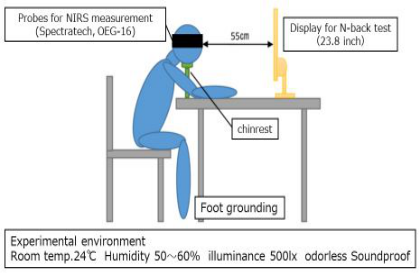
Figure 1: Experimental set up for CBF measurement.
were instructed to abstain from alcohol for 12 h and from eating and drin- king for 1 h before saliva collection to avoid contamination and dilution of the sample. Saliva was collected with a kit (Saliva Collection Aid, Salimetrics LLC, USA) using the passive drool method. The collected saliva was promptly frozen and analyzed at Kamakura Techno-Science, Inc. (Kanagawa, Japan) on the same day.
CBF Measurement We used an NIRS system (OEG-SpO2, Spectratech Inc., Tokyo, Japan). NIRS probes are each composed of an emitter and a detector unit. Twelve of each type of unit were placed at intervals of 3 cm in a lattice-like pattern, and the CBF within an area of 150 mm × 60 mm was measured. Figure 1 illustrates the procedure for measuring CBF. The participants were seated in a chair with their feet on the floor, 55 cm from a monitor (23.8 inch), with the NIRS probe array mounted on the forehead. A chin rest was used to prevent head movement. The participants were asked to rest their arms on a desk and use a cushion to allow them to perform the cognitive function task comfortably. They were then asked to perform the tasks presented on the monitor during NIRS recording. The NIRS signals were sampled every 0.65 s.
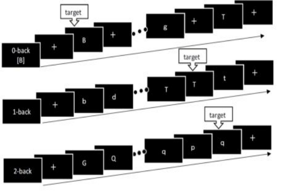
Design of the N-Back Task In this task, letters of the alphabet are presented in a random order, and participants are asked to respond when the presented letter matches the letter that appeared in letters earlier (Harvey et al., 2005). Figure 2 shows the task design.
NIRS Data Analysis Oxy-Hb values were recorded starting 5 seconds before the start of each session of the N-back task (t = −5 s) and continuing to the end of task pre- sentation (t = 60), using channels ch1–ch16 of the probe array. The amount of change in oxy-Hb (1oxy-Hb) was determined by subtracting the baseline from the values of the oxy-Hb. The slope from 0sec to 5sec was used as a
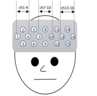
Figure 2: N-back task design. For 0-back task, click the mouse when the current chara- cter is the same as the first character displayed, 1-back task is one step before, 2-back task is the two step before.Figure 3: The layout of each channel and its designated area.
measure of the initial activation, which was then averaged over two sessi- ons of the same N-back task. Finally, the average initial activations in the 0-, 1-, and 2-back tasks were averaged over channels ch1–4 (typical of the left hemisphere), ch7–10 (typical of the center), and ch10–ch13 (typical of the right hemisphere) (Sasahara et al., 2020, Suto et al., 2004, Kameyama et al., 2006). Figure 3 shows the layout of each channel and its designated area.
Data Analysis Spearman’s rank correlation coefficients were calculated between testoste- rone level and the correct-response rate, the reaction time, and the initial brain activation measured in the N-back task.
Ethical Considerations Prior to study start, participants were given both written and verbal expla- nations of the study and were asked to sign an informed consent form.
Relationship between Activation of Prefrontal Cortex and Testosterone in N-Back Task 153
Table 1. Participant characteristics.
Table available in the original PDF paper.
All participants Mena) Womenb) Mean SD Mean SD Mean SD
Age, year 24.4 5.9 29.2 6.5 21.8 0.7 BMI, kg m−2 21.7 1.7 23.8 1.0 21.2 1.6 SDS 38.8 7.1 38.0 4.9 39.0 4.0 a) n = 6. b) n = 16. BMI; body mass index. SDS; Self-Rating Depression Scale score
Table 2. Correlations and descriptive statistics (N = 22).
This table shows relationship between testosterone and Oxy-Hb initial activation, N-back task correct response rate, and reaction time. Spearman’s rank correlation coefficients, TS; Testosterone, *p<.05, **p<0.01
This study was conducted in accordance with the principles of the Decla- ration of Helsinki and was approved by the Human Ethics Committee of the Kanagawa Institute of Technology (approval no. 20191011-01).
Results
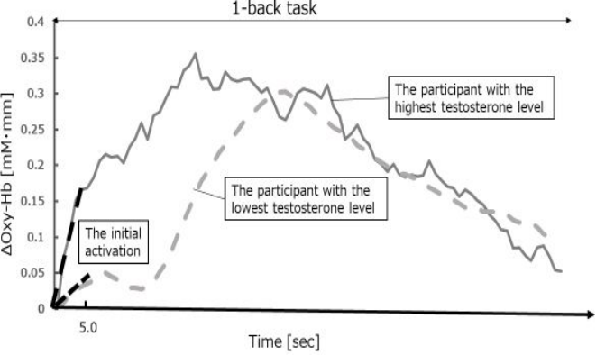
The number of participants is summarized in Table 1. The correlation coeffi- cients between testosterone and initial activation, correct-response rate, and reaction time during the N-back task are provided in Table 2. The items that were significantly correlated with testosterone were the initial activa- tion CH13-16 in the 0-back task (r = 48, p=.02). And the correct response rate (r=.42, p=.03), reaction time (p=.46, p=.03) and initial activation of CH13-16 (r=.68, p=.007) in the 1-back task. Figure 4 shows the 1Oxy-Hb at CH13-16 during the presentation of the 1-back task for the participants with the highest and lowest testosterone levels.
Discussion
The purpose of the present study was to clarify the relationship between cognitive function and testosterone and not to discuss cognitive function in terms of sex. Previous reports have shown an association between spatial cognitive ability and testosterone and an enhancement of working memory due to testosterone replacement therapy. Therefore, we employed here a working memory task, the N-back task. The present results showed that testosterone was associated with initial activation in the left prefrontal cor- tex in the 0-back and 1-back tasks. The N-back task is a working memory assessment task, and the percentage of correct responses assesses working memory, supporting the report that testosterone increases working memory
Figure 4: The 1 Oxy-Hb at CH13-16 during the presentation of the 1-back task for the participants with the highest and lowest testosterone levels. The initial activation is the slope from 0sec to 5sec.
(Janowsky et al., 2000). The initial activation is the average slope of the acti- vation in the first 5 seconds after the presentation of the task and is used as an index of brain responsiveness in the evaluation of mental disorders by NIRS to assess responsiveness (Takizawa et al., 2014) The 0-back and 1-back tasks are less difficult than the 2-back tasks. The present results suggest that the brain’s reactivity when presented with an N-back task of easy or moderate difficulty (Harvey et al., 2005) is related to testosterone. Although the relati- onship between reactivity and testosterone has also been reported for other cognitive tasks, it is possible that testosterone is also associated with reacti- vity in the N-back task. And the correct response rate and reaction time of the 1-back task also correlated with testosterone. Testosterone has been shown to increase the concentration of nerve growth factor in the hippocampus and to upregulate NGF receptors in the prefrontal cortex (Tirassa et al., 1997),. In addition, because of its protective effects on nerves, it has been reported to have a significant association with cognitive function, especially memory and learning (Beauchet, 2006). Thus, testosterone may be related to a series of processes by which people capture visual images, determine the correct answer, make decisions, and act. This was a pilot study; in the future, we intend to repeat the study with a greater number of participants.
CONCLUSION
In adults, testosterone was associated with initial activation of the NIRS Oxy- Hb signal in the 0-back and 1-back tasks. Furthermore, in the 1-back task, testosterone was associated with correct response rate, the reaction time, and initial activation of the NIRS Oxy-Hb signal of CH13-16. This result sugge- sts that testosterone may be related to left prefrontal cortex activation and responsiveness to presentation of the N-back task.
Relationship between Activation of Prefrontal Cortex and Testosterone in N-Back Task 155
ACKNOWLEDGMENT
We thank all participants involved in this study. This research was partially supported by the Kanagawa Institute of Technology Research grant.
REFERENCES
Adrian M. Owen Kathryn M. McMillan Angela R. Laird Ed Bullmore (2005), “N-back working memory paradigm: A meta-analysis of normative functional neuroimaging studies”, Human brain mapping, 25(1), 46–59. Akishita Masahiro (2014), Sex hormones and cognitive function. Japanese journal
of clinical medicine, 72(4), 657–661. Makiko Aoki, Masato Suzuki, Satoshi Suzuki, Hidenobu Takao, Hisayo Okayama (2022), “Cognitive function evaluation in premenstrual syndrome during the follicular and luteal phases using near-infrared spectroscopy”, Comprehensive Psychoneuroendocrinology, 10. Beauchet, O (2006), “Testosterone and cognitive function: current clinical evidence
of a relationship”, European journal of endocrinology,155(6), 773–781. Gaviglio, C.M., Crewther, B.T., Kilduff, L.P., Stokes, K. & Cook, C.J(2014), “Relationship between pregame concentrations of free testosterone and outcome in rugby union”. Harvey, P., Fossati, P., Pochon, J., Levy, R., LeBastard, G., Lehéricy, S., Allilaire, J.
& Dubois, B (2005), “Cognitive control and brain resources in major depression: An fMRI study using the n-back task”, NeuroImage, 26(3), 860–869. Janowsky, J.S., Chavez, B. & Orwoll, E. (2000), “Sex Steroids Modify Working Memory”, Journal of cognitive neuroscience, 12(3), 407–414. Kameyama, M., Fukuda, M., Yamagishi, Y., Sato, T., Uehara, T., Ito, M., Suto, T.
& Mikuni, M (2006), “Frontal lobe function in bipolar disorder: A multichannel near-infrared spectroscopy study”, NeuroImage , 29(1),172–184. Koolschijn, P. C. M. P, Peper, J.S. & Crone, E.A (2014), “The influence of sex steroids on structural brain maturation in adolescence”, PloS one, 9(1), e83929. Logie, R., Camos, V. & Cowan, N. 2021, Working Memory, Oxford University Press
USA - OSO, Oxford. Marquand, A., Mourão-Miranda, J., Brammer, M., Cleare, A. & Fu, C (2008), “Neuroanatomy of verbal working memory as a diagnostic biomarker for depression”, Neuroreport, 19(15), 1507–1511. Owen, A.M., McMillan, K.M., Laird, A.R. & Bullmore, E (2005), “N-back wor-
king memory paradigm: A meta-analysis of normative functional neuroimaging studies”, Human brain mapping, 25(1), 46–59. Sasahara, S., Andrea, C., Suzuki, G., Oi, Y., Doki, S., Hori, D., Ohira, T., Matsuda, C., Inoue, N., Takahashi, T., Ohtaki, Y., Saito, T., Furukawa, S., Ogata, K. & Mat- suzaki, I (2020), “Effect of exercise on brain function as assessed by functional near-infrared spectroscopy during a verbal fluency test in a simulated Internatio- nal Space Station environment: A single-case, experimental ABA study in Japan”, Acta astronautica, 166, 238–242. Shute, V.J., Pellegrino, J.W., Hubert, L. & Reynolds, R.W (1983), “The relationship
between androgen levels and human spatial abilities”, Bulletin of the Psychonomic Society, 21(6), 465–468. Suto, T., Fukuda, M., Ito, M., Uehara, T. & Mikuni, M (2004), “Multichannel nearinfrared spectroscopy in depression and schizophrenia: cognitive brain activation study”, Biological psychiatry, 55(5), 501–511.
Takizawa, R., Fukuda, M., Kawasaki, S., Kasai, K., Mimura, M., Pu, S., Noda, T., Niwa, S. & Okazaki, Y (2014), “Neuroimaging-aided differential diagnosis of the depressive state”, NeuroImage , 85, 498–507. Tirassa, P., Thiblin, I., Ågren, G., Vigneti, E., Aloe, L. & Stenfors, C (1997), “High-
dose anabolic androgenic steroids modulate concentrations of nerve growth factor and expression of its low affinity receptor (p75-NGFr) in male rat brain”, Journal of neuroscience research, 47 (2), 198–207. Torres, A., Gómez-Gil, E., Vidal, A., Puig, O., Boget, T. & Salamero, M (2006), “Gender differences in cognitive functions and influence of sex hormones”, Actas españolas de psiquiatría, 34(6), 408–415.
LISTED IN THE AHFE DIGITAL BOOKSTORE CATALOGUE
Explore the complete digital volume
Neuroergonomics and Cognitive Engineering
Read this individual research paper free through the original publisher, or explore the full digital proceedings book.
ISBN 978-1-958651-18-6
Explore this ISBN in the AHFE Bookstore ↗Listed in the July 2026 bookstore catalogue; search by ISBN to confirm current availability · Digital book purchase optional