George Murrell, Antonette Bilog, Yash Agrawal, Mina Shenouda, Lisa Hackett (2026). Elastography of the Shoulder – A Game Changer for Diagnosis, Prognosis and Monitoring. In: Human Interaction and Emerging Technologies (IHIET-AI 2026): Artificial Intelligence and Future Applications. AHFE Open Access, vol. 201, pp. 603–609. https://doi.org/10.54941/ahfe1007213
The supraspinatus tendon is a key component of the rotator cuff and plays a critical role in overhead function of the shoulder. Overuse tendinopathy and tears of the supraspinatus are is a common cause of shoulder pain and functional limitation. The aim of this paper is to summarise the body of work undertaken by our group evaluating supraspinatus tendon shear wave elastography (SWE) in shoulder pathology, with a focus on diagnosis, prognosis, and monitoring. In adhesive capsulitis, a condition characterised by painful restriction of both active and passive glenohumeral motion, supraspinatus tendon stiffness increased by approximately 50–100%. In supraspinatus tendinopathy, stiffness was reduced by approximately 25–35%. In rotator cuff tear cohorts, increasing age was associated with a 10–15% reduction in stiffness per decade, and larger tears demonstrated stiffness values approximately 20–40% lower than smaller tears. Following rotator cuff repair, supraspinatus tendon stiffness increased progressively by approximately 20–25% over 12 months. Increased elastographic stiffness in adhesive capsulitis explains the decreased re-tear rate in patients with rotator cuff repairs and stiffness going into surgery or coming out of surgery
The supraspinatus tendon is a principal stabiliser of the shoulder and a critical component of the rotator cuff, contributing to the initiation of arm elevation and dynamic centring of the humeral head within the glenoid during functional and overhead activities. Disorders affecting the supraspinatus, including tendinopathy and rotator cuff tears, are among the most common causes of shoulder pain and disability in adult populations.
Shear wave elastography (SWE) is an ultrasound-based no-invasive technique that enables quantitative assessment of tissue mechanical properties in vivo by measuring shear wave propagation through tissue, reported as shear wave velocity and/or elastic modulus. In musculoskeletal applications, SWE provides objective numerical measures of tendon stiffness that can be assessed cross-sectionally and longitudinally using standardised acquisition protocols.
Studies from our group have evaluated supraspinatus tendon SWE in several scenarios. In adhesive capsulitis, a condition characterised by painful restriction of both active and passive glenohumeral motion, substantial alterations in tendon stiffness have been observed. In non-torn supraspinatus tendinopathy, stiffness reductions have been demonstrated relative to normal tendons. Following rotator cuff repair, tendon stiffness changes systematically over time and demonstrates associations with functional recovery.
The aim of this paper is to summarise the body of work undertaken by our group evaluating supraspinatus tendon shear wave elastography, focusing on diagnostic stiffness phenotypes, prognostic associations with age and tear characteristics, and longitudinal changes during tendon healing following rotator cuff repair.
METHODS
Study Design and Included Studies This paper synthesises a series of completed prospective, longitudinal, and cross-sectional studies conducted at a single tertiary referral shoulder unit. Each study evaluated supraspinatus tendon shear wave elastography (SWE) using a consistent acquisition and analysis protocol, enabling comparison across diagnostic states, prognostic subgroups, and longitudinal follow-up.
Study Populations Participants were adults with adhesive capsulitis, non-torn supraspinatus tendinopathy, or rotator cuff tears treated surgically. Individual studies included approximately 40–120 shoulders. Exclusion criteria included previous shoulder surgery, moderate to severe glenohumeral osteoarthritis, inflammatory arthropathy, neurological disorders, and inability to obtain reliable elastographic measurements.
Ultrasound and Elastography Equipment All ultrasound and shear wave elastography examinations were performed by experienced musculoskeletal sonographers using the ACUSON Sequoia Select Ultrasound System (Siemens Medical Solutions, Mountain View, California, USA). The system was operated in two-dimensional shear wave elastography mode with a high-frequency linear-array transducer suitable for superficial musculoskeletal imaging (Maloof et al., 2023; Playford et al., 2021; Hackett et al., 2020; Williams et al., 2023; Shenouda et al., 2026).
Elastography Acquisition Protocol Participants were examined in a standardised seated position. The supraspinatus tendon was assessed in the longitudinal plane proximal to the footprint with careful transducer alignment parallel to tendon fibres. A single standardised region of interest was placed within the tendon substance, avoiding artefact (Maloof et al., 2023; Playford et al., 2021; Hackett et al., 2020).
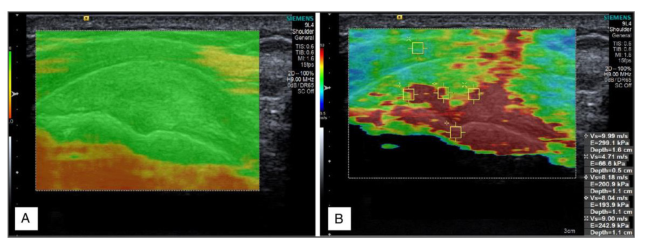
Figure 1: Standard B-mode ultrasound (A) compared with shear-wave elastography of the supraspinatus tendon (B).
Outcome Measures The primary outcome was supraspinatus tendon elastographic stiffness. Secondary outcomes included associations with age, tear characteristics, longitudinal changes following repair, regional variation, and functional outcomes.
Statistical Analysis Associations were assessed using correlation and regression analyses. Longitudinal changes were evaluated using repeated-measures approaches. Effect sizes are reported as percentage change or fold change with correlation coefficients. Statistical significance was defined as p < 0.05.
RESULTS
Findings were derived from multiple completed studies encompassing diagnostic, prognostic, and longitudinal analyses. A summary of included cohorts and key elastographic outcomes is presented in Table 1.
Table 1.
Table available in the original PDF paper.
Key Elastography Finding
Condition Study Type Approx. n (Shoulders) Adhesive capsulitis Cross-sectional ~40–60 50–100% higher supraspinatus stiffness
Supraspinatus tendinopathy
Cross-sectional ~60–100 25–35% lower stiffness (Playford et al., 2021; Hackett et al., 2020) Rotator cuff tears Prospective ~80–120 Inverse relationship with age and tear size (Playford et al., 2021; Solari et al., 2024; Hackett et al., 2023)
Post–rotator cuff repair
Longitudinal ~50–90 20–25% stiffness increase over 12 months (Solari et al., 2024; Hackett et al., 2023)
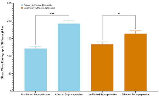
Diagnostic elastography findings (Figure 2), In adhesive capsulitis, supraspinatus tendon stiffness increased by approximately 50–100% compared with the contralateral shoulder (p < 0.001).
Figure 2: Comparison of the mean elastographic stiffness of supraspinatus tendon in patients with primary and secondary adhesive capsulitis *p < 0.05; *** p < 0.001 Non-torn supraspinatus tendinopathy demonstrated stiffness reductions of approximately 25–35% (Hackett et al., 2020).
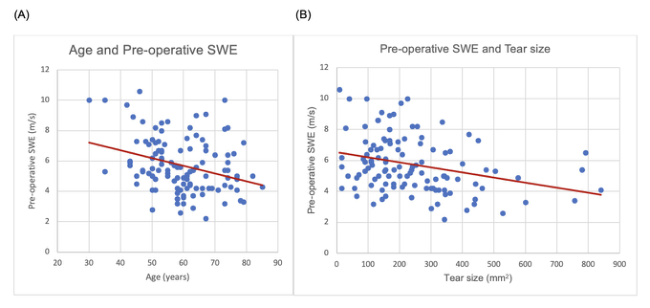
Tear-related associations (Figure 3).
Figure 3: The effect of (A) patient age (Spearman’s rho correlation R= –0.3, p= 0.002) and (B) tear size (Spearman’s rho correlation R= –0.3, p <0.0001) on the pre-operative elastographic stiffness of torn supraspinatus tendons prior to surgery.
Larger and thicker tears demonstrated stiffness values approximately 20–40% lower than smaller tears (r ≈ −0.35 to −0.50, p < 0.001). Early post-operative changes (Figure 4).
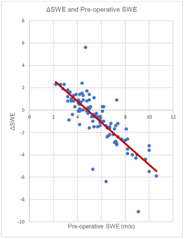
Elastography of the Shoulder – A Game Changer 607 Lower pre-operative stiffness was associated with early increases of 30–50%, whereas higher baseline stiffness showed changes <10–15%.
Figure 4: The relationship between the change in shear wave elastography of the supraspinatus tendon from before surgery to 6 weeks after surgery (ΔSWE) and pre- operative elastographic stiffness of the torn supraspinatus tendon (Spearman’s rho correlation R= –0.8.
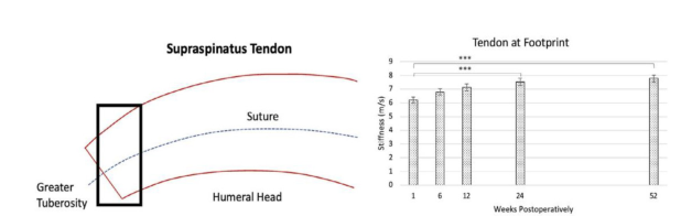
Longitudinal monitoring (Figure 5). Following repair, tendon thickness decreased by approximately 10–12% within six weeks, while stiffness increased progressively by approximately 20–25% over 12 months (Hackett et al., 2023).
Figure 5: Temporal changes in the elastographic stiffness of supraspinatus tendon stiffness following rotator cuff repair.
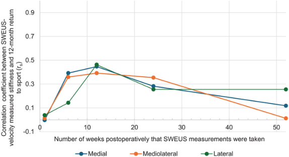
Functional associations: Early post-operative stiffness was 20–30% higher in patients who returned to work and 15–25% higher in those who returned to sport (Maloof et al., 2025) (Figure 6).
Figure 6: Timepoint of supraspinatus elastographic stiffness measurement versus correlation to return to sports at 12 months post repair. SWEU, shear wave elastography ultrasound.
DISCUSSION
This synthesis demonstrates three principal findings. First, supraspinatus tendon shear wave elastographic stiffness differs across shoulder conditions, with increased stiffness in adhesive capsulitis and reduced stiffness in non- torn supraspinatus tendinopathy. Second, in rotator cuff tears, elastographic stiffness is inversely associated with age and tear characteristics, with larger and thicker tears in older patients demonstrating lower stiffness values. Third, following rotator cuff repair, elastographic stiffness increases progressively over time and is associated with functional recovery, despite early stabilisation.
Interpretation of findings: Across the completed studies summarised here, supraspinatus tendon elastographic stiffness provided quantitative information that complemented conventional morphological assessment. The observed stiffness phenotypes likely reflect differences in tendon composition, loading history, and biological adaptation, and they were consistent across diagnostic, prognostic, and longitudinal analyses.
Relationship between tendon structure and mechanical properties: A consistent observation in the post-repair studies was the dissociation between changes in tendon thickness and elastographic stiffness. Tendon thickness decreased early after surgery and then stabilised, whereas elastographic stiffness increased progressively during follow-up. This pattern suggests ongoing mechanical maturation of the repaired tendon beyond early morphological recovery.
Prognostic and monitoring implications: Associations between early post- operative elastographic stiffness and return to work or sport indicate that SWE may assist in monitoring recovery trajectories during rehabilitation. These relationships should be interpreted as associative rather than predictive, but they support the potential value of objective mechanical assessment when combined with clinical examination and patient-reported outcomes.
CONCLUSION
The work outlined in this paper demonstrates that shear wave elastography enables quantitative assessment of supraspinatus tendon mechanical properties that complement conventional morphological imaging. Elastographic stiffness was shown to differ between adhesive capsulitis, supraspinatus tendinopathy, and rotator cuff tears, and was associated with patient age, tear size and morphology, and longitudinal changes following rotator cuff repair. Progressive increases in tendon stiffness were observed during follow-up, while tendon thickness stabilised early, indicating that elastographic measures captured aspects of tendon healing not reflected by structural imaging alone. These results suggest that elastography of the supraspinatus tendon may provide a useful adjunct to clinical history, clinical examination, functional assessment, and standard imaging in the diagnosis, prognostic assessment, and longitudinal monitoring of shoulder pathology.
REFERENCES
Chen V, Beretov J, Murrell GAC. Postoperative stiffness and rotator cuff tendon healing: a narrative review. JSES Rev Rep Tech. 2025 Nov;5(4):910–5. Guo AA, Ting RS, Lam PH, Murrell GAC. An assessment of factors associated with
early postoperative stiffness in arthroscopic rotator cuff repair. ANZ J Surg. 2023 Dec;93(12):2958–63. Hackett L, Aveledo R, Lam PH, Murrell GA. Reliability of shear wave elastography ultrasound to assess the supraspinatus tendon: An intra and inter-rater in vivo study. Shoulder Elbow. 2020 Feb;12(1):18–23. Hackett L, Ting RS, Lam PH, Murrell GAC. A Systematic Temporal Assessment of
Changes in Tendon Stiffness Following Rotator Cuff Repair. J Ultrasound Med. 2023 Aug;42(8):1799–808. *Maloof AG, *Hackett L, Lam PH, Murrell GAC. (*co-first author) Man versus Machine: surgeon vs. elastography assessment of the quality of the rotator cuff. JSES Int. 2023 May;7(3):450–63. McNamara WJ, Lam PH, Murrell GA. The relationship between shoulder stiffness and rotator cuff healing: a study of 1,533 consecutive arthroscopic rotator cuff repairs. J Bone Joint Surg Am. 2016 Nov 16;98(22):1879–89. Murrell GAC. Editorial Commentary: Shoulder Stiffness Enhances Repair Integrity
After Rotator Cuff Repair. Arthroscopy. 2024 Aug;40(8):2195–6. Playford SE, Hackett L, Murrell GAC. Application of shear wave elastography in diagnosing tendinopathies. Int J Physical Med Rehab. 2021;9(S4):1–4. Shenouda M, Bouletos N, Bilbrough J, Chen V, Hawa A, Hayek C, Murrell GAC.
Is elastography feasible in torn rotator cuffs before surgery? JSES Int. 2026 Jan;10(1):101402. *Solari HE, *Hackett LM, Lam P, Murrell GAC. (*co-first author) The Fate of the Shoulder Post Rotator Cuff Repair: Biomechanical Properties of the Supraspinatus Tendon and Surrounding Structures. Sports Med Arthrosc Rev. 2024 Mar 1;32(1):51–7. Williams TG, Hackett LM, Lam PH, Stitz DJ, Al-Housni H, Murrell DF, Murrell GAC. Fibrillar matrix, Echogenicity, Contour, Thickness, and Suture (FECTS) vs. Global: A comparison of 2 scales developed to assess ultrasound images post rotator cuff repair. J Shoulder Elbow Surg. 2023 Apr;32(4):713–28.
LISTED IN THE AHFE DIGITAL BOOKSTORE CATALOGUE
Explore the complete digital volume
Human Interaction and Emerging Technologies (IHIET-AI 2026): Artificial Intelligence and Future Applications
Read this individual research paper free through the original publisher, or explore the full digital proceedings book.
ISBN 978-1-964867-77-9
Explore this ISBN in the AHFE Bookstore ↗Listed in the July 2026 bookstore catalogue; search by ISBN to confirm current availability · Digital book purchase optional