Novák Jaroslav, Milan Stork (2024). Indirectly determined maximal cardiac output in men and women with different physical activity. In: Intelligent Human Systems Integration (IHSI 2024): Integrating People and Intelligent Systems. AHFE Open Access, vol. 119, pp. 204–212. https://doi.org/10.54941/ahfe1004484
Introduction: Maximal cardiac output (COmax) is one of the most important functional parameters related to cardiorespiratory capacity (VO2max) and physical fitness. In clinical medicine, low COmax values are a risk factor for imminent heart failure. Conversely, high COmax values are a prerequisite for achieving high performance in sports, especially in endurance disciplines. Methods: To determine the COmax, we used our own modification of the COmax calculation from the VO2max values obtained during the spiroergometric examination (Stork et al. - 1). Our work is based on the findings of Stringer et al (2), which documented a very close correlation between COmax values obtained by calculation and COmax values obtained by the classical method according to Fick's principle. Subjects: From our 20-year examination database, we included in this study 641 men (M) and 164 women (F) over 19 years of age who completed a stress test on a bicycle ergometer with a graded load to the maximum. To calculate COmax, we used the equation with which we have already worked in several of our earlier studies. We have divided the files into three groups according to different physical activity: group A included well-trained endurance-athletes, who had several years of sports training at the time of the examination. Group B included subjects who engaged in physical activity only in their free time without competing in any sports competition. Subjects with various health problems who either never played any sport at all or ended their sports career many years ago, were included in group C. Results: COmax in group M-A (n=462, COmax=28.38±4.47 l/min) was significantly higher than in groups M-B (n=143, COmax=19.4±3.7 l/min) and M-C (n=36, COmax=18.4±5.9 l/min). It was the same with the women's groups. COmax values in group F-A (n=58, COmax=20.7±3.7 l/min) were significantly higher than in groups F-B (n=88, COmax=12.8±2.3 l/min) and F-C (n=18, COmax=12.1±2.0 l/min). There were no significant differences in both M and F between groups C and D. The same findings were found when comparing maximal stroke volume (SVmax) values. Conclusions: Movement activity is a condition for the development and maintenance of the adaptive abilities of the cardiorespiratory system. The parameters measured in recreational sportsmen correspond to the average of a healthy average fit population. If physical activity is not part of a healthy lifestyle and, in addition, there are health problems (in our groups C obesity and resting and exertional chest pains are the most common), in many subjects from our groups C the COmax parameters are significantly below average and close to the level characterizing the NYHA-1 to NYHA-2 group according to cardiology classification. The results show, on the one hand, how important factor in health prevention is regular physical activity, and, on the other hand, the applicability of our modified method to determine non-invasively the maximum cardiac output. References: 1. Stork M., Novak J., Zeman V.: Noninvasive cardiac output estimation evaluated from spiroergometric stress test. International Conference on Applied Electronics. Zapadoceska Univerzita, Plzen, 2010: 331-334. 2. Stringer W.W., Hansen J.E., Wasserman K.: Cardiac output estimated noninvasively from oxygen uptake during exercise. J. Appl. Physiol. 82(3): 908–912, 1997.
Indirectly Determined Maximal Cardiac Output in Men and Women 205
INTRODUCTION
Cardiac output (CO) is a measure of the amount of blood pumped by either ventricle. In resting steady state, the outputs of both ventricles are the same. In a healthy adult male, CO is approximately 5 l/min [Bock, 2006]. CO can vary, however, according to the body’s physiological needs; for example, a well-trained athlete, while exercising, can increase CO to up to 30 L/min to increase the rate of transport of oxygen, nutrients, and wastes [Guyton, 2006]. Abnormally low levels of COmax can also be an indication of pathology.
The primary function of the heart is to import energy to blood to gen- erate and sustain an arterial blood pressure sufficient to adequately perfuse organs. The heart achieves this by contracting its muscular walls around a closed chamber to generate sufficient pressure to propel blood from the left ventricle, through the aortic valve, and into the aorta. Each time the left ven- tricle contract, a volume of blood is ejected into the aorta. This SV, multiplied by the number of beats per minute (HR), equals the CO: CO = SV * HR [l/min, ml/min/beat, beats/min] (1)
CO indicates how well the heart is performing this function. CO is reg- ulated principally by the demand for oxygen by the cells of the body. If the cells are working hard, with a high metabolic oxygen demand then the CO is raised to increase the supply of oxygen to the cells, while at rest when the cellular demand is low, the CO returns to baseline. CO is regulated not only by the heart as it pumps, but also by the function of the vessels of the body as they actively relax and contract thereby increasing and decreasing the resistance to flow.
When CO increases in a healthy but untrained individual, most of the increase can be attributed to an increase in HR. Increased sympathetic ner- vous system activity, and decreased parasympathetic nervous system activity can also increase CO. HR in average fit adult subject can vary by a ratio fac- tor of approximately 3, between 60 and 180 beats per minute, while SV can vary between 70 and 120 ml, a ratio factor of only 1.7.
The ability to accurately measure CO is important in clinical medicine as it provides for improved diagnosis of abnormalities, and can be used to guide appropriate management. CO measurements, if it were accurate and non- invasive, would be adopted as part of every clinical examination from general observations to the intensive care ward, and would be as common as simple blood pressure measurements are now. Such practice, if it were adopted, may revolutionize the treatment of many cardiovascular diseases including hyper- tension and heart failure. This is the reason why CO measurement is now an important research and clinical focus in cardiovascular medicine.
Current invasive procedures for monitoring CO increase the potential for complications, including the higher risk of infection and sepsis. Other methods of measuring CO exist, but require additional measurements, tests, and/or equipment: The impedance cardiography is a non-invasive method of measuring CO. Impedance changes are due to changes in intrathoracic fluid volume and
206 Novák and Stork
respiration, so changes in blood volume per cardiac cycle can be measured and used to estimate SV and CO [White 1990].
The Doppler ultrasound method (Doppler echocardiography) uses reflected sound waves to calculate flow velocity and volume to obtain CO. From the product of aortic blood velocity and cross-sectional area of the aorta, stroke volume (SV) can be calculated (Rowland 2002).
The Pulse Pressure (PP) methods measure the pressure in an artery over time to derive a waveform and use this information to calculate cardiac per- formance. The problem is that any measure from the artery includes the changes in pressure associated with changes in arterial function (compliance, impedance, etc.).
The Fick method derives CO through calculating oxygen consumed over a given period of time by measuring oxygen consumption per minute with a spirometer, oxygen concentration of venous blood from the pulmonary artery, and oxygen concentration of arterial blood from a peripheral artery [Fick, 1870]. CO can be calculated from these measurements: VO2 consump- tion per minute using an O2 gas-analyzer (with the subject re-breathing air) and a CO2 absorber, the oxygen content of blood taken from the pulmonary artery (representing mixed venous blood) and the oxygen content of blood from a cannula in a peripheral artery (representing arterial blood) From these values, it is known (2):
VO2 = (CO*CA) - (CO*CV) (2)
where CA is oxygen content of arterial blood and CV is oxygen content of venous blood and CO is:
Equation reproduced from the original PDF.
While considered to be the most accurate method for CO measurement, Fick method is invasive, requires time for the sample analysis, and accu- rate oxygen consumption samples are difficult to acquire. There have also been modifications to the Fick method where respiratory oxygen content is measured as part of a closed system and the consumed oxygen calculated using an assumed oxygen consumption index which is then used to calculate CO. Other modifications use inert gas as tracers and measure the change in inspired and expired gas concentrations to calculate CO.
Athletic training, mainly endurance training, evokes significant adaptive changes both in cardiac output and in many other body functions. Maximal oxygen uptake (VO2max) as the highest rate of oxygen consumption during maximal or exhaustive exercise, is considered as the best objective laboratory measure of endurance capacity. It markedly depends on the oxygen transport capacity (cardiac output, red blood cell count, plasma volume) and oxidative capacity of muscles. While measuring of heart rate is practicable method how to evaluate response and adaptability to physical load, assessment of cardiac output was not so easy.
Indirectly Determined Maximal Cardiac Output in Men and Women 207
MATERIALS AND METHODS
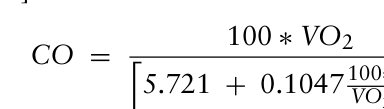
Study 1 Because both HR and VO2 can be easily measured during standard incre- mental cardio-pulmonary exercise testing [Guyton, 2005], both CO and SV could be accurately quantified if the simultaneous arteriovenous O2 content difference (CA – CV) could be estimated [Stringer, 1997; Sullivan, 1989]. For noninvasive CO estimation, exercise tests were performed on an electroni- cally braked cycle ergometer (or treadmill) controlled by computer. Subjects were familiarized with the apparatus and performed a continuous incremen- tal symptom-limited maximal test for determination of VO2max and lactic acidosis threshold (LAT). The samples of expired gas were connected to gas analyzer (O2 and CO2 analyzer). All electrical signals from sensors and from gas analyzer were processed in personal computer. From the measured values workload [W] for cycle ergometer or running velocity [km/h] for tread- mill, HR [beats/min] and VO2 [l/min] the CO and also SV were calculated [Stringer, 1998]. The CO was estimated according formula (4):
Equation reproduced from the original PDF.
i [l/min] (4)
5.721 + 0.1047100∗VO2 VO2MAX
Six subjects (3 men and 3 women) of our data-base with very different athletic background were used to demonstrate the dynamics of CO and SV during standard spiroergometric stress test (Tab. 1). M1 – top-class cross-country skier, M2 – former top-class cross-country skier, recently jogger, M3 – leisure “hobby” athlete, F1 – top-class triathlete, F2 – young competitive swimmer, F3 – leisure “hobby” athlete.
Sub Age He We VO2m VO2mk M1 28 177 70.3 5.75 81.9 M2 49 170 66.7 4.21 63.1 M3 65 166.5 65.2 3.15 39.7 F1 31 168.5 63.2 4.52 71.5 F2 18 171 64.8 4.32 66.7 F3 40 165.5 72 2.69 37.4
Study 2 From our 20-year examination database, we included in this study 641 men (M) and 164 women (F) over 19 years of age who completed a stress test on a bicycle ergometer with a graded load to the maximum. From an extensive database of all persons examined at the Department of Sports Medicine of the University Hospital in Pilsen and at the Department of Sports Medicine of the Medical Faculty of Charles University in Pilsen between 1994 and 2015, the protocols of those examined were selected that provided the necessary data.
208 Novák and Stork
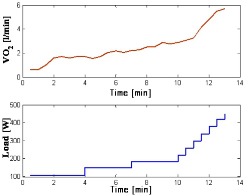
Figure 1: VO2 [l/min] and workloads [in W] during stress-test in M1 (top-class cross- country skier) on cycle ergometer.
During each of these complex sports medical examinations, the examined person underwent a stress test on a bicycle ergometer. It consisted of the workload on the bicycle ergometer gradually increased until exhaustion. The procedure was similar to that of the International Biological Program in the 70s of the last century, which was the basis for determining the normatives of physical fitness of the Czechoslovak population [Seliger, 1976]. These nor- matives are still used in our country, as a similar study has not been carried out since.
The subjects were pedaling on the ergometer 3 times 3 minutes (light, mod- erate and sub-maximum load), followed by increasing the load every half minute to the maximum, ie until exhaustion. At maximum load, at least two of the following criteria must have been met: the age norm for maximal heart rate (HRmax), RER (respiratory exchange ratio) greater than 1.10, maxi- mum ventilatory equivalent for O2 (VEqO2) greater than 30 l and maximum lactate concentration (LAmax) greater than 10 mmol/l. To calculate COmax, we used the same equation like in Study 1: CO = 100 ∗VO2 h
i [l/min]
5.721 + 0.1047100∗VO2 VO2MAX
The files were divided into three groups according to different physical activity: group A included well-trained endurance-athletes, who had several years of sports training at the time of the examination. Group B included subjects who engaged in physical activity only in their leisure time without competing in any sports competition. Subjects with various health problems who either never played any sport at all or ended their sports career many years ago, were included in group C.
Indirectly Determined Maximal Cardiac Output in Men and Women 209
RESULTS
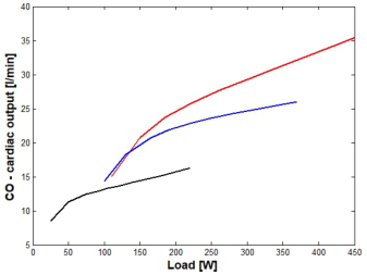
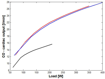
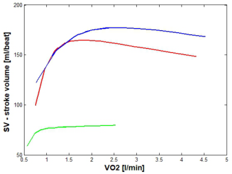
Study 1 The VO2 values, measured during the graded load to the maximum on bicycle ergometer, were used to calculate estimated CO and SV values. Results of those calculations are presented in Fig. 2 for men and in Fig. 3 for women. VO2 isopleths according to Stringer et al. [1997] experience were used to illustrate the difference in highly trained endurance athlete and less trained or even untrained subjects. The data clearly documented the difference in the heart’s pumping efficiency during increasing peripheral tissue needs. Higher cardiac output as a function of higher stroke volume plays important role in increased transporting capacity of blood for oxygen and enables well trained subject to achieve significantly higher physical performance.
Figure 2: CO and SV changes during the stress-test in men. Left: CO related to work- load intensity (Watts) during stress-test. Right: SV related to VO2 during stress-test (red - top class cross-country skier, blue – former top class cross-country skier, now “masters” age group; black (green) - leisure “hobby” athlete).Figure 3: CO and SV changes during the stress-test in women. Left: CO related to workload intensity (Watts) during stress-test. Right: SV related to VO2 during stress- test (blue – top class triathlete, red – young competitive swimmer, F3 – black (green) – leisure time “hobby” athlete).
210 Novák and Stork
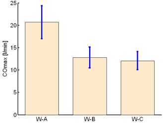
Study 2 COmax in group M-A (trained endurance athletes) (n = 462, COmax = 28.38 ±4.47 l/min) was significantly higher than in groups M-B (leisure-time ath- letes) (n = 143, COmax = 19.4±3.7 l/min) and M-C (untrained subjects) (n = 36, COmax = 18.4±5.9 l/min). It was the same with the women’s groups. COmax value in group F-A (n = 58, COmax = 20.7±3.7 l/min) was significantly higher than that in groups F-B (n = 88, COmax = 12.8±2.3 l/min) and F-C (n = 18, COmax = 12.1±2.0 l/min). There were no significant differences in both M and F between groups C and D.
The same findings were obtained when comparing maximal stroke volume (SVmax) values. SVmax parameters were significantly higher in the group M-A (trained endurance athletes) compared to the groups M-B (recreational athletes) and M-C (untrained subjects).
When comparing SV in the three groups of women, the results were very similar. The significantly highest values of and SVmax were evident in group F-A in comparison with groups F-B and F.C (see Fig. 4 and Fig. 5).
Figure 4: COmax and SVmax values in men with different physical activity (M-A - endurance athletes, M-B - leisure time athletes, M-C - former athletes and subjects with health problems).Figure 5: COmax and SVmax values in women with different physical activity (W-A - endurance athletes, W-B - leisure time athletes, W-C - former athletes and subjects with health problems).
Indirectly Determined Maximal Cardiac Output in Men and Women 211
DISCUSSION
A totally noninvasive determination of CO and SV during exercise would be very useful in healthy subjects as well as in patients with various degrees of cardiac insufficiency [8, 10]. Having estimated CO and corresponding HR, SV can be calculated. This can provide a simple and low-cost assessment of cardiac function in response to exercise.
Although it is generally assumed that CO increases linearly with VO2, the pattern of variation in VO2 and CO as maximal O2 consumption is approached has not been extensively investigated and may vary among indi- viduals. According to Frank-Starling mechanism the amount of blood that the heart pumps works up to a limit of 3 times the resting normal cardiac output. When the peripheral tissues demand excessive amounts of blood flow, the nervous signals increase cardiac output [Guyton, 1979]. Our examples doc- ument that the time course of these changes is similar in the subjects of very different cardio-respiratory fitness level (see Fig. 2 and 3). However, these findings still need to be proved in the groups of subjects of different lifestyle, different athletic background, men and women, and even patients. Top level endurance athletes can reach outstanding values of CO and SV at about 40 l/min and 200 ml/beat respectively [Ekblom, 1968]. Hence, this method seems to offer another useful data to evaluate cardio-respiratory capacity and adaptation to physical activity and/or inactivity.
CONCLUSION
Movement activity is a condition for the development and maintenance of the adaptive abilities of the cardiorespiratory system. The parameters measured in recreational sportsmen correspond to the average of a healthy average fit population. If physical activity is not part of a healthy lifestyle and, in addition, there are health problems (in our groups C obesity and resting and exertional chest pains are the most common), in many subjects from our groups C the COmax parameters are significantly below average and close to the level characterizing the NYHA-1 to NYHA-2 group according to cardiology classification. The results show, on the one hand, how impor- tant factor in health prevention is regular physical activity, and, on the other hand, the applicability of our modified method to determine non-invasively the maximum cardiac output.
CONFLICT OF INTEREST STATEMENT The authors have no conflicts to report relative to this work.
ACKNOWLEDGMENT
This work was supported by the grant of Ministry of Health of the Czech Republic–Conceptual Development of Research Organization (University Hospital Pilsen – FNPl, 00669806), by the Cooperatio Program, research area Immunity and Infection and by Department of Electronics and Informa- tion Technology/RICE, University of West Bohemia, Plzen, Czech Republic and by the Ministry of Education, Youth and Sports of the Czech Republic
212 Novák and Stork
under the project OP VVV Electrical Engineering Technologies with High- Level of Embedded Intelligence, CZ.02.1.01/0.0/0.0/18_069/0009855 and by the Internal Grant Agency of University of West Bohemia in Plzen, the project SGS - 2021- 005.
REFERENCES
Beck, K. C., Randolph, L. N., Bailey, K. R, Wood, C. M, Snyder, E. M., Johnson, B. D. (2006): Relationship between cardiac output and oxygen consumption dur- ing upright cycle exercise in healthy humans. J Appl Physiol 101: 1474–1480 (2006). Costanzo, L. S. (2006): Physiology. Saunders and Company, 3rd edition. Ekblom, B., Hermansen, L. (1968): Cardiac output in athletes. J. Appl. Physiol. 25,
619–625. Fick, A. (1870): Uber die messung des Blutquantums in den Hertzventrikeln. Sitzber Physik Med Ges, Wurzburg, 36, pp. 290–291. Guyton, A. C., Hall, J. E., Lohmeier, T. E., Jackson, T. E. (1979): Modern con-
cepts of circulatory function. In: Hayase, S., Murao, S.: Cardiology. Excer. Med, Amsterdam- Oxford-Princeton. Guyton, A. C., Hall, J. E. (2005): Textbook of Medical Physiology. Saunders and Company, 11th edition. Rowland, T., Obert P. (2002): Doppler echocardiography for the estimation of
cardiac output with exercise. Sports Med. 32 (15): 973–986. Seliger, V., Bart˚unˇek Z. (eds.) Mean values of various indices of physical fitness of Czechoslovak population aged 12–55 years. CSTV, Praha 1976, 117 pp.. Stork, M., Novak, J., Zeman, V. (2010): Noninvasive cardiac output estimation
evaluated from spiroergometric stress test. International Conference on Applied Electronics. Zapadoceska Univerzita, Plzen, pp. 331–334. Stringer, W. W., Hansen, J. E., Wasserman, K. (1997): Cardiac output estimated noninvasively from oxygen uptake during exercise. J. Appl. Physiol. 82(3): 908–912. Sullivan, M. J., Knight, J. D., Higginbotham, M. B., Cobb, F. R. (1989): Relation
between central and peripheral hemodynamics during exercise in patients with chronic heart failure. Circulation 80: 769–781. Wasserman, K., Hansen, J. E., Sue, D. Y., Whipp, B. J. and Casaburi, R. (1999): Principles of exercise testing and interpretation, including pathophysiology and clinical applications, Lippincott Williams and Wilkins, Philadelphia, Penn. White, S. W., Quail, A. W,De Leeuw, P. W,Traugott, F.M., Brown, W. J., Porges, W. L.,
Cottee, D. B. (1990): Impedance cardiography for cardiac output measurement: an evaluation of accuracy and limitations. Europ Heart J, 11, Issue Suppl. 1: 79–92.
LISTED IN THE AHFE DIGITAL BOOKSTORE CATALOGUE
Explore the complete digital volume
Intelligent Human Systems Integration (IHSI 2024): Integrating People and Intelligent Systems
Read this individual research paper free through the original publisher, or explore the full digital proceedings book.
ISBN 978-1-958651-95-7
Explore this ISBN in the AHFE Bookstore ↗Listed in the July 2026 bookstore catalogue; search by ISBN to confirm current availability · Digital book purchase optional