Monica Daniela Gómez Rios, Nestor Raul Martillo Martinez, Miguel Angel Quiroz Martinez, Maikel Yelandi Leyva Vazquez (2021). Tracking Knowledge Evolution, Hotspots and future directions of Breast Cancer Detection using Deep Learning: A bibliometrics Review. In: Human Systems Engineering and Design (IHSED2021): Future Trends and Applications. AHFE Open Access, vol. 21. https://doi.org/10.54941/ahfe1001163
In the medical field, it has been necessary to provide resources to detect ear-ly-stage diseases, including breast cancer. Deep learning is immersed in all aspects of medical image analysis, catapulting it as a possible dominant autonomous technology. In this systematic review, a total of 250 results were located, of which 40 were selected, for which a quantitative methodology with a descriptive basis was chosen. The objective of this bibliometric review is to analyze models in image processing for the early detection of breast cancer using deep learning. As result, digital mammography is the most effective method for detecting abnormalities in images. The research concludes that the application of CNN (Convolutional Neural Networks) is the most preferred choice of experts for medical image analysis due to its powerful pattern recognition and feature classifier.
Breast cancer is generated by a mutation in the genes of the nucleus of cells, which, instead of remaking dead cells by other healthy cells, will generate an exponential and uncontrolled number of equal cells forming a mass of tissue called a tumor (American Cancer Society, s.f.). Although breast cancer is much more common in women, it also occurs in men between the ages of 60 and 70 more often (Sha, 2020). The importance of effective prognosis and treatment of early-stage disease is vital (Li, 2019). The identification of cancer depends on the doctor's interpretation from information obtained from the patients, this information is presented by different detection methods, for example, the processing of medical images. The most successful Artificial Intelligent Architectures for image analysis to date are Convolutional Neural Networks (CNN). In particular, they lead to a notable impact on image examination and understanding, especially segmentation, classification, and image analysis (Litjens, 2017). These approaches are very successful in automating complex tasks, especially in the medicinal field, including diabetes, using technology for drug control, and patient food consumption (Swapna, 2018). In medical imaging, deep learning is used for breast density segmentation and texture scoring and extracting features from the image (Kallenberg, 2016). Automatic detection of breast abnormalities from various imaging modalities is a challenging task due to the variation in size, texture, and contrast of breast abnormalities (Rashed, 2019). To carry out this study, a systematic review is carried out in the main search engines based on different inclusion and exclusion criteria, the goal is to analyze what type of image processing models exist for the early detection of breast cancer using deep learning. The results indicate that digital mammography is the most efficient modality in the detection of anomalies. The research concludes that CNN is the most popular alternative for researchers for the analysis of medical images.
DEEPENING To carry out this analysis, it’s important to know the main components and methods that support the study of images for the early detection of breast cancer.
Starting from the concept of deep learning as the set of computational, algorithmic processes that allow a computerized system to learn by itself and develop its criteria for making and executing decisions (Universidad de Alcalá, s.f.). This technique has the
Human Systems Engineering and Design (IHSED 2021) https://openaccess.cms-conferences.org/#/publications/book/978-1-7923-8987-0 utility of recognizing objects in images, transferring dissertations in writings, selecting relevant results in searches (LeCun, 2015).
Convolutional Layer
Output
Input
Pooling
Fully Connected Layer
Figure. 1. Deep learning algorithm.
The image arrives in the form of an arrangement of pixels; in the first layer, the characteristics that one aspires to learn are the absence or presence of edges and specific locations in the image. The second layer detects particular edge arrangements until the output is passed to a "fully connected" layer that performs the final sorting (Ou, 2021). NN (Neural Networks) are processing systems that consist of a large number of neurons that are organized in layers. CNN consists of adding more layers at the beginning of the network, which will be in charge of manipulating the pixels of the images (Ahmir Malik, s.f.).
RESULTS
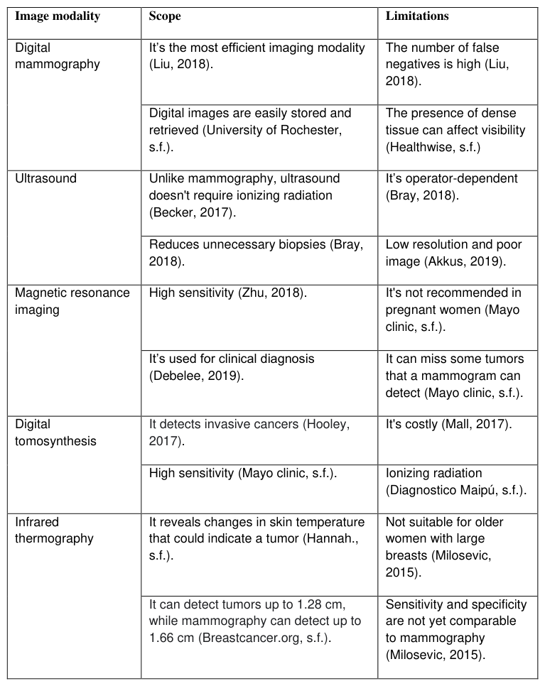
Experts have studied many diagnostic methods, including mammography, MRI, and ultrasound, among others. Next, Table 1 presents the scope and limitations of the different imaging modalities in detecting breast cancer.
Human Systems Engineering and Design (IHSED 2021) https://openaccess.cms-conferences.org/#/publications/book/978-1-7923-8987-0
Human Systems Engineering and Design (IHSED 2021) https://openaccess.cms-conferences.org/#/publications/book/978-1-7923-8987-0
In the following image, you can see the application of deep learning in medical images, establishing the tasks where CNN achieves successful results.
Deep Learning in the analysis of medical images
Classification Location Detection
Deconvolution
Binary labels
Location of
Sampling
object (s)
Combination of
Identification
Avoid
machine learning techniques
Location
omission of suspicious objects
Results
Interpretation
Figure 2. Analysis of medical images through deep learning.
Figure available in the original PDF paper.
Tree important groups were established that make up the elementary phases within the analysis of medical images: the "Classification" of the medical image is the main task in deep learning. Medical imaging normal and abnormal binary labels are set. The "Location" consists of extracting the importance of each object to focus only on the interesting object and discard the noise. Deconvolution restores and corrects degraded data extracted and classified to provide a better description of the analysis, while sampling is responsible for selecting the objects that need to be interpreted. "Detection" refers to the location of one or more target objects that belong to different categories in an image and marking them with a bounding box (Yunchao, 2019).
Below, different CNN architectural models designed over the years are presented.
Table 2. CNN architectures
Human Systems Engineering and Design (IHSED 2021) https://openaccess.cms-conferences.org/#/publications/book/978-1-7923-8987-0
Table 2 illustrates the various architectural models of CNN over the last 23 years; advances and efficiency are reflected as the technology is improved and updated. The number of layers that each algorithm uses for learning stands out. The term top-5 error rate refers to the method of comparing machine learning models, where performance is evaluated or labelled by percentages, the exposed architectures, utilizing contests. The parameters establish the amount of input data (set of images) required for training the models.
CONCLUSIONS
According to the literature reviewed, deep learning has permeated all aspects of the analysis of medical images; the shortcomings are evident, mainly, the number of parameters (samples) that are required for the processing and analysis of medical images. Digital mammography is presented as the best alternative and medical imaging modality for the training of networks and algorithmic processing in deep learning. This study concludes that CNNs were the most popular choice of researchers because it has the best performance among all the architectures reviewed. Given that deep neural architectures are advanced and optimizing their learning methods over the years, reaching lower rates and error rates. It suggested including the exploration of optical imaging, a modality that is under investigation.
Human Systems Engineering and Design (IHSED 2021) https://openaccess.cms-conferences.org/#/publications/book/978-1-7923-8987-0
ACKNOWLEDGMENTS
This work has been supported by the GIIAR research group and the Universidad Politécnica Salesiana.
REFERENCES
Ahmir Malik, M. (s.f.). (2020). Predicción con redes neuronales convolucionales (CNN).
Nebulova. Website: https://www.nebulova.es/blog/redes-neuronales-convolucionales Akkus, Z. C. (2019). A survey of deep-learning applications in ultrasound: Artificial intelligence–powered ul-trasound for improving clinical workflow. Journal of the American College of Radiology. doi:https://doi.org/10.1016/j.jacr.2019.06.004. American Cancer Society. (s.f.). Cáncer de seno. American Cancer Society Website:
https://www.cancer.org/es/cancer/cancer-de-seno American Cancer Society. (s.f.). Estudios por imágenes (Radiología) y cáncer. American Cancer Society Website: https://www.cancer.org/es/tratamiento/como-comprender-su- diagnostico/pruebas/estudios-por-imagenes-y-el-cancer.html Becker, A. M. (2017). Classification of breast cancer from ultrasound imaging using a
generic deep learning analysis software: a pilot study. The British Journal of Radiology.. doi:https://doi.org/10.1259/bjr.20170576. Bray, F. F. (2018). Global cancer sta-tistics 2018: GLOBOCAN estimates of incidence and mortality worldwide for 36 cancers in 185 countries. CA: A Cancer Journal for Clinicians. doi:https://doi.org/10.3322/caac.21492. Breastcancer.org. (s.f.). Thermography. Breast Cancer Website: https://www.breastcancer.org/symptoms/testing/types/thermography. Debelee, T. S. (2019). Survey of deep learning in breast cancer image analysis. Evolving
Systems. doi:https://doi.org/10.1007/s12530-019-09297-2. Diagnostico Maipú. (s.f.). Tomosíntesis: el revolucionario método para la detección precoz delcáncer website:https://www.diagnosticomaipu.com/institucional/novedades/item/769- tomosintesis.html. Diplomado Medico. (s.f.). Diplomado en Ultrasonografía médica.. Ultrasonido de mama.
Website: https://diplomadomedico.com/ultrasonido-de-mama/. Hannah., N. (s.f.). Tomosynthesis vs. other screening methods: Benefits and risks. Website: https://www.medicalnewstoday.com/articles/314780. Healthwise. (s.f.). Detección del cáncer de seno y senos densos. Website:
https://www.cigna.com/individuals-families/health-wellness/hw-en-espanol/temas-de- salud/deteccion-del-cancer-de-seno-y-senos-densos-abp1927 Hooley, R. D. (2017). Unsupervised Deep Learning Applied to Breast Density Segmentation and Mammographic Risk Scoring. IEEE Transactions on Medical Imaging. doi:https://doi.org/10.2214/AJR.16.17127. Kallenberg, M. P.-l. (2016). Advances in Digital Breast Tomosynthesis. American Journal
of Roentgenology. doi:https://doi.org/10.1109/TMI.2016.2532122.
Human Systems Engineering and Design (IHSED 2021) https://openaccess.cms-conferences.org/#/publications/book/978-1-7923-8987-0
LeCun, Y. B. (2015). Deep learning. Nature. doi:https://doi.org/10.1038/nature14539. Li, Y. W. (2019). Classification of Breast Cancer Histology Images Using Multi-Size and Discriminative Patches Based on Deep Learning.. doi:https://doi.org/10.1109/ACCESS.2019.2898044. Litjens, G. K. (2017). A survey on deep learning in medical image anal-ysis. Medical Image
Analysis. https://doi.org/10.1016/j.media.2017.07.005. Liu, J. Z. (2018). (2015). Radiation dose reduction in digital breast tomosynthesis (DBT) by means of deep-learning-based supervised image processing. doi:https://doi.org/10.1117/12.2293125 Mall, S. L.-T. (2017). The role of digital breast tomosynthesis in the breast assessment clinic:
a review. doi:https://doi.org/10.1002/jmrs.230 Mayo clinic. (s.f.). Breast MRI. Website https://www.mayoclinic.org/tests- procedures/breast-mri/about/pac-20384809 Milosevic, M. D. (2015). Comparative analysis of breast cancer detection in mammograms and thermograms. doi:https://doi.org/10.1515/bmt-2014-0047 Ou, W. P. (2021 Deep learning in breast radiology: current progress and future directions.
European Radiology. https://doi.org/10.1007/s00330-020-07640-9. Rashed, E. E. (2019). Deep learning approach for breast cancer diagnosis. Proceedings of the 2019 8th International Conference on Software and Information Engineering. doi:https://doi.org/10.1145/3328833.3328867. Sha, Z. L. (2020). Deep learning and optimization algo-rithms for automatic breast cancer detection. International Journal of Imaging Systems and Technology. doi:https://doi.org/10.1002/ima.22400 Swapna, G. R. (2018). Diabetes detection using deep learning algo-rithms. doi:https://doi.org/10.1016/j.icte.2018.10.005. Universidad de Alcalá, M. (s.f.). Importancia del Deep Learning. Website https://master-
deeplearning.com/importancia-del-deep-learning/ University of Rochester, M. C. (s.f.). Breast imaging. Website https://www.urmc.rochester.edu/highland/departments-centers/breast-imaging/tests- procedures/advantages-digital-mammography.aspx Yunchao, G. J. (2019). Application of Computer Vision and Deep Learning in Breast Cancer Assisted Diagnosis. doi:https://doi.org/10.1145/3310986.3311010 Zhu, Z. H. (2018). Deep learning-based features of breast MRI for prediction of occult
invasive disease following a di-agnosis of ductal carcinoma in situ: preliminary data.. doi:https://doi.org/10.1117/12.2295470.
Human Systems Engineering and Design (IHSED 2021) https://openaccess.cms-conferences.org/#/publications/book/978-1-7923-8987-0
LISTED IN THE AHFE DIGITAL BOOKSTORE CATALOGUE
Explore the complete digital volume
Human Systems Engineering and Design (IHSED2021): Future Trends and Applications
Read this individual research paper free through the original publisher, or explore the full digital proceedings book.
ISBN 978-1-7923-8987-0
Explore this ISBN in the AHFE Bookstore ↗Listed in the July 2026 bookstore catalogue; search by ISBN to confirm current availability · Digital book purchase optional